

The DOI matches its Crossref record; 62 of the 63 references it cites were found in a registry or an index.
Mutations in CFAP43 and CFAP44 cause male infertility and flagellum defects in Trypanosoma and human
Open accessAbstract
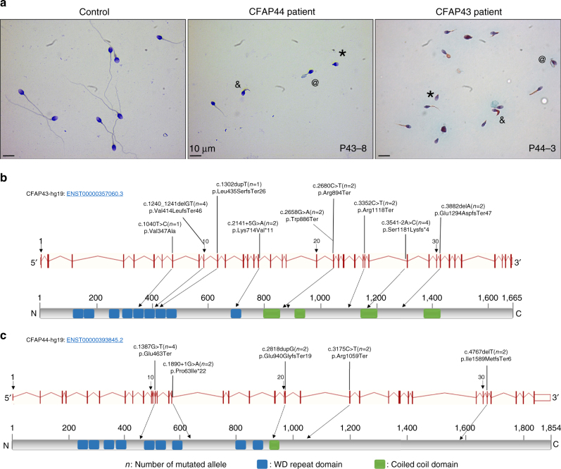
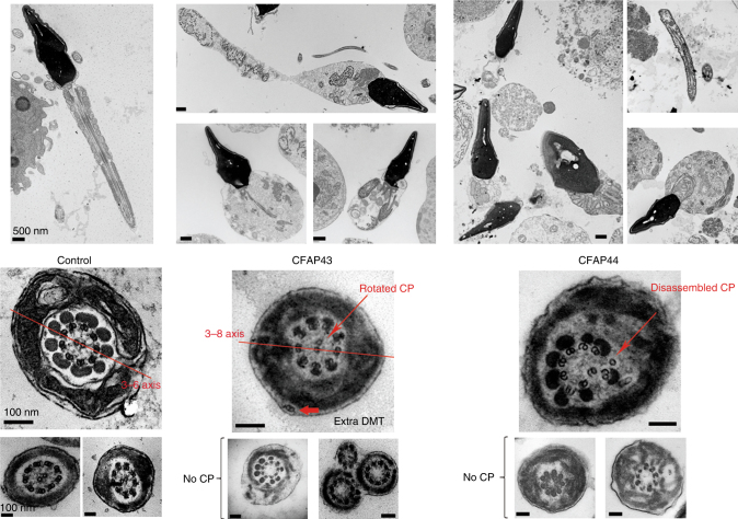
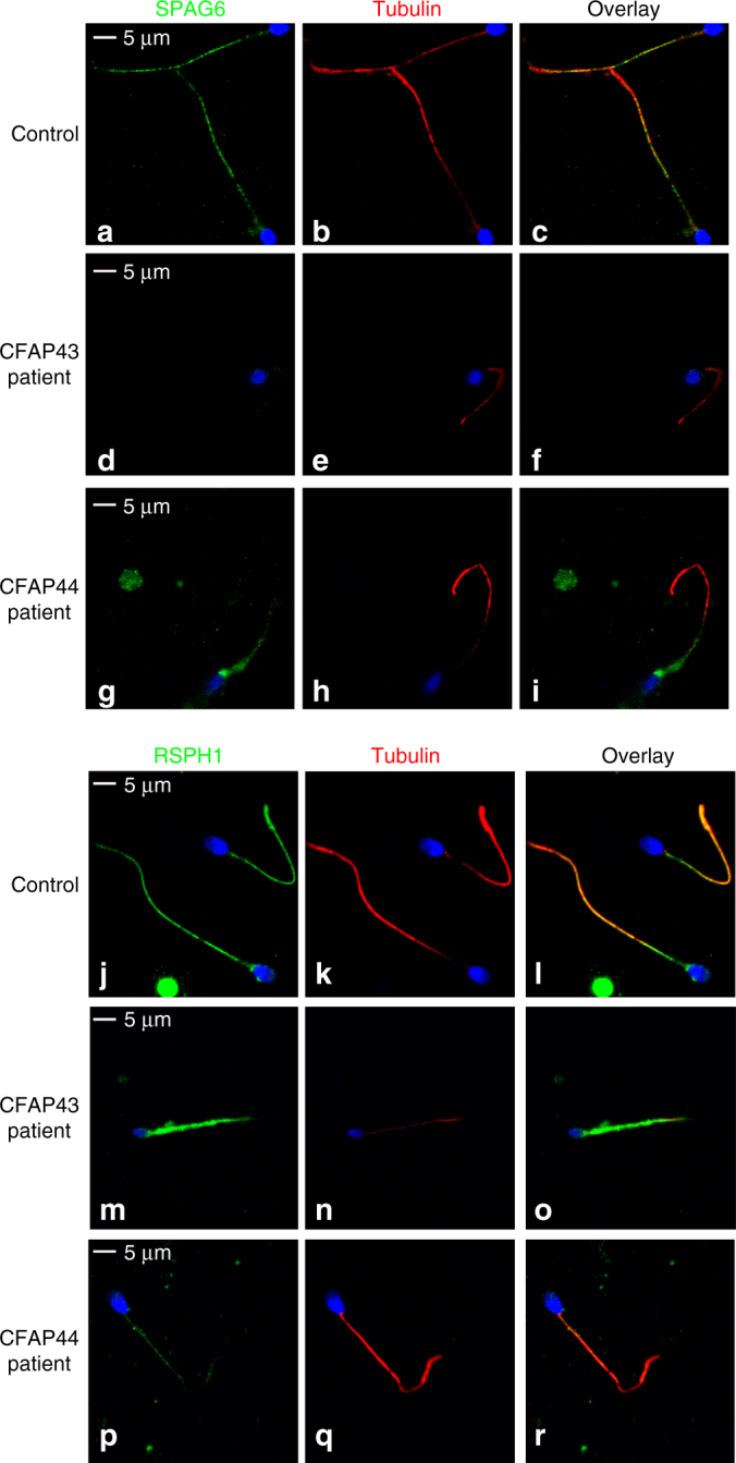
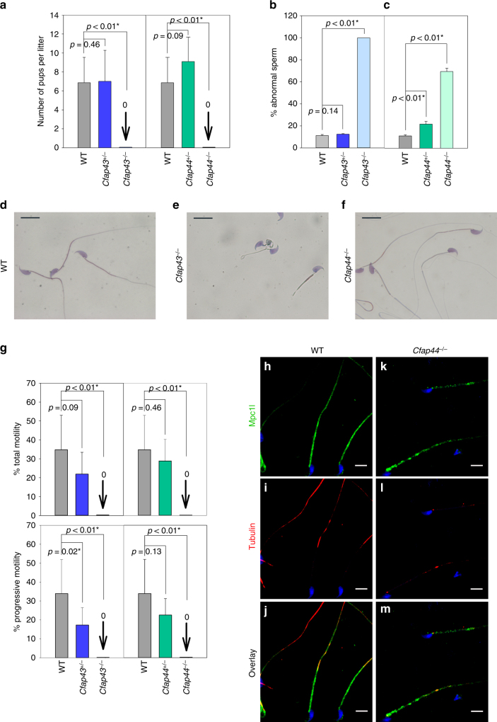
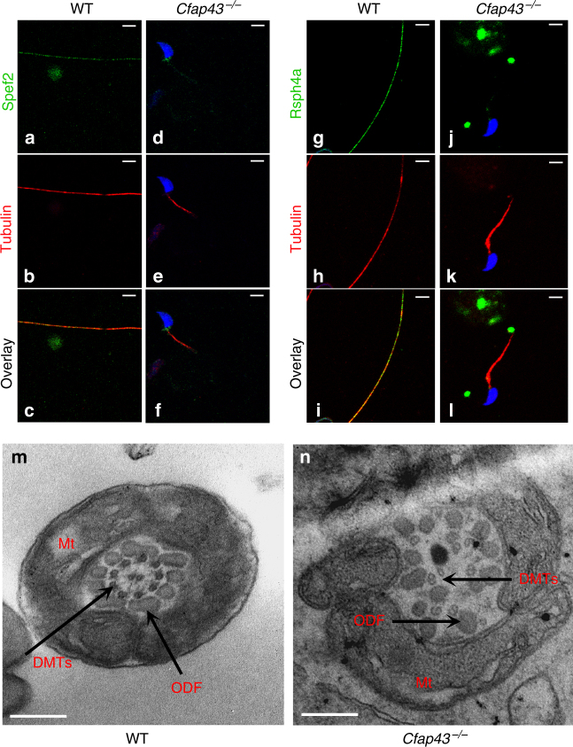
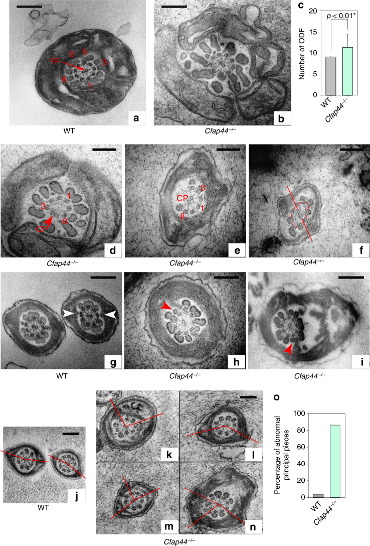
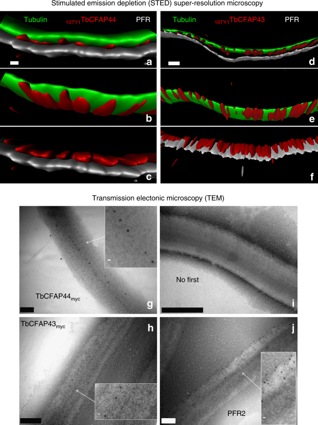
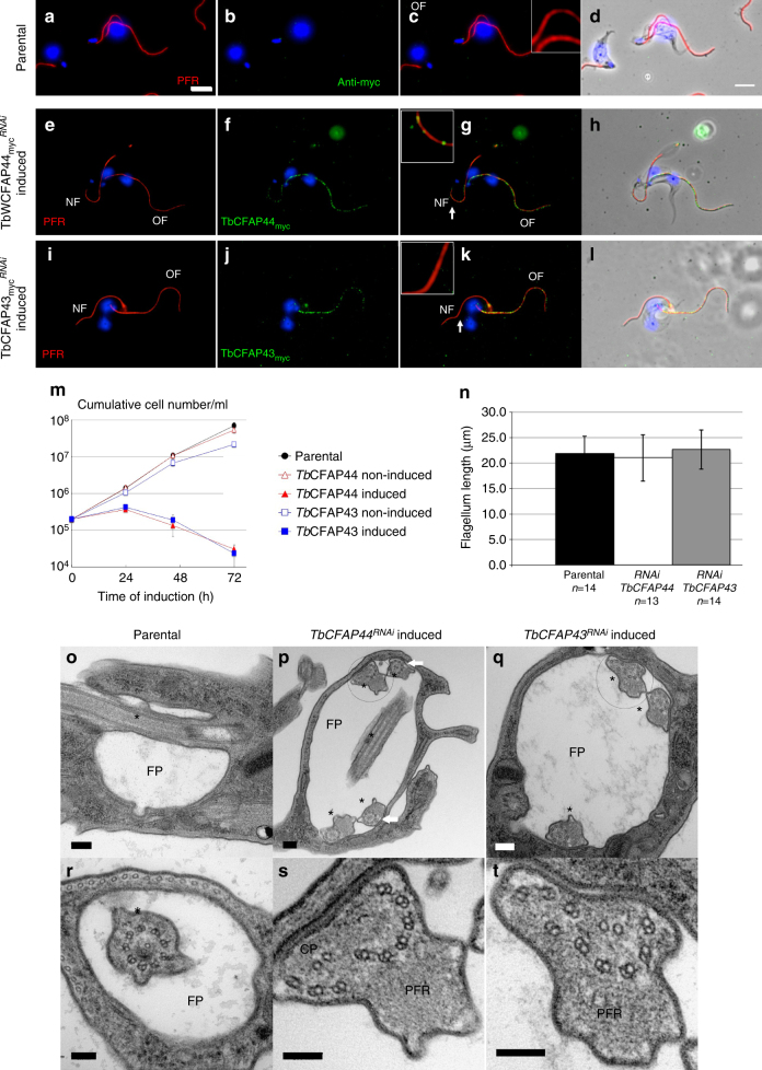
Spermatogenesis defects concern millions of men worldwide, yet the vast majority remains undiagnosed. Here we report men with primary infertility due to multiple morphological abnormalities of the sperm flagella with severe disorganization of the sperm axoneme, a microtubule-based structure highly conserved throughout evolution. Whole-exome sequencing was performed on 78 patients allowing the identification of 22 men with bi-allelic mutations in DNAH1 ( n = 6), CFAP43 ( n = 10), and CFAP44 ( n = 6). CRISPR/Cas9 created homozygous CFAP43 / 44 male mice that were infertile and presented severe flagellar defects confirming the human genetic results. Immunoelectron and stimulated-emission-depletion microscopy performed on CFAP43 and CFAP44 orthologs in Trypanosoma brucei evidenced that both proteins are located between the doublet microtubules 5 and 6 and the paraflagellar rod. Overall, we demonstrate that CFAP43 and CFAP44 have a similar structure with a unique axonemal localization and are necessary to produce functional flagella in species ranging from Trypanosoma to human.
Overview
The claims and statistics of this paper haven’t been checked yet.
Run the check — free, no account needed. It reads the open-access text and checks the sources it cites.
Integrity signals
Registry
Cites
Statistics
Figures
Verification
Claims the paper makes
The claims and statistics of this paper haven’t been checked yet.
Run the check — free, no account needed. It reads the open-access text and checks the sources it cites.
Claims other papers make about this one
Each is checked against this paper’s own text: does it say what the citing paper says it does?
The cited source says what the claim attributes to it.
In the citing paper
“CFAP43 (orthologous to CG17687) is mutated in patients with multiple morphological abnormalities of the sperm flagella (MMAF), and CFAP52 (orthologous to CG10064) is mutated in laterality disorder, a condition caused by defects in ciliary beating during development [45,46].”
From Functional unknomics: Systematic screening of conserved genes of unknown function (2023) · cites this paper as [45]
Self-check
Percentages, sums and reported differences re-computed from the paper’s own numbers.
The self-check needs the paper’s full text.
Citations
Cites
Analysis of Relative Gene Expression Data Using Real-Time Quantitative PCR and the 2−ΔΔCT Method
Livak, K. J., & Schmittgen, T. D. (2001) · Methods · doi:10.1006/meth.2001.1262 · Cited by 180,860
Fast, scalable generation of high‐quality protein multiple sequence alignments using Clustal Omega
Sievers, F., Wilm, A., Dineen, D. G., Gibson, T. J., Karplus, K. J., Li, W., López, R., McWilliam, H., Remmert, M., Söding, J., et al. (2011) · Molecular Systems Biology · doi:10.1038/msb.2011.75 · Cited by 15,426
A method and server for predicting damaging missense mutations.
Adzhubei, I. A., Schmidt, S., Peshkin, L., Ramensky, V. E., Gerasimova, A., Bork, P., Kondrashov, A. S., & Sunyaev, S. R. (2010) · Nature Methods · doi:10.1038/nmeth0410-248 · Cited by 13,194
Genome engineering using the CRISPR-Cas9 system
Ran, F. A., Hsu, P. D., Wright, J., Agarwala, V., Scott, D. A., & Zhang, F. (2013) · Nature Protocols · doi:10.1038/nprot.2013.143 · Cited by 11,133
Analysis of protein-coding genetic variation in 60,706 humans
Exome Aggregation Consortium, Lek, M., Karczewski, K. J., Minikel, E. V., Samocha, K. E., Banks, E., Fennell, T., O’Donnell-Luria, A. H., Ware, J. S., Hill, A. J., et al. (2016) · Nature · doi:10.1038/nature19057 · Cited by 9,587
The Ensembl Variant Effect Predictor
McLaren, W., Gil, L., Hunt, S. E., Riat, H. S., Ritchie, G. R. S., Thormann, A., Flicek, P., & Cunningham, F. (2016) · Genome Biology · doi:10.1186/s13059-016-0974-4 · Cited by 8,099
An integrated map of genetic variation from 1,092 human genomes
The 1000 Genomes Project Consortium (2012) · Nature · doi:10.1038/nature11632 · Cited by 7,711
Kumar, P., Henikoff, S., & Ng, P. C. (2009) · Nature Protocols · doi:10.1038/nprot.2009.86 · Cited by 6,521
Murzin, A. G., Brenner, S. E., Hubbard, T., & Chothia, C. (1995) · Journal of Molecular Biology · doi:10.1016/s0022-2836(05)80134-2 · Cited by 6,445
Mutation of the mouse klotho gene leads to a syndrome resembling ageing
Kuro-o, M., Matsumura, Y., Aizawa, H., Kawaguchi, H., Suga, T., Utsugi, T., Ohyama, Y., Kurabayashi, M., Kaname, T., Kume, E., et al. (1997) · Nature · doi:10.1038/36285 · Cited by 3,837
Wirtz, E., Leal, S., Ochatt, C. M., & Cross, G. (1999) · Molecular and Biochemical Parasitology · doi:10.1016/s0166-6851(99)00002-x · Cited by 1,381
Gough, J., Karplus, K. J., Hughey, R., & Chothia, C. (2001) · Journal of Molecular Biology · doi:10.1006/jmbi.2001.5080 · Cited by 1,200
TriTrypDB: a functional genomic resource for the Trypanosomatidae
Aslett, M. A., Aurrecoechea, C., Berriman, M., Brestelli, J. E., Brunk, B. P., Carrington, M., Depledge, D. P., Fischer, S., Gajria, B., Gao, X., et al. (2009) · Nucleic Acids Research · doi:10.1093/nar/gkp851 · Cited by 988
SEQC/MAQC-III Consortium (2014) · Nature Biotechnology · doi:10.1038/nbt.2957 · Cited by 983
Fawcett, D. W. (1975) · Developmental Biology · doi:10.1016/0012-1606(75)90411-x · Cited by 846
Woods, A., Sherwin, T., Sasse, R. Z., MacRae, T. H., Baines, A. J., & Keith Gull, B. (1989) · Journal of Cell Science · doi:10.1242/jcs.93.3.491 · Cited by 662
Electron Microscopy of the Sperm Tail Results Obtained with a New Fixative
Afzelius, B. A. (1959) · The Journal of Cell Biology · doi:10.1083/jcb.5.2.269 · Cited by 499
SUPERFAMILY—sophisticated comparative genomics, data mining, visualization and phylogeny
Wilson, D. A., Pethica, R., Zhou, Y., Talbot, C. C., Vogel, C., Madera, M., Chothia, C., & Gough, J. (2008) · Nucleic Acids Research · doi:10.1093/nar/gkn762 · Cited by 493
Flagellar motility is required for the viability of the bloodstream trypanosome
Broadhead, R., Dawe, H. R., Farr, H., Griffiths, S. J., Hart, S. R., Portman, N., Shaw, M. K., Ginger, M. L., Gaskell, S. J., McKean, P. G., et al. (2006) · Nature · doi:10.1038/nature04541 · Cited by 488
Ben Khelifa, M., Coutton, C., Zouari, R., Karaouzène, T., Rendu, J., Bidart, M., Yassine, S., Pierre, V., Delaroche, J., Hennebicq, S., et al. (2014) · The American Journal of Human Genetics · doi:10.1016/j.ajhg.2013.11.017 · Cited by 474
Self-citationSherwin, T., & Gull, K. (1989) · Philosophical transactions of the Royal Society of London. Series B, Biological sciences · doi:10.1098/rstb.1989.0037 · Cited by 352
Teratozoospermia: spotlight on the main genetic actors in the human
Coutton, C., Escoffier, J., Martinez, G., Arnoult, C., & Ray, P. F. (2015) · Human Reproduction Update · doi:10.1093/humupd/dmv020 · Cited by 352
Self-citationA novel epitope tag system to study protein targeting and organelle biogenesis in Trypanosoma brucei
Bastin, P., Bagherzadeh, A., Matthews, K. R., & Keith Gull, B. (1996) · Molecular and Biochemical Parasitology · doi:10.1016/0166-6851(96)02598-4 · Cited by 319
Assembly and Motility of Eukaryotic Cilia and Flagella. Lessons from Chlamydomonas reinhardtii
Silflow, C. D., & Lefebvre, P. A. (2001) · PLANT PHYSIOLOGY · doi:10.1104/pp.127.4.1500 · Cited by 246
A toolkit enabling efficient, scalable and reproducible gene tagging in trypanosomatids
Dean, S., Sunter, J. D., Wheeler, R. J., Hodkinson, I., Gluenz, E., & Keith Gull, B. (2015) · Open Biology · doi:10.1098/rsob.140197 · Cited by 243
Male infertility caused by spermiogenic defects: Lessons from gene knockouts
Yan, W. (2009) · Molecular and Cellular Endocrinology · doi:10.1016/j.mce.2009.03.003 · Cited by 207
A vector series for rapid PCR-mediated C-terminal in situ tagging of Trypanosoma brucei genes
Oberholzer, M., Morand, S., Kunz, S., & Seebeck, T. (2005) · Molecular and Biochemical Parasitology · doi:10.1016/j.molbiopara.2005.09.002 · Cited by 206
Motility and more: the flagellum of Trypanosoma brucei
Langousis, G., & Hill, K. L. (2014) · Nature Reviews Microbiology · doi:10.1038/nrmicro3274 · Cited by 205
Paraflagellar rod is vital for trypanosome motility
Bastin, P., Sherwin, T., & Keith Gull, B. (1998) · Nature · doi:10.1038/35300 · Cited by 198
Trypanosoma brucei FLA1 Is Required for Flagellum Attachment and Cytokinesis
LaCount, D. J., Barrett, B., & Donelson, J. E. (2002) · Journal of Biological Chemistry · doi:10.1074/jbc.m200873200 · Cited by 186
Genetic abnormalities leading to qualitative defects of sperm morphology or function
Ray, P. F., Toure, A., Metzler‐Guillemain, C., Mitchell, M. J., Arnoult, C., & Coutton, C. (2017) · Clinical Genetics · doi:10.1111/cge.12905 · Cited by 179
Self-citationNeesen, J. (2001) · Human Molecular Genetics · doi:10.1093/hmg/10.11.1117 · Cited by 176
1001 model organisms to study cilia and flagella
Vincensini, L., Blisnick, T., & Bastin, P. (2011) · Biology of the Cell · doi:10.1042/bc20100104 · Cited by 169
Diversity of WD-Repeat proteins
Smith, T. F. (2008) · Sub-cellular biochemistry/Subcellular biochemistry · doi:10.1007/978-0-387-09595-0_3 · Cited by 163
Subota, I., Julkowska, D., Vincensini, L., Reeg, N., Buisson, J., Blisnick, T., Huet, D., Perrot, S., Santi‐Rocca, J., Duchateau, M., et al. (2014) · Molecular & Cellular Proteomics · doi:10.1074/mcp.m113.033357 · Cited by 138
Kherraf, Z., Christou‐Kent, M., Karaouzène, T., Amiri‐Yekta, A., Martinez, G., Vargas, A., Lambert, E., Borel, C., Dorphin, B., Aknin-Seifer, I. E., et al. (2017) · EMBO Molecular Medicine · doi:10.15252/emmm.201607461 · Cited by 131
Self-citationAmiri‐Yekta, A., Coutton, C., Kherraf, Z., Karaouzène, T., Tanno, P. L., Sanati, M. H., Sabbaghian, M., Almadani, N., Gilani, M. A. S., Hosseini, S., et al. (2016) · Human Reproduction · doi:10.1093/humrep/dew262 · Cited by 123
Self-citationMirabello, C., & Pollastri, G. (2013) · Bioinformatics · doi:10.1093/bioinformatics/btt344 · Cited by 122
Wambergue, C., Zouari, R., Mustapha, S. F. B., Martinez, G., Devillard, F., Hennebicq, S., Satre, V., Brouillet, S., Halouani, L., Marrakchi, O., et al. (2016) · Human Reproduction · doi:10.1093/humrep/dew083 · Cited by 118
Self-citationSantrich, C., Moore, L. L., Sherwin, T., Bastin, P., Brokaw, C. J., Keith Gull, B., & LeBowitz, J. H. (1997) · Molecular and Biochemical Parasitology · doi:10.1016/s0166-6851(97)00149-7 · Cited by 112
Baron, D. M., Ralston, K. S., Kabututu, Z. P., & Hill, K. L. (2007) · Journal of Cell Science · doi:10.1242/jcs.03352 · Cited by 99
Sha, Y., Yang, X., Mei, L., Ji, Z., Wang, X., Ding, L., Li, P., & Yang, S. (2017) · Fertility and Sterility · doi:10.1016/j.fertnstert.2017.04.007 · Cited by 97
Escoffier, J., Yassine, S., Lee, H. C., Martinez, G., Delaroche, J., Coutton, C., Karaouzène, T., Zouari, R., Metzler‐Guillemain, C., Pernet‐Gallay, K., et al. (2014) · Molecular Human Reproduction · doi:10.1093/molehr/gau098 · Cited by 97
Self-citationLindemann, C. B., Orlando, A., & Kanous, K. S. (1992) · Journal of Cell Science · doi:10.1242/jcs.102.2.249 · Cited by 95
Wang, X., Jin, H., Han, F., Cui, Y., Chen, J., Yang, C., Zhu, P., Wang, W., Jiao, G., Wang, W., et al. (2016) · Clinical Genetics · doi:10.1111/cge.12857 · Cited by 94
Exploring the Transcriptome of Ciliated Cells Using In Silico Dissection of Human Tissues
Ivliev, A. E., Hoen, P. A. C. ’., van Roon‐Mom, W., Peters, D. J. M., & Sergeeva, M. G. (2012) · PLoS ONE · doi:10.1371/journal.pone.0035618 · Cited by 89
Selection of reference genes for mRNA quantification in Trypanosoma brucei
Brenndörfer, M., & Boshart, M. (2010) · Molecular and Biochemical Parasitology · doi:10.1016/j.molbiopara.2010.03.007 · Cited by 86
Unique among ciliopathies: primary ciliary dyskinesia, a motile cilia disorder
Praveen, K., Davis, E. E., & Katsanis, N. (2015) · F1000Prime Reports · doi:10.12703/p7-36 · Cited by 80
The GTPase IFT27 is involved in both anterograde and retrograde intraflagellar transport
Huet, D., Blisnick, T., Perrot, S., & Bastin, P. (2014) · eLife · doi:10.7554/elife.02419 · Cited by 76
Protein Targeting of an Unusual, Evolutionarily Conserved Adenylate Kinase to a Eukaryotic Flagellum
Pullen, T. J., Ginger, M. L., Gaskell, S. J., & Keith Gull, B. (2004) · Molecular Biology of the Cell · doi:10.1091/mbc.e04-03-0217 · Cited by 74
Structure of Trypanosoma brucei flagellum accounts for its bihelical motion
Koyfman, A. Y., Schmid, M. F., Gheiratmand, L., Fu, C., Khant, H. A., Huang, D., He, C. Y., & Chiu, W. (2011) · Proceedings of the National Academy of Sciences · doi:10.1073/pnas.1103634108 · Cited by 69
The Paraxial Structure of the Flagellum of Trypanosomatidae
de Souza, W., Souto-Padrón, T. C. B. S., & Souto-Padrón, T. C. B. S. (1980) · Journal of Parasitology · doi:10.2307/3280809 · Cited by 69
Lin, J., Heuser, T., Song, K., Fu, X., & Nicastro, D. (2012) · PLoS ONE · doi:10.1371/journal.pone.0046494 · Cited by 60
Morga, B., & Bastin, P. (2013) · Cilia · doi:10.1186/2046-2530-2-16 · Cited by 54
Vanderperre, B., Cermakova, K., Escoffier, J., Kaba, M., Bender, T., Nef, S., & Martinou, J. (2016) · Journal of Biological Chemistry · doi:10.1074/jbc.m116.733840 · Cited by 52
Self-citationCilia and Models for Studying Structure and Function
Ostrowski, L. E., Dutcher, S. K., & Lo, C. W. (2011) · Proceedings of the American Thoracic Society · doi:10.1513/pats.201103-027sd · Cited by 46
Male Infertility: Genetics, Mechanism, and Therapies
Coutton, C., Fissore, R. A., Palermo, G. D., Stouffs, K., & Touré, A. (2016) · BioMed Research International · doi:10.1155/2016/7372362 · Cited by 46
Self-citationAlbisetti, A., Florimond, C., Landrein, N., Vidilaseris, K., Eggenspieler, M., Lesigang, J., Dong, G., Robinson, D. R., & Bonhivers, M. (2017) · PLoS Pathogens · doi:10.1371/journal.ppat.1006710 · Cited by 42
Self-citationTrypanosomes and mammalian sperm: one of a kind?
Oberholzer, M., Bregy, P., Marti, G., Minca, M., Peier, M., & Seebeck, T. (2006) · Trends in Parasitology · doi:10.1016/j.pt.2006.12.002 · Cited by 41
Ralston, K. S., Kisalu, N. K., & Hill, K. L. (2011) · Eukaryotic Cell · doi:10.1128/ec.00298-10 · Cited by 41
Calmodulin-binding properties of the paraflagellar rod complex from Trypanosoma brucei
Ridgley, E. L., Webster, P. C., Patton, C. L., & Ruben, L. (2000) · Molecular and Biochemical Parasitology · doi:10.1016/s0166-6851(00)00246-2 · Cited by 40
Trypanosoma brucei CYP51: Essentiality and Targeting Therapy in an Experimental Model
Dauchy, F., Bonhivers, M., Landrein, N., Dacheux, D., Courtois, P., Lauruol, F., Daulouède, S., Vincendeau, P., & Robinson, D. R. (2016) · PLoS neglected tropical diseases · doi:10.1371/journal.pntd.0005125 · Cited by 32
Self-citation
Research works printed but not linked: 1
- Coutton, C., Escoffier, J., Martinez, G., Arnoult, C. & Ray, P. F. Teratozoospermia: spotlight on the main genetic actors in the human. Hum. Reprod. Update 21, 455–485 (2015). · DOI · Paper page
Cited by
Sorted by citations
Wallmeier, J., Nielsen, K. G., Kuehni, C. E., Lucas, J. S., Leigh, M. W., Zariwala, M. A., & Omran, H. (2020) · Nature Reviews Disease Primers · doi:10.1038/s41572-020-0209-6 · Cited by 403
Jiao, S.-Y., Yang, Y.-H., & Chen, S. (2020) · Human Reproduction Update · doi:10.1093/humupd/dmaa034 · Cited by 286
Sperm defects in primary ciliary dyskinesia and related causes of male infertility
Sironen, A. I., Shoemark, A., Patel, M., Loebinger, M. R., & Mitchison, H. M. (2019) · Cellular and Molecular Life Sciences · doi:10.1007/s00018-019-03389-7 · Cited by 255
The genetic architecture of morphological abnormalities of the sperm tail
Touré, A., Martinez, G., Kherraf, Z., Cazin, C., Beurois, J., Arnoult, C., Ray, P. F., & Coutton, C. (2020) · Human Genetics · doi:10.1007/s00439-020-02113-x · Cited by 235
Self-citationLiu, C., Miyata, H., Gao, Y., Sha, Y., Tang, S., Xu, Z., Whitfield, M., Patrat, C., Wu, H., Dulioust, E., et al. (2020) · The American Journal of Human Genetics · doi:10.1016/j.ajhg.2020.06.004 · Cited by 191
Self-citation
Metadata
- Authors
- Coutton, C., Vargas, A. S., Amiri-Yekta, A., Kherraf, Z.-E., Ben Mustapha, S. F., Le Tanno, P., Wambergue-Legrand, C., Karaouzène, T., Martinez, G., Crouzy, S., Daneshipour, A., Hosseini, S. H., Mitchell, V., Halouani, L., Marrakchi, O., Makni, M., Latrous, H., Kharouf, M., Deleuze, J.-F., Boland, A., Hennebicq, S., Satre, V., Jouk, P.-S., Thierry-Mieg, N., Conne, B., Dacheux, D., Landrein, N., Schmitt, A., Stouvenel, L., Lorès, P., El Khouri, E., Bottari, S. P., Fauré, J., Wolf, J.-P., Pernet-Gallay, K., Escoffier, J., Gourabi, H., Robinson, D. R., Nef, S., Dulioust, E., Zouari, R., Bonhivers, M., Touré, A., Arnoult, C., & Ray, P. F.
- Affiliations
- Centre National de la Recherche Scientifique (FR)
- Royan Institute (IR)
- Commissariat à l'Énergie Atomique et aux Énergies Alternatives (FR)
- Centre Hospitalier Universitaire de Grenoble (FR)
- Inserm (FR)
- Sorbonne Paris Cité (FR)
- University of Geneva (CH)
- Corresponding author
- Ray, Pierre F.
- ORCID
- Coutton, Charles
- Vargas, Alexandra S.
- Amiri-Yekta, Amir
- Kherraf, Zine-Eddine
- Martinez, Guillaume
- Crouzy, Serge
- Hosseini, Seyedeh Hanieh
- Deleuze, Jean-François
- Boland, Anne
- Hennebicq, Sylviane
- Satre, Véronique
- Jouk, Pierre-Simon
- Dacheux, Denis
- Landrein, Nicolas
- Schmitt, Alain
- Lorès, Patrick
- Bottari, Serge P.
- Fauré, Julien
- Wolf, Jean-Philippe
- Escoffier, Jessica
- Gourabi, Hamid
- Robinson, Derrick R.
- Nef, Serge
- Bonhivers, Mélanie
- Touré, Aminata
- Arnoult, Christophe
- Ray, Pierre F.
- Venue
- Nature Communications
- Published
- 2018-02-15
- Volume
- 9
- Issue
- 1
- Pages
- 686-686
- ISSN
- 2041-1723 (ISSN-L)
- PMC
- PMC5814398
- OpenAlex
- W2806261901
- Cited by
- 245
- References
- 63
- Citations by year
- 2026: 22 · 2025: 19 · 2024: 30 · 2023: 34 · 2022: 35 · 2021: 27 · 2020: 31 · 2019: 29 · 2018: 18
- FWCI
- 28.16
- Type
- journal article
- Language
- en
- Open access
- gold
- doi.org
- Locations
- Nature Communications (publishedVersion, cc-by)
- PubMed Central (publishedVersion, cc-by)
- PubMed (publishedVersion)
- Archive ouverte UNIGE (University of Geneva) (publishedVersion, other-oa)
- HAL (submittedVersion, cc-by)
- Oskar-Bordeaux (Universite de Bordeaux) (submittedVersion, cc-by)
- RePEc: Research Papers in Economics (submittedVersion)
- DOAJ (DOAJ: Directory of Open Access Journals) (submittedVersion, cc-by-sa)
- DOAJ (DOAJ: Directory of Open Access Journals) (submittedVersion)
- Archive ouverte UNIGE (University of Geneva) (submittedVersion, cc-by)
- Greater South Information System
- Funders
- Fondation Maladies Rares
- University of Oxford
- Agence Nationale de la Recherche (ANR-15-CE17-0008, 10-INBS-04, ANR‐10_INBS‐04, ANR-11-LABX-0024, ANR-10, ANR-10-INBS-0004, ANR-14-CE15-0002)
- Institut National de la Santé et de la Recherche Médicale (ANR-10-INBS-04)
- Centre National de la Recherche Scientifique (ANR-10-INBS-04)
- Université de Genève
- Direction Générale de l’offre de Soins (PRTS 2014)
- Topics
- Health Sciences › Medicine › Reproductive Medicine › Sperm and Testicular Function
- Health Sciences › Medicine › Pulmonary and Respiratory Medicine › Cystic Fibrosis Research Advances
- Life Sciences › Biochemistry, Genetics and Molecular Biology › Genetics › Genetic and Kidney Cyst Diseases
- Keywords
- DNAH1, male infertility, axoneme, paraflagellar rod, doublet microtubules, spermatogenesis, Trypanosoma brucei, axonemal proteins, CRISPR/Cas9 mouse model
- MeSH terms
- Exome Sequencing, Adult, Animals, Cytoskeletal Proteins, Fertility, Flagella, Homozygote, Humans, Infertility, Male, Male, Microtubule Proteins, Middle Aged, Mutation, Nuclear Proteins, Peptide Hydrolases, Sperm Motility, Spermatozoa, Trypanosoma, Cohort Studies, Microscopy, Immunoelectron, Mice, Knockout, Mice, Axoneme, Clustered Regularly Interspaced Short Palindromic Repeats
About these checks
Registry, retraction, Cites and statistics results are computed, not judged, with one exception: a printed reference that no index lists is read once by a model before it counts as a citation error. Claims are extracted by a model and kept only when their quote appears verbatim in the text; each cited source is then checked, with a model’s help, for whether it says what the claim relies on. Those verdicts can be wrong — read the claim’s quote and the cited source before relying on one.
Statistics and arithmetic are recomputed, not judged.
Confidence bands are shown only for fields calibrated on our annotated test set. None is calibrated yet, so no band is shown.
What counts as a claim ›How we measure confidence ›How we check this ›
Are you an author, or do you see a problem with this page? Request a review
