

The DOI matches its Crossref record; 73 of the 73 references it cites were found in a registry or an index.
Non-coding variability at the APOE locus contributes to the Alzheimer’s risk
Open accessAbstract
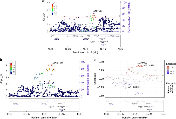
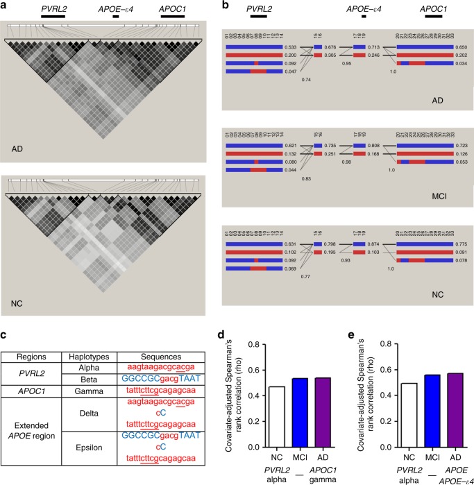
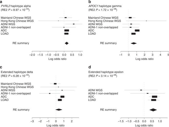
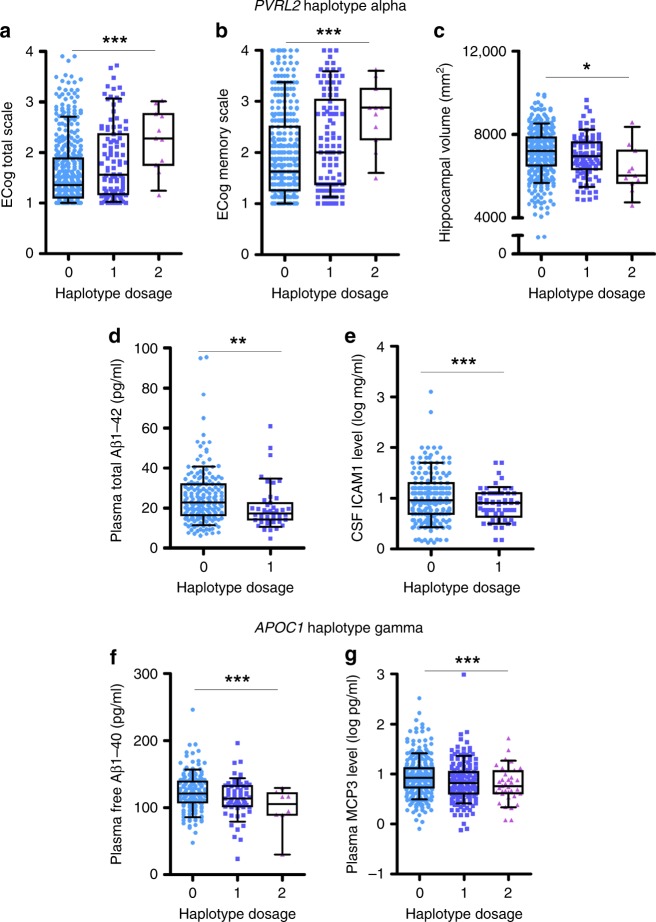
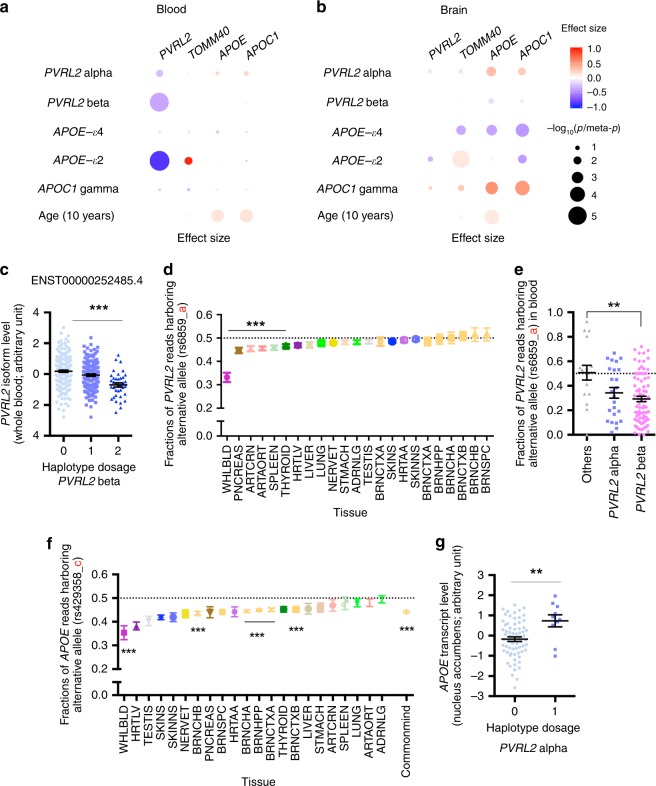
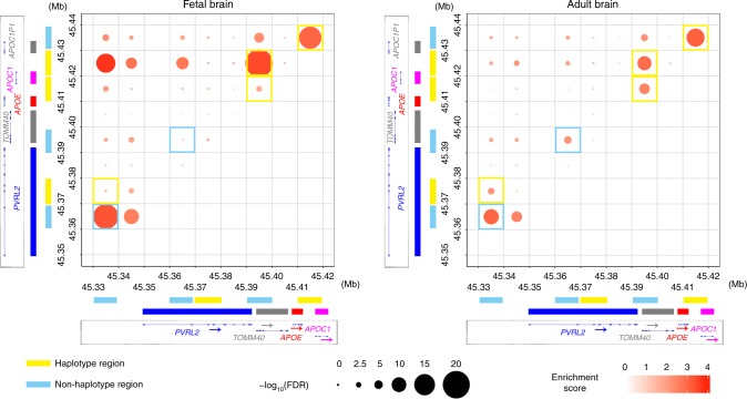
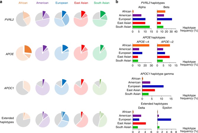
Alzheimer's disease (AD) is a leading cause of mortality in the elderly. While the coding change of APOE-ε4 is a key risk factor for late-onset AD and has been believed to be the only risk factor in the APOE locus, it does not fully explain the risk effect conferred by the locus. Here, we report the identification of AD causal variants in PVRL2 and APOC1 regions in proximity to APOE and define common risk haplotypes independent of APOE-ε4 coding change. These risk haplotypes are associated with changes of AD-related endophenotypes including cognitive performance, and altered expression of APOE and its nearby genes in the human brain and blood. High-throughput genome-wide chromosome conformation capture analysis further supports the roles of these risk haplotypes in modulating chromatin states and gene expression in the brain. Our findings provide compelling evidence for additional risk factors in the APOE locus that contribute to AD pathogenesis.
Overview
The claims and statistics of this paper haven’t been checked yet.
Run the check — free, no account needed. It reads the open-access text and checks the sources it cites.
Integrity signals
Registry
Cites
Statistics
Figures
Verification
Claims the paper makes
The claims and statistics of this paper haven’t been checked yet.
Run the check — free, no account needed. It reads the open-access text and checks the sources it cites.
Claims other papers make about this one
Each is checked against this paper’s own text: does it say what the citing paper says it does?
The cited source says what the claim attributes to it.
In the citing paper
“Meanwhile, a haplotype analysis in the APOE locus identified risk haplotypes that are independent of the APOE ɛ4 allele [48].”
From Association of SPI1 haplotypes with altered SPI1 gene expression and Alzheimer’s disease risk (2022) · cites this paper as [48]
The cited source says what the claim attributes to it.
In the citing paper
“It is also supported by APOE4 risk modifying variants identified in APOE local admixture backgrounds in Hispanics and African Americans (Rajabli et al., 2018) and by APOE expression modifiers such as variants in PVRL2 and APOC1 loci (Zhou et al., 2019).”
From Cholesterol and matrisome pathways dysregulated in astrocytes and microglia (2022) · cites this paper as Zhou et al., 2019
The cited source says what the claim attributes to it.
In the citing paper
“The ApoC1 gamma haplotype has also been found to increase AD risk independent of ApoE [135].”
From ApoE lipoproteins in the central nervous system under homeostasis and role in Alzheimer’s disease and related disorders (2026) · cites this paper as [135]
The cited source says what the claim attributes to it.
In the citing paper
“emerging evidence suggests that APOC1 may exert independent effects on AD pathogenesis [12].”
From Association of APOC1 with cortical atrophy during conversion to Alzheimer’s disease (2025) · cites this paper as [12]
The cited source says what the claim attributes to it.
In the citing paper
“and have been reported in previous studies to exhibit strong associations with AD risk [4, 12, 77–80].”
From Association of APOC1 with cortical atrophy during conversion to Alzheimer’s disease (2025) · cites this paper as [12]
Self-check
Percentages, sums and reported differences re-computed from the paper’s own numbers.
The self-check needs the paper’s full text.
Citations
Cites
Diagnostic and Statistical Manual of Mental Disorders
American Psychiatric Association (2013) · doi:10.1176/appi.books.9780890425596 · Cited by 116,369
Fast gapped-read alignment with Bowtie 2
Langmead, B., & Salzberg, S. L. (2012) · Nature Methods · doi:10.1038/nmeth.1923 · Cited by 56,400
PLINK: a tool set for whole-genome association and population-based linkage analyses.
Purcell, S., Neale, B., Todd-Brown, K., Thomas, L., Ferreira, M. A. R., Bender, D., Maller, J., Sklar, P., de Bakker, P. I. W., Daly, M. J., et al. (2007) · The American Journal of Human Genetics · doi:10.1086/519795 · Cited by 34,089
McKhann, G., Drachman, D., Folstein, M., Katzman, R., Price, D., & Stadlan, E. M. (1984) · Neurology · doi:10.1212/wnl.34.7.939 · Cited by 27,915
The Genome Analysis Toolkit: A MapReduce framework for analyzing next-generation DNA sequencing data
McKenna, A., Hanna, M., Banks, E., Sivachenko, A., Cibulskis, K., Kernytsky, A., Garimella, K., Altshuler, D., Gabriel, S., Daly, M., et al. (2010) · Genome Research · doi:10.1101/gr.107524.110 · Cited by 27,895
The Montreal Cognitive Assessment, MoCA: a brief screening tool for mild cognitive impairment
Nasreddine, Z. S., Phillips, N. A., Bédirian, V., Charbonneau, S., Whitehead, V., Collin, I., Cummings, J. L., & Chertkow, H. (2005) · Journal of the American Geriatrics Society · doi:10.1111/j.1532-5415.2005.53221.x · Cited by 25,553
Conducting Meta-Analyses in R with the metafor Package
Viechtbauer, W. (2010) · Journal of Statistical Software · doi:10.18637/jss.v036.i03 · Cited by 20,464
McKhann, G. M., Knopman, D. S., Chertkow, H., Hyman, B. T., Jack, C. R., Kawas, C. H., Klunk, W. E., Koroshetz, W. J., Manly, J. J., Mayeux, R., et al. (2011) · Alzheimer's & Dementia · doi:10.1016/j.jalz.2011.03.005 · Cited by 18,657
Self-citationA framework for variation discovery and genotyping using next-generation DNA sequencing data
DePristo, M. A., Banks, E., Poplin, R. E., Garimella, K. V., Maguire, J., Hartl, C., Philippakis, A., del Angel, G., Rivas, M. A., Hanna, M., et al. (2011) · Nature Genetics · doi:10.1038/ng.806 · Cited by 11,457
The Human Genome Browser at UCSC
Kent, W. J., Sugnet, C. W., Furey, T. S., Roskin, K. M., Pringle, T. H., Zahler, A. M., & Haussler, A. D. (2002) · Genome Research · doi:10.1101/gr.229102 · Cited by 10,400
Research works printed but not linked: 2
- Zook, J. et al. Reproducible integration of multiple sequencing datasets to form high-confidence SNP, indel, and reference calls for five human genome reference materials. bioRxiv 281006 (2018). · DOI
- Karssen, L. & Team, G. New software and developments in the GenABEL project. Hum. Hered. 76, 111–111 (2013).
Cited by
Sorted by citations
Items 101–119 of 119
Clinical Epidemiology and APOE Gene Etiology: An Update on Alzheimer’s Disease.
Elhawary, N. A., & Almuqati, R. M. (2024) · Medical & Clinical Case Reports Journal · doi:10.51219/mccrj/nasser-a-elhawary/87 · Cited by 1
Prevailing Antagonistic Risks in Pleiotropic Associations with Alzheimer’s Disease and Diabetes
Kulminski, A. M., Feng, F., Loiko, E., Nazarian, A., Loika, Y., & Culminskaya, I. V. (2023) · Journal of Alzheimer s Disease · doi:10.3233/jad-230397 · Cited by 2
Identification of epigenome-wide DNA methylation differences between carriers of APOE ε4 and APOE ε2Preprint
Walker, R. M., Vaher, K., Bermingham, M. L., Morris, S. W., Bretherick, A. D., Zeng, Y., Rawlik, K., Amador, C., Campbell, A., Haley, C. S., et al. (2019) · bioRxiv (Cold Spring Harbor Laboratory) · doi:10.1101/815035 · Cited by 2
A multiple-causal-gene-cluster model underlying GWAS signals of Alzheimer’s diseasePreprint
Xu, M., Liu, Q., Bi, R., Li, Y., Zeng, C., Yan, Z., Zheng, Q., Li, X., Sun, C., Ye, M., et al. (2021) · bioRxiv (Cold Spring Harbor Laboratory) · doi:10.1101/2021.05.14.444131 · Cited by 1
Self-citationNew Genetic Loci Discovery for Alzheimer’s Disease Using Explainable Deep Neural NetworksPreprint
Gkertsou, C., Parameshwarappa, A. K., Shiyanbola, A., Balkhiyarova, Z., Kouchaki, S., Prokopenko, I., Lagou, V., & Demirkan, A. (2023) · Preprints.org · doi:10.1101/2023.12.11.23299839
Advancing Human Population Genomics with DNA Foundation ModelsPreprint
Zhu, R., Zhou, X., de Aquino, M. M., Engchuan, W., Zhou, H., Cheng, H., Initiative, A. D. N., Hardy, J., Tang, H., Scherer, S. W., et al. (2025) · medRxiv · doi:10.1101/2025.09.09.25335438 · Cited by 1
Self-citationXu, C. D., Acevedo, P., Lu, Y., Su, B., Padilla, V., Ozuna, K., Moreno, K., Karithara, A., Mao, C. X., Navia, O., et al. (2021) · doi:10.21203/rs.3.rs-194964/v1
xiaoyan, L., Liu, C., Liu, K., Cong, L., Wang, Y., Liu, R., Fa, W., Tian, N., Cheng, Y., Wang, N., et al. (2021) · Research Square · doi:10.21203/rs.3.rs-832373/v1
Liang, X., Liu, C., Liu, K., Cong, L., Wang, Y., Liu, R., Fa, W., Tian, N., Cheng, Y., Wang, N., et al. (2021) · SSRN Electronic Journal · doi:10.2139/ssrn.3914659
The systematic comparison between Gaussian mirror and Model-X knockoff models
Chen, S., Li, Z., Liu, L., & Wen, Y. (2023) · Scientific Reports · doi:10.1038/s41598-023-32605-5
Nazarian, A., Morado, M., & Kulminski, A. M. (2025) · GeroScience · doi:10.1007/s11357-024-01477-6
Chen, S., Lee, E.-G., Leong, L., Tulloch, J., & Yu, C. (2025) · Research Square · doi:10.21203/rs.3.rs-7634101/v1
Walker, R. M., Vaher, K., Bermingham, M. L., Morris, S. W., Bretherick, A. D., Zeng, Y., Rawlik, K., Amador, C., Campbell, A., Haley, C. S., et al. (2021) · Figshare · doi:10.6084/m9.figshare.13521105
Wang, Y., Gao, R., Wei, T., Johnston, L., Yuan, X., Zhang, Y., & Yu, Z. (2024) · Figshare · doi:10.6084/m9.figshare.25389437
Walker, R. M., Vaher, K., Bermingham, M. L., Morris, S. W., Bretherick, A. D., Zeng, Y., Rawlik, K., Amador, C., Campbell, A., Haley, C. S., et al. (2021) · Figshare · doi:10.6084/m9.figshare.13521102
Pelletier, A., Tuck, T., & Julia, T. C. W. (2026) · bioRxiv (Cold Spring Harbor Laboratory) · doi:10.64898/2026.01.02.697432
Li, C., Wu, D., & Lu, Q. L. (2026) · Briefings in Bioinformatics · doi:10.1093/bib/bbag155
Wang, Z., Ding, J., Hu, X., Bai, J., Li, J., Pang, J., Ren, L., Dong, M., Wang, L., & Sun, J. (2026) · Brain Research Bulletin · doi:10.1016/j.brainresbull.2026.112017
Strickland, M. R., & Holtzman, D. M. (2026) · Molecular Neurodegeneration · doi:10.1186/s13024-026-00970-0
Self-citation
Metadata
- Authors
- Zhou, X., Chen, Y., Mok, K. Y., Kwok, T. C. Y., Mok, V. C. T., Guo, Q., Ip, F. C., Chen, Y., Mullapudi, N., Initiative, A. D. N., Weiner, M. W., Aisen, P., Petersen, R., Jack, C. R., Jagust, W., Trojanowski, J. Q., Toga, A. W., Beckett, L., Green, R. C., Saykin, A. J., Morris, J., Shaw, L. M., Khachaturian, Z., Sorensen, G., Kuller, L., Raichle, M., Paul, S., Davies, P., Fillit, H., Hefti, F., Holtzman, D., Mesulam, M. M., Potter, W., Snyder, P., Schwartz, A., Montine, T., Thomas, R. G., Donohue, M., Walter, S., Gessert, D., Sather, T., Jiminez, G., Harvey, D., Bernstein, M., Thompson, P., Schuff, N., Borowski, B., Gunter, J., Senjem, M., Vemuri, P., Jones, D., Kantarci, K., Ward, C., Koeppe, R. A., Foster, N., Reiman, E. M., Chen, K., Mathis, C., Landau, S., Cairns, N. J., Householder, E., Taylor-Reinwald, L., Lee, V., Korecka, M., Figurski, M., Crawford, K., Neu, S., Foroud, T. M., Potkin, S. G., Shen, L., Faber, K., Kim, S., Nho, K., Thal, L., Buckholtz, N., Albert, M., Frank, R., Hsiao, J., Kaye, J., Quinn, J., Lind, B., Carter, R., Dolen, S., Schneider, L. S., Pawluczyk, S., Beccera, M., Teodoro, L., Spann, B. M., Brewer, J., Vanderswag, H., Fleisher, A., Heidebrink, J. L., Lord, J. L., Mason, S. S., Albers, C. S., Knopman, D., Johnson, K., Doody, R. S., Villanueva-Meyer, J., Chowdhury, M., Rountree, S., Dang, M., Stern, Y., Honig, L. S., Bell, K. L., Ances, B., Carroll, M., Leon, S., Mintun, M. A., Schneider, S., Oliver, A., Marson, D., Griffith, R., Clark, D., Geldmacher, D., Brockington, J., Roberson, E., Grossman, H., Mitsis, E., de Toledo-Morrell, L., Shah, R. C., Duara, R., Varon, D., Greig, M. T., Roberts, P., Onyike, C., D’Agostino, D., Kielb, S., Galvin, J. E., Cerbone, B., Michel, C. A., Rusinek, H., de Leon, M. J., Glodzik, L., De Santi, S., Doraiswamy, P. M., Petrella, J. R., Wong, T. Z., Arnold, S. E., Karlawish, J. H., Wolk, D., Smith, C. D., Jicha, G., Hardy, P., Sinha, P., Oates, E., Conrad, G., Lopez, O. L., Oakley, M., Simpson, D. M., Porsteinsson, A. P., Goldstein, B. S., Martin, K., Makino, K. M., Ismail, M. S., Brand, C., Mulnard, R. A., Thai, G., McAdams-Ortiz, C., Womack, K., Mathews, D., Quiceno, M., Diaz-Arrastia, R., King, R., Weiner, M., Martin-Cook, K., DeVous, M., Levey, A. I., Lah, J. J., Cellar, J. S., Burns, J. M., Anderson, H. S., Swerdlow, R. H., Apostolova, L., Tingus, K., Woo, E., Silverman, D. H. S., Lu, P. H., Bartzokis, G., Graff-Radford, N. R., Parfitt, F., Kendall, T., Johnson, H., Farlow, M. R., Hake, A. M., Matthews, B. R., Herring, S., Hunt, C., van Dyck, C. H., Carson, R. E., MacAvoy, M. G., Chertkow, H., Bergman, H., Hosein, C., Hsiung, G.-Y. R., Feldman, H., Mudge, B., Assaly, M., Bernick, C., Munic, D., Kertesz, A., Rogers, J., Trost, D., Kerwin, D., Lipowski, K., Wu, C.-K., Johnson, N., Sadowsky, C., Martinez, W., Villena, T., Turner, R. S., Johnson, K., Reynolds, B., Sperling, R. A., Johnson, K. A., Marshall, G., Frey, M., Lane, B., Rosen, A., Tinklenberg, J., Sabbagh, M. N., Belden, C. M., Jacobson, S. A., Sirrel, S. A., Kowall, N., Killiany, R., Budson, A. E., Norbash, A., Johnson, P. L., Allard, J., Lerner, A., Ogrocki, P., Hudson, L., Fletcher, E., Carmichael, O., Olichney, J., DeCarli, C., Kittur, S., Borrie, M., Lee, T.-Y., Bartha, R., Johnson, S., Asthana, S., Carlsson, C. M., Preda, A., Nguyen, D., Tariot, P., Reeder, S., Bates, V., Capote, H., Rainka, M., Scharre, D. W., Kataki, M., Adeli, A., Zimmerman, E. A., Celmins, D., Brown, A. D., Pearlson, G. D., Blank, K., Anderson, K., Santulli, R. B., Kitzmiller, T. J., Schwartz, E. S., Sink, K. M., Williamson, J. D., Garg, P., Watkins, F., Ott, B. R., Querfurth, H., Tremont, G., Salloway, S., Malloy, P., Correia, S., Rosen, H. J., Miller, B. L., Mintzer, J., Spicer, K., Bachman, D., Pasternak, S., Rachinsky, I., Drost, D., Pomara, N., Hernando, R., Sarrael, A., Schultz, S. K., Boles Ponto, L. L., Shim, H., Smith, K. E., Relkin, N., Chaing, G., Raudin, L., Smith, A., Fargher, K., Raj, B. A., Neylan, T., Grafman, J., Davis, M., Morrison, R., Hayes, J., Finley, S., Friedl, K., Fleischman, D., Arfanakis, K., James, O., Massoglia, D., Fruehling, J. J., Harding, S., Peskind, E. R., Petrie, E. C., Li, G., Yesavage, J. A., Taylor, J. L., Furst, A. J., Giusti-Rodríguez, P., Sullivan, P. F., Hardy, J., Fu, A. K. Y., Li, Y., & Ip, N. Y.
- Affiliations
- Hong Kong University of Science and Technology (HK)
- Chinese Academy of Sciences (CN)
- Chinese University of Hong Kong (HK)
- Fudan University (CN)
- University of California, San Francisco (US)
- Mayo Clinic (US)
- University of Pennsylvania (US)
- University of Southern California (US)
- Brigham and Women's Hospital (US)
- Washington University in St. Louis (US)
- Siemens (Germany) (DE)
- University of Pittsburgh (US)
- Albert Einstein College of Medicine (US)
- Alzheimer's Drug Discovery Foundation (US)
- Northwestern University (US)
- Brown University (US)
- Eli Lilly (United States) (US)
- University of Washington (US)
- University of California, Los Angeles (US)
- University of Utah (US)
- Banner Alzheimer’s Institute (US)
- University of California, Irvine (US)
- Indiana University Bloomington (US)
- University of California San Diego (US)
- National Institutes of Health (US)
- Johns Hopkins University (US)
- Oregon Health & Science University (US)
- University of Michigan (US)
- Baylor College of Medicine (US)
- University of Alabama (US)
- Icahn School of Medicine at Mount Sinai (US)
- Rush University Medical Center (US)
- New York University (US)
- Duke Medical Center (US)
- University of Kentucky (US)
- University of Rochester Medicine (US)
- The University of Texas Southwestern Medical Center (US)
- Emory University (US)
- University of Kansas Medical Center (US)
- Mayo Clinic in Florida (US)
- Yale University (US)
- Jewish General Hospital (CA)
- University of British Columbia (CA)
- Cleveland Clinic (US)
- St Joseph's Health Care (CA)
- Palm Beach Neurology (US)
- Georgetown University Medical Center (US)
- Banner Sun Health Research Institute (US)
- Boston University (US)
- Howard University (US)
- Case Western Reserve University (US)
- University of California, Davis (US)
- University of Wisconsin–Madison (US)
- Dent Neurologic Institute (US)
- The Ohio State University (US)
- Albany Medical College (US)
- Hartford Hospital (US)
- Dartmouth–Hitchcock Medical Center (US)
- Wake Forest University Health Sciences (US)
- Rhode Island Hospital (US)
- Butler Hospital (US)
- Medical University of South Carolina (US)
- Nathan Kline Institute for Psychiatric Research (US)
- University of Iowa (US)
- Cornell University (US)
- University of South Florida (US)
- United States Department of Defense (US)
- Stanford University (US)
- University of North Carolina at Chapel Hill (US)
- Corresponding author
- Ip, Nancy Y.
- ORCID
- Zhou, Xiaopu
- Chen, Yu
- Mok, Kin Y.
- Kwok, Timothy C. Y.
- Mok, Vincent C. T.
- Guo, Qihao
- Ip, Fanny C.
- Chen, Yuewen
- Mullapudi, Nandita
- Weiner, Michael W.
- Petersen, Ronald
- Jack, Clifford R.
- Jagust, William
- Trojanowski, John Q.
- Toga, Arthur W.
- Beckett, Laurel
- Green, Robert C.
- Saykin, Andrew J.
- Morris, John
- Shaw, Leslie M.
- Raichle, Marcus
- Paul, Steven
- Davies, Peter
- Fillit, Howard
- Hefti, Franz
- Holtzman, David
- Snyder, Peter
- Thomas, Ronald G.
- Donohue, Michael
- Walter, Sarah
- Thompson, Paul
- Vemuri, Prashanthi
- Jones, David
- Kantarci, Kejal
- Koeppe, Robert A.
- Reiman, Eric M.
- Chen, Kewei
- Lee, Virginia
- Korecka, Magdalena
- Crawford, Karen
- Foroud, Tatiana M.
- Potkin, Steven G.
- Shen, Li
- Faber, Kelley
- Kim, Sungeun
- Nho, Kwangsik
- Hsiao, John
- Kaye, Jeffrey
- Quinn, Joseph
- Schneider, Lon S.
- Fleisher, Adam
- Heidebrink, Judith L.
- Lord, Joanne L.
- Knopman, David
- Johnson, Kris
- Stern, Yaakov
- Honig, Lawrence S.
- Bell, Karen L.
- Ances, Beau
- Carroll, Maria
- Oliver, Angela
- Marson, Daniel
- Clark, David
- Geldmacher, David
- Roberson, Erik
- Shah, Raj C.
- Duara, Ranjan
- Onyike, Chiadi
- D’Agostino, Daniel
- Kielb, Stephanie
- Galvin, James E.
- Cerbone, Brittany
- Rusinek, Henry
- de Leon, Mony J.
- Glodzik, Lidia
- Doraiswamy, P Murali
- Petrella, Jeffrey R.
- Wong, Terence Z.
- Arnold, Steven E.
- Karlawish, Jason H.
- Wolk, David
- Smith, Charles D.
- Hardy, Peter
- Sinha, Partha
- Porsteinsson, Anton P.
- Ismail, M Saleem
- Thai, Gaby
- Womack, Kyle
- Quiceno, Mary
- Diaz-Arrastia, Ramon
- Weiner, Myron
- DeVous, Michael
- Levey, Allan I
- Lah, James J.
- Burns, Jeffrey M.
- Swerdlow, Russell H.
- Apostolova, Liana
- Silverman, Daniel H. S.
- Lu, Po H.
- Johnson, Heather
- Farlow, Martin R.
- Hake, Ann Marie
- van Dyck, Christopher H.
- Carson, Richard E.
- Chertkow, Howard
- Bergman, Howard
- Hsiung, Ging-Yuek Robin
- Feldman, Howard
- Bernick, Charles
- Kerwin, Diana
- Turner, Raymond Scott
- Sperling, Reisa A.
- Johnson, Keith A.
- Marshall, Gad
- Frey, Meghan
- Lane, Barton
- Rosen, Allyson
- Sabbagh, Marwan N.
- Belden, Christine M.
- Jacobson, Sandra A.
- Kowall, Neil
- Killiany, Ronald
- Budson, Andrew E.
- Norbash, Alexander
- Lerner, Alan
- Carmichael, Owen
- Olichney, John
- DeCarli, Charles
- Johnson, Sterling
- Asthana, Sanjay
- Carlsson, Cynthia M.
- Preda, Adrian
- Nguyen, Dana
- Tariot, Pierre
- Scharre, Douglas W.
- Kataki, Maria
- Pearlson, Godfrey D.
- Santulli, Robert B.
- Sink, Kaycee M.
- Williamson, Jeff D.
- Garg, Pradeep
- Ott, Brian R.
- Tremont, Geoffrey
- Salloway, Stephen
- Correia, Stephen
- Miller, Bruce L.
- Mintzer, Jacobo
- Bachman, David
- Pasternak, Stephen
- Pomara, Nunzio
- Schultz, Susan K.
- Shim, Hyungsub
- Relkin, Norman
- Smith, Amanda
- Neylan, Thomas
- Grafman, Jordan
- Hayes, Jacqueline
- Friedl, Karl
- Fleischman, Debra
- Arfanakis, Konstantinos
- Peskind, Elaine R.
- Petrie, Eric C.
- Yesavage, Jerome A.
- Taylor, Joy L.
- Furst, Ansgar J.
- Giusti-Rodríguez, Paola
- Sullivan, Patrick F.
- Hardy, John
- Fu, Amy K. Y.
- Li, Yun
- Ip, Nancy Y.
- Venue
- Nature Communications
- Published
- 2019-07-25
- Volume
- 10
- Issue
- 1
- Pages
- 3310-3310
- ISSN
- 2041-1723 (ISSN-L)
- PMC
- PMC6658518
- OpenAlex
- W2962898942
- Cited by
- 120
- References
- 73
- Citations by year
- 2026: 5 · 2025: 8 · 2024: 11 · 2023: 20 · 2022: 29 · 2021: 27 · 2020: 17 · 2019: 3
- FWCI
- 13.03
- Type
- journal article
- Language
- en
- Open access
- gold
- doi.org
- Locations
- Nature Communications (publishedVersion, cc-by)
- PubMed Central (publishedVersion, cc-by)
- PubMed (publishedVersion)
- Rare & Special e-Zone (The Hong Kong University of Science and Technology) (submittedVersion)
- Carolina Digital Repository (University of North Carolina at Chapel Hill) (submittedVersion)
- RePEc: Research Papers in Economics (submittedVersion)
- DOAJ (DOAJ: Directory of Open Access Journals) (submittedVersion)
- DOAJ (DOAJ: Directory of Open Access Journals) (submittedVersion, cc-by-sa)
- UCL Discovery (University College London) (submittedVersion)
- eScholarship (California Digital Library) (submittedVersion)
- eScholarship (California Digital Library) (submittedVersion, cc-by)
- Scholarship@Western (Western University) (submittedVersion)
- UNC Libraries
- Funders
- Alzheimer's Disease Neuroimaging Initiative
- Parkinson's UK (G-0907)
- Rosetrees Trust
- National Natural Science Foundation of China (ITCPD/17-9, 2018B030336001, T13-607/12R, 973 Program, JCYJ20170413173717055, 31400923, 2013CB530900, JCYJ20170413165053031, 31671047, AoE/M-604/16)
- University Grants Committee (T13-607/12R, AoE/M-604/16)
- Chinese University of Hong Kong
- Shenzhen Knowledge Innovation Program (JCYJ20170413173717055, JCYJ20170413165053031)
- Nature
- National Institutes of Health (UL1TR002369)
- Medical Research Council (MR/K01417X/1, MR/L501542/1, MR/N026004/1)
- National Key Research and Development Program of China (SQ2018YFE020417, 2013CB530900, 2017YFE0190000, 2018B030336001)
- National Institute on Aging (P30AG010124, P30AG059307)
- National Institute of Mental Health (K01MH109772)
- National Center for Advancing Translational Sciences
- Topics
- Life Sciences › Neuroscience › Cellular and Molecular Neuroscience › Alzheimer's disease research and treatments
- Health Sciences › Medicine › Psychiatry and Mental health › Dementia and Cognitive Impairment Research
- Life Sciences › Biochemistry, Genetics and Molecular Biology › Genetics › Genetic Associations and Epidemiology
- Keywords
- Alzheimer's disease, APOE ε4, non-coding variants, risk haplotype, PVRL2, APOC1, late-onset Alzheimer's disease, chromatin looping, chromosome conformation capture, gene expression, cognitive performance, brain gene expression, blood gene expression, genetic risk factors
- MeSH terms
- Nectins, Aged, Aged, 80 and over, Alzheimer Disease, Apolipoproteins C, Apolipoproteins E, Brain, Cognition, Female, Haplotypes, Humans, Male, Middle Aged, Genetic Variation, Gene Expression, Case-Control Studies, Genetic Predisposition to Disease, Polymorphism, Single Nucleotide
About these checks
Registry, retraction, Cites and statistics results are computed, not judged, with one exception: a printed reference that no index lists is read once by a model before it counts as a citation error. Claims are extracted by a model and kept only when their quote appears verbatim in the text; each cited source is then checked, with a model’s help, for whether it says what the claim relies on. Those verdicts can be wrong — read the claim’s quote and the cited source before relying on one.
Statistics and arithmetic are recomputed, not judged.
Confidence bands are shown only for fields calibrated on our annotated test set. None is calibrated yet, so no band is shown.
What counts as a claim ›How we measure confidence ›How we check this ›
Are you an author, or do you see a problem with this page? Request a review
