Cited by 120Advanced Nanomaterials in CatalysisNanoplatforms for cancer theranosticsNanoparticle-Based Drug Delivery

The DOI matches its Crossref record; the references it cites are not checked yet.

Small molecule-assisted assembly of multifunctional ceria nanozymes for synergistic treatment of atherosclerosis

Open access

Abstract

Considering that intravascular reactive oxygen species (ROS) and inflammation are two characteristic features of the atherosclerotic microenvironment, developing an appropriate strategy to treat atherosclerosis by synergistically regulating ROS and inflammation has attracted widespread attention. Herein, a special molecule, zoledronic acid, containing imidazole and bisphosphonate groups, was selected for the first time to assist the assembly of cerium ions and produce functionalized ceria-zoledronic acid nanocomposites (CZ NCs). It not only serves as a new carrier for different kinds of drugs (e.g. probucol, PB) but also exerts an efficient multienzyme activity to achieve collaborative therapy. More importantly, platelet membrane-coated biomimetic nanoplatform (PCZ@PB NCs) specifically accumulate at inflammatory atherosclerotic lesions, synergistically regulate ROS levels and inflammation, and efficiently inhibit foam cell formation. This novel assembly method can also be applied in the treatment of many other diseases associated with oxidative stress and inflammation.

Overview

The claims and statistics of this paper haven’t been checked yet.

Run the check — free, no account needed. It reads the open-access text and checks the sources it cites.

A few free checks a day · papers already verified open instantly · results are public

Integrity signals

Registry

Crossref · 10.1038/s41467-022-34248-y

Cites

Statistics

Figures

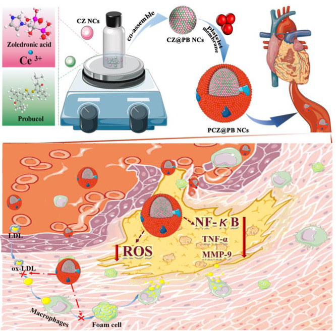
Fig. 1 The preparation and application of the nanozyme (platelet membrane-coated biomimetic nanoplatform, PCZ@PB NCs). Biomimetic probucol-loaded nanozyme is obtained by assembly of zoledronic acid, cerium ions and probucol and encapsulated in the platelet membranes. After administration, the PCZ@PB NCs were enriched to plaques through passive accumulation as well as active targeting of inflammatory lesions, reducing foam cell formation, resisting inflammation and removing excess ROS.
Fig. 1 The preparation and application of the nanozyme (platelet membrane-coated biomimetic nanoplatform, PCZ@PB NCs). Biomimetic probucol-loaded nanozyme is obtained by assembly of zoledronic acid, cerium ions and probucol and encapsulated in the platelet membranes. After administration, the PCZ@PB NCs were enriched to plaques through passive accumulation as well as active targeting of inflammatory lesions, reducing foam cell formation, resisting inflammation and removing excess ROS.CC BY · PubMed Central
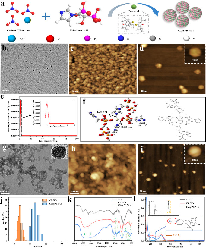
Fig. 2 Preparation and characterization of CZ NCs and CZ@PB NCs. a Schematic illustrating the preparation of CZ@PB NCs. b TEM image of CZ NCs, and c, d AFM images of CZ NCs. e Pore size distribution of CZ NCs. f Molecular simulation of CZ NCs. g TEM image of CZ@PB NCs, and h, i AFM images of CZ@PB NCs. j Size distribution. k Infrared spectra. l UV–vis absorption spectra. CZ NCs ceria-zoledronic acid nanocomposites, CZ@PB NCs CZ NCs assembled with probucol, TEM transmission electron microscopy, AFM atomic force microscopy. All the experiments were repeated for three times (n = 3) with similar results.
Fig. 2 Preparation and characterization of CZ NCs and CZ@PB NCs. a Schematic illustrating the preparation of CZ@PB NCs. b TEM image of CZ NCs, and c, d AFM images of CZ NCs. e Pore size distribution of CZ NCs. f Molecular simulation of CZ NCs. g TEM image of CZ@PB NCs, and h, i AFM images of CZ@PB NCs. j Size distribution. k Infrared spectra. l UV–vis absorption spectra. CZ NCs ceria-zoledronic acid nanocomposites, CZ@PB NCs CZ NCs assembled with probucol, TEM transmission electron microscopy, AFM atomic force microscopy. All the experiments were repeated for three times (n = 3) with similar results.CC BY · PubMed Central
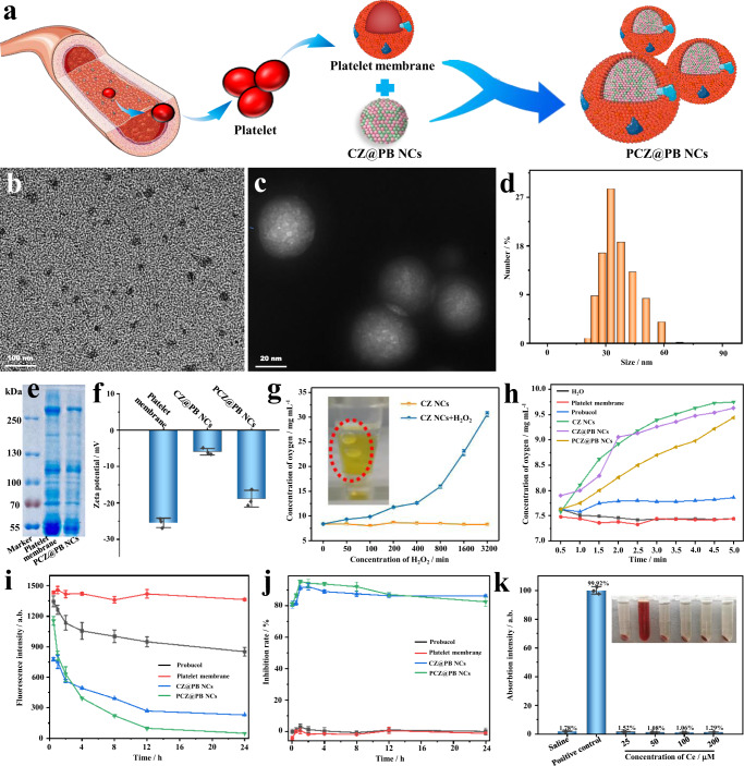
Fig. 3 Characterization of the biomimetic nanodrugs PCZ@PB NCs, and verification of their multienzyme activity. a Schematic illustrating the preparation of PCZ@PB NCs with a platelet membrane coating on CZ@PB NCs. b TEM image of PCZ@PB NCs. c TEM image of PCZ@PB NCs negatively stained with uranyl acetate. d Size distribution. e Representative protein bands stained with Coomassie brilliant blue. f Zeta potential. g Characterization of the ability of CZ NCs to catalyze oxygen production from different concentrations of hydrogen peroxide (H2O2). h Determination of different components catalyzing H2O2 (1 mM) into oxygen within 5 min. i Staining with the fluorescent probe 2’,7’-dichlorofluorescein-diacetate (DCFH-DA) to characterize the concentration of H2O2 in the solution over 24 h. j Sodium dismutase (SOD) activity measurement. k Characterization of the blood compatibility of PCZ@PB NCs. CZ NCs ceria-zoledronic acid nanocomposites, CZ@PB NCs CZ NCs assembled with probucol, TEM transmission electron microscopy. All the experiments were repeated for three times (n = 3) and data were presented as mean ± s.d. Analysis of mean zeta potential, dissolved oxygen concentration, mean DCFH fluorescence, superoxide anion inhibition rate and hemolysis rate were performed using one-way ANOVA. *P ≤ 0.05, **P ≤ 0.01, and ***P ≤ 0.001. Specific P-values are shown in the source data. Source data are provided as source data files.
Fig. 3 Characterization of the biomimetic nanodrugs PCZ@PB NCs, and verification of their multienzyme activity. a Schematic illustrating the preparation of PCZ@PB NCs with a platelet membrane coating on CZ@PB NCs. b TEM image of PCZ@PB NCs. c TEM image of PCZ@PB NCs negatively stained with uranyl acetate. d Size distribution. e Representative protein bands stained with Coomassie brilliant blue. f Zeta potential. g Characterization of the ability of CZ NCs to catalyze oxygen production from different concentrations of hydrogen peroxide (H2O2). h Determination of different components catalyzing H2O2 (1 mM) into oxygen within 5 min. i Staining with the fluorescent probe 2’,7’-dichlorofluorescein-diacetate (DCFH-DA) to characterize the concentration of H2O2 in the solution over 24 h. j Sodium dismutase (SOD) activity measurement. k Characterization of the blood compatibility of PCZ@PB NCs. CZ NCs ceria-zoledronic acid nanocomposites, CZ@PB NCs CZ NCs assembled with probucol, TEM transmission electron microscopy. All the experiments were repeated for three times (n = 3) and data were presented as mean ± s.d. Analysis of mean zeta potential, dissolved oxygen concentration, mean DCFH fluorescence, superoxide anion inhibition rate and hemolysis rate were performed using one-way ANOVA. *P ≤ 0.05, **P ≤ 0.01, and ***P ≤ 0.001. Specific P-values are shown in the source data. Source data are provided as source data files.CC BY · PubMed Central
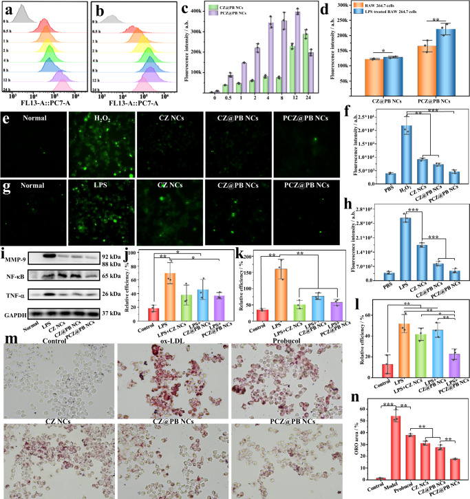
Fig. 4 In vitro effects of the biomimetic nanodrug PCZ@PB NCs. a Uptake of CZ@PB NCs and b PCZ@PB NCs by RAW 264.7 cells within 24 h. c Quantification of uptake based on fluorescence intensity. d Uptake of CZ@PB NCs and PCZ@PB NCs in RAW 264.7 cells and inflammatory RAW 264.7 cells. Intracellular hydrigen peroxide (H2O2) levels were characterized using the DCFH-DA probe. e Cellular fluorescence imaging. f Fluorescence intensity quantified using flow cytometry. Intracellular reactive oxygen species generation following treatment with different formulations and stimulation with Lipopolysaccharide (LPS). g Cellular fluorescence imaging. h Graph of the fluorescence intensity quantified using flow cytometry. i–l Western blot analysis of the intracellular expression levels of proinflammatory factors. j Tumor necrosis factor-alpha (TNF-α) expression level, k Matrix metalloproteinase-9 (MMP-9) expression level, l Nuclear factor kappa-B (NF-κB) expression level. m Oil red O (ORO) staining to characterize lipid droplets in foam cells. n Quantification of foam cells. Scale bars, 20 μm. DCFH-DA 2′,7′-dichlorofluorescein-diacetate, CZ@PB NCs ceria-zoledronic acid nanocomposites assembled with probucol, PCZ@PB NCs platelet membrane coating on CZ@PB NCs. All the experiments were repeated for three times (n = 3) and data were presented as mean ± s.d. Analysis of mean fluorescence intensity, relative grayscale, and ORO staining area was performed using one-way ANOVA. *P ≤ 0.05, **P ≤ 0.01, and ***P ≤ 0.001. Specific P-values are shown in the source data. Source data are provided as source data files.
Fig. 4 In vitro effects of the biomimetic nanodrug PCZ@PB NCs. a Uptake of CZ@PB NCs and b PCZ@PB NCs by RAW 264.7 cells within 24 h. c Quantification of uptake based on fluorescence intensity. d Uptake of CZ@PB NCs and PCZ@PB NCs in RAW 264.7 cells and inflammatory RAW 264.7 cells. Intracellular hydrigen peroxide (H2O2) levels were characterized using the DCFH-DA probe. e Cellular fluorescence imaging. f Fluorescence intensity quantified using flow cytometry. Intracellular reactive oxygen species generation following treatment with different formulations and stimulation with Lipopolysaccharide (LPS). g Cellular fluorescence imaging. h Graph of the fluorescence intensity quantified using flow cytometry. i–l Western blot analysis of the intracellular expression levels of proinflammatory factors. j Tumor necrosis factor-alpha (TNF-α) expression level, k Matrix metalloproteinase-9 (MMP-9) expression level, l Nuclear factor kappa-B (NF-κB) expression level. m Oil red O (ORO) staining to characterize lipid droplets in foam cells. n Quantification of foam cells. Scale bars, 20 μm. DCFH-DA 2′,7′-dichlorofluorescein-diacetate, CZ@PB NCs ceria-zoledronic acid nanocomposites assembled with probucol, PCZ@PB NCs platelet membrane coating on CZ@PB NCs. All the experiments were repeated for three times (n = 3) and data were presented as mean ± s.d. Analysis of mean fluorescence intensity, relative grayscale, and ORO staining area was performed using one-way ANOVA. *P ≤ 0.05, **P ≤ 0.01, and ***P ≤ 0.001. Specific P-values are shown in the source data. Source data are provided as source data files.CC BY · PubMed Central
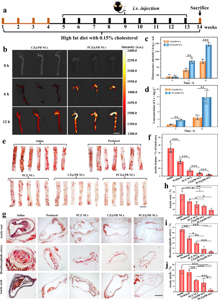
Fig. 5 In vivo therapeutic effect of the biomimetic nanodrug, PCZ@PB NCs. a Schematic illustrating the treatment protocols. b Fluorescence images of CZ@PB NCs and PCZ@PB NCs accumulation in the aortas of atherosclerotic model mice at 0, 6, and 12 h. Scale bars, 50 mm. c Quantification of the fluorescence intensity of CZ@PB NCs and PCZ@PB NCs that accumulated in the aortas of atherosclerotic model mice at 0, 6, and 12 h, n = 3. d Inductively coupled plasma-mass spectrometry for quantifying cerium ion content in the aortas of atherosclerotic model mice treated with CZ@PB NCs and PCZ@PB NCs at 12 h, n = 3. e Representative photographs of oil red O (ORO)-stained aortas from mice following treatment with different formulations. Scale bars, 50 mm. f Quantitative analysis of the lesion area in aortas. n = 7 lesion area in aortas of saline and probucol treated mice. n = 6 lesion area in aortas of CZ@PB NCs and PCZ@PB NCs-treated mice. n = 5 lesion area in aortas of PCZ NCs-treated mice. g–j ORO-stained cryosections of the aortic root, aortic arch, and brachiocephalic artery. Scale bars, 200 μm. Quantitative analysis of the relative plaque area in sections of the aortic root (h), brachiocephalic artery (i), and aortic arch (j), n = 3. Data were presented as mean ± s.d. Analysis of mean fluorescence intensity, cerium ion concentration, and aortic lesion area was performed using one-way ANOVA. *P ≤ 0.05, **P ≤ 0.01, and ***P ≤ 0.001. Specific P-values are shown in the source data. Source data are provided as source data files.
Fig. 5 In vivo therapeutic effect of the biomimetic nanodrug, PCZ@PB NCs. a Schematic illustrating the treatment protocols. b Fluorescence images of CZ@PB NCs and PCZ@PB NCs accumulation in the aortas of atherosclerotic model mice at 0, 6, and 12 h. Scale bars, 50 mm. c Quantification of the fluorescence intensity of CZ@PB NCs and PCZ@PB NCs that accumulated in the aortas of atherosclerotic model mice at 0, 6, and 12 h, n = 3. d Inductively coupled plasma-mass spectrometry for quantifying cerium ion content in the aortas of atherosclerotic model mice treated with CZ@PB NCs and PCZ@PB NCs at 12 h, n = 3. e Representative photographs of oil red O (ORO)-stained aortas from mice following treatment with different formulations. Scale bars, 50 mm. f Quantitative analysis of the lesion area in aortas. n = 7 lesion area in aortas of saline and probucol treated mice. n = 6 lesion area in aortas of CZ@PB NCs and PCZ@PB NCs-treated mice. n = 5 lesion area in aortas of PCZ NCs-treated mice. g–j ORO-stained cryosections of the aortic root, aortic arch, and brachiocephalic artery. Scale bars, 200 μm. Quantitative analysis of the relative plaque area in sections of the aortic root (h), brachiocephalic artery (i), and aortic arch (j), n = 3. Data were presented as mean ± s.d. Analysis of mean fluorescence intensity, cerium ion concentration, and aortic lesion area was performed using one-way ANOVA. *P ≤ 0.05, **P ≤ 0.01, and ***P ≤ 0.001. Specific P-values are shown in the source data. Source data are provided as source data files.CC BY · PubMed Central

Verification

Claims the paper makes

The claims and statistics of this paper haven’t been checked yet.

Run the check — free, no account needed. It reads the open-access text and checks the sources it cites.

A few free checks a day · papers already verified open instantly · results are public

Claims other papers make about this one

Each is checked against this paper’s own text: does it say what the citing paper says it does?

Self-check

Percentages, sums and reported differences re-computed from the paper’s own numbers.

The self-check needs the paper’s full text.

Citations

The citation lists are queued

They are fetched later and appear here then.

References 43 · Cited by 120

Research works printed but not linked: 1

  1. Basnet, R., Khadka, S., Wang, Y. & Gupta, R. Research progress on diagnosis and treatment of atherosclerosis. Sci. Lett. 8, 18–22 (2020).

Metadata

Authors
Fu, X., Yu, X., Jiang, J., Yang, J., Chen, L., Yang, Z., & Yu, C.
Affiliations
Chongqing Medical University (CN)
Corresponding author
  • Yang, Zhangyou
  • Yu, Chao
Venue
Nature Communications
Published
2022-11-01
Volume
13
Issue
1
Pages
6528-6528
ISSN
2041-1723 (ISSN-L)
OpenAlex
W4308027181
Cited by
120
References
43
Citations by year
2026: 26 · 2025: 45 · 2024: 31 · 2023: 18
FWCI
6.59
Type
journal article
Language
en
Open access
Locations
  • Nature Communications (publishedVersion, cc-by)
  • PubMed Central (publishedVersion, cc-by)
  • PubMed (publishedVersion)
  • RePEc: Research Papers in Economics (submittedVersion)
  • DOAJ (DOAJ: Directory of Open Access Journals) (submittedVersion, cc-by-sa)
Funders
  • National Natural Science Foundation of China (81703156)
  • Youth Talent Foundation in the College of Pharmacy, Chongqing Medical University (No. YXY2019QNGD1).
  • Chongqing Special Project for Technological Innovation and Application Development
Topics
  • Physical Sciences › Materials Science › Materials Chemistry › Advanced Nanomaterials in Catalysis
  • Physical Sciences › Engineering › Biomedical Engineering › Nanoplatforms for cancer theranostics
  • Physical Sciences › Materials Science › Biomaterials › Nanoparticle-Based Drug Delivery
Keywords
atherosclerosis, zoledronic acid, reactive oxygen species, inflammation, probucol, platelet membrane coating, foam cell formation, drug delivery, oxidative stress, synergistic therapy
MeSH terms
Zoledronic Acid, Humans, Inflammation, Reactive Oxygen Species, Atherosclerosis, Nanoparticles

About these checks

Registry, retraction, Cites and statistics results are computed, not judged, with one exception: a printed reference that no index lists is read once by a model before it counts as a citation error. Claims are extracted by a model and kept only when their quote appears verbatim in the text; each cited source is then checked, with a model’s help, for whether it says what the claim relies on. Those verdicts can be wrong — read the claim’s quote and the cited source before relying on one.

Statistics and arithmetic are recomputed, not judged.

Confidence bands are shown only for fields calibrated on our annotated test set. None is calibrated yet, so no band is shown.

What counts as a claim ›How we measure confidence ›How we check this ›

Are you an author, or do you see a problem with this page? Request a review

How to cite

Cite CoChat for the checks on this page, and the paper for its own findings.

This page
CoChat. (2026). Citation check: Small molecule-assisted assembly of multifunctional ceria nanozymes for synergistic treatment of atherosclerosis. https://app.cochat.ai/papers/10.1038/s41467-022-34248-y
The paper
Fu, X., Yu, X., Jiang, J., Yang, J., Chen, L., Yang, Z., & Yu, C. (2022). Small molecule-assisted assembly of multifunctional ceria nanozymes for synergistic treatment of atherosclerosis. Nature Communications. https://doi.org/10.1038/s41467-022-34248-y