Cited by 24Bacterial Genetics and BiotechnologyMicrotubule and mitosis dynamicsGenomics and Chromatin Dynamics

The DOI matches its Crossref record; 1 of the 1 references it cites were found in a registry or an index.

Multiple ParA/MinD ATPases coordinate the positioning of disparate cargos in a bacterial cell

Open access

Published version of a preprint

Abstract

In eukaryotes, linear motor proteins govern intracellular transport and organization. In bacteria, where linear motors involved in spatial regulation are absent, the ParA/MinD family of ATPases organize an array of genetic- and protein-based cellular cargos. The positioning of these cargos has been independently investigated to varying degrees in several bacterial species. However, it remains unclear how multiple ParA/MinD ATPases can coordinate the positioning of diverse cargos in the same cell. Here, we find that over a third of sequenced bacterial genomes encode multiple ParA/MinD ATPases. We identify an organism ( Halothiobacillus neapolitanus ) with seven ParA/MinD ATPases, demonstrate that five of these are each dedicated to the spatial regulation of a single cellular cargo, and define potential specificity determinants for each system. Furthermore, we show how these positioning reactions can influence each other, stressing the importance of understanding how organelle trafficking, chromosome segregation, and cell division are coordinated in bacterial cells. Together, our data show how multiple ParA/MinD ATPases coexist and function to position a diverse set of fundamental cargos in the same bacterial cell.

Overview

The claims and statistics of this paper haven’t been checked yet.

Run the check — free, no account needed. It reads the open-access text and checks the sources it cites.

A few free checks a day · papers already verified open instantly · results are public

Integrity signals

Registry

Crossref · 10.1038/s41467-023-39019-x

Cites

1 of 1 references verified0 citation errors · 0 not checkable

Statistics

Figures

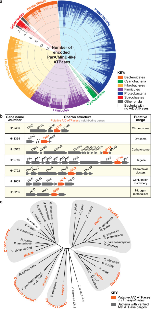
Fig. 1 ParA/MinD-like ATPases are widespread in bacteria. a 96% of sequenced bacterial genomes encode at least one A/D ATPase and 35% encode multiple. Each spike represents a bacterial species and spike length indicates the number of unique A/D hits per bacterium. b H. neapolitanus encodes seven putative ParA/MinD-like positioning systems. Gene neighborhood analysis implicates the putative cargos associated with each putative A/D ATPase. c Multiple sequence alignment of each A/D ATPase from H. neapolitanus against experimentally-verified ParA/MinD family members further implicates the putative cargos identified by gene neighborhood analysis. Each of the putative A/D ATPases in H. neapolitanus cluster with family members shown to position the indicated cellular cargos (orange) in other bacteria.
Fig. 1 ParA/MinD-like ATPases are widespread in bacteria. a 96% of sequenced bacterial genomes encode at least one A/D ATPase and 35% encode multiple. Each spike represents a bacterial species and spike length indicates the number of unique A/D hits per bacterium. b H. neapolitanus encodes seven putative ParA/MinD-like positioning systems. Gene neighborhood analysis implicates the putative cargos associated with each putative A/D ATPase. c Multiple sequence alignment of each A/D ATPase from H. neapolitanus against experimentally-verified ParA/MinD family members further implicates the putative cargos identified by gene neighborhood analysis. Each of the putative A/D ATPases in H. neapolitanus cluster with family members shown to position the indicated cellular cargos (orange) in other bacteria.CC BY · PubMed Central
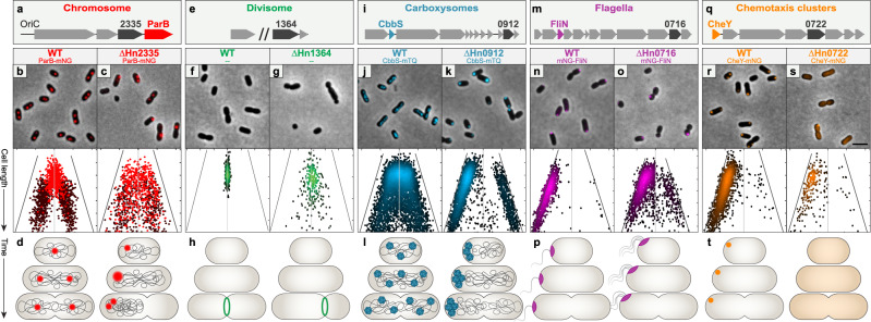
Fig. 2 Each A/D ATPase in H. neapolitanus positions a specific cargo. a–d Hn2335 is required for chromosome segregation. a Hn2335 is found near OriC and has a ParB-homolog encoded downstream. b, c The origin region of the chromosome was tagged by labelling the ParB homolog. Light red: 1 focus/cell; dark red: 2 foci/cell. Short WT cells had a single focus at mid-cell, whereas longer cells had two foci at the quarter positions. ΔHn2335 cells displayed random positioning of ParB foci regardless of cell length. d Cartoon diagrams depict chromosome segregation in WT and ΔHn2335 cells. e–h Hn1364 is required for divisome positioning. e Hn1364 is found upstream of minE. f, g Divisome positioning was determined by the location of constriction sites. Each dot on the density graph represents one identified constriction site. In WT cells, constriction sites were at mid-cell. In ΔHn1364, constriction sites were found across the cell length. h Cartoon diagrams depict cell division in WT and ΔHn1364 cells. i–l Carboxysome positioning is determined by McdA. i Hn0912 (mcdA) is found near genes encoding carboxysome shell proteins and Rubisco. j, k Carboxysomes were visualized by labelling the small subunit of the Rubisco enzyme, cbbS. Each dot on the density graph represents one carboxysome focus. In WT cells, carboxysomes are distributed along the cell length. In ΔmcdA, carboxysomes formed large polar foci at one or both poles. l Cartoon diagrams depict carboxysome distribution in WT and ΔHn0912 cells. m–p Hn0716 is required for regulating flagella position and copy number. m Hn0716 is found near flagella-associated genes. n, o Flagella localization was visualized by labelling a component of the flagellar basal body, fliN. Each dot on the density graph represents one FliN focus. WT cells had a single polar FliN focus. In ΔHn0716 cells, FliN foci were more randomly positioned along the cell length. p Cartoon diagrams depict flagella localization and number in WT and ΔHn0716 cells.q–t Hn0722 is required for chemotaxis cluster positioning. q Hn0722 is found near chemotaxis-associated genes. r, s Chemotaxis clusters were visualized by labelling the response regulator, cheY. Each dot on the density graph represents one CheY focus. WT cells had a single CheY polar focus. ΔHn0722 mutant cells typically had no CheY foci. t Cartoon diagrams depict chemotaxis clusters in WT and ΔHn0722 cells. (All images) Micrographs shown are representative images from at least 3 experiments. Scale bar: 2 μm. (All graphs) Cells were analyzed and quantified using MicrobeJ. On the y-axis, cells are sorted by increasing cell length, with shorter cells at the top and longer cells at the bottom. The x-axis represents the distance from mid-cell in microns; the center vertical line equates to a distance of zero from mid-cell. Each dot represents where a focus was found along the length of the cell. For all density plots, lighter colors represent higher density, darker colors represent lower density. For flagella, chemotaxis, and carboxysome graphs, the cell pole closest to a focus was oriented to the left; foci in the right half of the graph indicate the presence of a second focus. Graph axes for chromosome, carboxysome, flagella, and chemotaxis-labelled mutants:y-axis range (cell length): 0.8–2.1 μm; x-axis range (distance from mid-cell): −1.1–1.1 μm. X-axis of ΔflhG cells is 0.5–1.8 μm. Graph axes for divisome: y-axis range (cell length): 1.5–3.0 μm; x-axis range (distance from mid-cell): −1.5–1.5 μm. Source data are provided as a Source Data file.
Fig. 2 Each A/D ATPase in H. neapolitanus positions a specific cargo. a–d Hn2335 is required for chromosome segregation. a Hn2335 is found near OriC and has a ParB-homolog encoded downstream. b, c The origin region of the chromosome was tagged by labelling the ParB homolog. Light red: 1 focus/cell; dark red: 2 foci/cell. Short WT cells had a single focus at mid-cell, whereas longer cells had two foci at the quarter positions. ΔHn2335 cells displayed random positioning of ParB foci regardless of cell length. d Cartoon diagrams depict chromosome segregation in WT and ΔHn2335 cells. e–h Hn1364 is required for divisome positioning. e Hn1364 is found upstream of minE. f, g Divisome positioning was determined by the location of constriction sites. Each dot on the density graph represents one identified constriction site. In WT cells, constriction sites were at mid-cell. In ΔHn1364, constriction sites were found across the cell length. h Cartoon diagrams depict cell division in WT and ΔHn1364 cells. i–l Carboxysome positioning is determined by McdA. i Hn0912 (mcdA) is found near genes encoding carboxysome shell proteins and Rubisco. j, k Carboxysomes were visualized by labelling the small subunit of the Rubisco enzyme, cbbS. Each dot on the density graph represents one carboxysome focus. In WT cells, carboxysomes are distributed along the cell length. In ΔmcdA, carboxysomes formed large polar foci at one or both poles. l Cartoon diagrams depict carboxysome distribution in WT and ΔHn0912 cells. m–p Hn0716 is required for regulating flagella position and copy number. m Hn0716 is found near flagella-associated genes. n, o Flagella localization was visualized by labelling a component of the flagellar basal body, fliN. Each dot on the density graph represents one FliN focus. WT cells had a single polar FliN focus. In ΔHn0716 cells, FliN foci were more randomly positioned along the cell length. p Cartoon diagrams depict flagella localization and number in WT and ΔHn0716 cells.q–t Hn0722 is required for chemotaxis cluster positioning. q Hn0722 is found near chemotaxis-associated genes. r, s Chemotaxis clusters were visualized by labelling the response regulator, cheY. Each dot on the density graph represents one CheY focus. WT cells had a single CheY polar focus. ΔHn0722 mutant cells typically had no CheY foci. t Cartoon diagrams depict chemotaxis clusters in WT and ΔHn0722 cells. (All images) Micrographs shown are representative images from at least 3 experiments. Scale bar: 2 μm. (All graphs) Cells were analyzed and quantified using MicrobeJ. On the y-axis, cells are sorted by increasing cell length, with shorter cells at the top and longer cells at the bottom. The x-axis represents the distance from mid-cell in microns; the center vertical line equates to a distance of zero from mid-cell. Each dot represents where a focus was found along the length of the cell. For all density plots, lighter colors represent higher density, darker colors represent lower density. For flagella, chemotaxis, and carboxysome graphs, the cell pole closest to a focus was oriented to the left; foci in the right half of the graph indicate the presence of a second focus. Graph axes for chromosome, carboxysome, flagella, and chemotaxis-labelled mutants:y-axis range (cell length): 0.8–2.1 μm; x-axis range (distance from mid-cell): −1.1–1.1 μm. X-axis of ΔflhG cells is 0.5–1.8 μm. Graph axes for divisome: y-axis range (cell length): 1.5–3.0 μm; x-axis range (distance from mid-cell): −1.5–1.5 μm. Source data are provided as a Source Data file.CC BY · PubMed Central
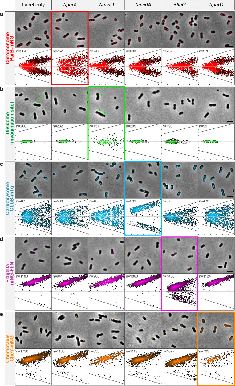
Fig. 3 Cargo positioning is not directly controlled by A/D ATPases encoded at distant loci. Each of the five cellular cargos were fluorescently tagged as indicated (Left): a chromosome (ParB-mNG), b divisome (invagination site), c carboxysomes (CbbS-mTQ), d flagella (mNG-FliN), and e chemotaxis clusters (CheY-mNG). (All images) Micrographs shown are representative images from at least 3 experiments. Scale bar: 2 μm. The “Label Only” column shows the WT positioning of each of the fluorescent cargos. Deletion of an A/D ATPase resulted in the mislocalization of only its specific cargo (bolded rectangles). Graphs in the “label only” column and graphs in the bolded rectangles are duplicated from Fig. 2. Graph axes for chromosome, carboxysome, flagella, and chemotaxis-labelled mutants: x-axis range (cell length): 0.8–2.1 μm; y-axis range (distance from mid-cell): −1.1–1.1 μm. All x-axes of ΔflhG cells are 0.5–1.8 μm. Graph axes for divisome: x-axis range (cell length): 1.5–3.0 μm; y-axis range (distance from mid-cell): −1.5–1.5 μm. Source data are provided as a Source Data file.
Fig. 3 Cargo positioning is not directly controlled by A/D ATPases encoded at distant loci. Each of the five cellular cargos were fluorescently tagged as indicated (Left): a chromosome (ParB-mNG), b divisome (invagination site), c carboxysomes (CbbS-mTQ), d flagella (mNG-FliN), and e chemotaxis clusters (CheY-mNG). (All images) Micrographs shown are representative images from at least 3 experiments. Scale bar: 2 μm. The “Label Only” column shows the WT positioning of each of the fluorescent cargos. Deletion of an A/D ATPase resulted in the mislocalization of only its specific cargo (bolded rectangles). Graphs in the “label only” column and graphs in the bolded rectangles are duplicated from Fig. 2. Graph axes for chromosome, carboxysome, flagella, and chemotaxis-labelled mutants: x-axis range (cell length): 0.8–2.1 μm; y-axis range (distance from mid-cell): −1.1–1.1 μm. All x-axes of ΔflhG cells are 0.5–1.8 μm. Graph axes for divisome: x-axis range (cell length): 1.5–3.0 μm; y-axis range (distance from mid-cell): −1.5–1.5 μm. Source data are provided as a Source Data file.CC BY · PubMed Central
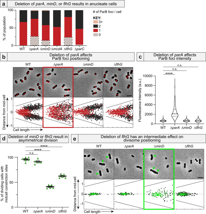
Fig. 4 Anucleate cells form via three different mechanisms. a Deletion of the A/D ATPase required for chromosome (parA), divisome (minD), or flagellar (flhG) positioning resulted in anucleate cells (grey boxes). b Only ΔparA cells had mispositioned ParB foci. Cells with a single ParB focus are red. Cells with two ParB foci are black. ΔflhG cells maintained a single ParB focus in long cells, suggesting a DNA replication defect. c ParB foci are brighter only in ΔparA cells. WT: n = 1289; ∆parA: n = 773; ∆minD: n = 990; ∆flhG: n = 747 biologically independent cells. Kruskal-Wallis p-values: ∆parA: <0.0001; ∆minD: 0.97; ∆flhG: 1. d Deletion of minD or flhG resulted in divisome mispositioning. Constriction sites were considered “mid-cell” when found within 5% of the cell center along the long axis. WT: n = 6; ∆parA, ∆minD, and ∆flhG: n = 5 biologically independent samples. Brown-Forsythe and Welch ANOVA test p-values: ∆parA: 0.28; ∆minD: <0.0001; ∆flhG: <0.0001 e Deletion of flhG resulted in an intermediate effect on divisome positioning when compared to ΔminD. Each dot on the density graph represents one identified constriction site. Lighter colors represent higher density, darker colors represent lower density. Panels b and e are summarized from Fig. 3. (All images) Scale bar: 2 μm. Source data are provided as a Source Data file.
Fig. 4 Anucleate cells form via three different mechanisms. a Deletion of the A/D ATPase required for chromosome (parA), divisome (minD), or flagellar (flhG) positioning resulted in anucleate cells (grey boxes). b Only ΔparA cells had mispositioned ParB foci. Cells with a single ParB focus are red. Cells with two ParB foci are black. ΔflhG cells maintained a single ParB focus in long cells, suggesting a DNA replication defect. c ParB foci are brighter only in ΔparA cells. WT: n = 1289; ∆parA: n = 773; ∆minD: n = 990; ∆flhG: n = 747 biologically independent cells. Kruskal-Wallis p-values: ∆parA: <0.0001; ∆minD: 0.97; ∆flhG: 1. d Deletion of minD or flhG resulted in divisome mispositioning. Constriction sites were considered “mid-cell” when found within 5% of the cell center along the long axis. WT: n = 6; ∆parA, ∆minD, and ∆flhG: n = 5 biologically independent samples. Brown-Forsythe and Welch ANOVA test p-values: ∆parA: 0.28; ∆minD: <0.0001; ∆flhG: <0.0001 e Deletion of flhG resulted in an intermediate effect on divisome positioning when compared to ΔminD. Each dot on the density graph represents one identified constriction site. Lighter colors represent higher density, darker colors represent lower density. Panels b and e are summarized from Fig. 3. (All images) Scale bar: 2 μm. Source data are provided as a Source Data file.CC BY · PubMed Central
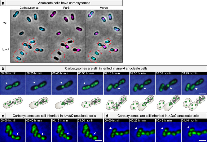
Fig. 5 Anucleate cells inherit carboxysomes. a Carboxysomes are present in anucleate cells (dashed circle). Micrographs shown are representative images from 2 experiments. b Time-lapse microscopy shows that carboxysomes (green) in ΔparA cells are bound to the nucleoid, but are inherited in anucleate cells via release from the extruded chromosome. Carboxysomes are also inherited in c ΔminD anucleate cells and d ΔflhG anucleate cells via the same mechanism. (All videos) White arrows highlight carboxysome bundling and subsequent release. Scale bar: 1 μm.
Fig. 5 Anucleate cells inherit carboxysomes. a Carboxysomes are present in anucleate cells (dashed circle). Micrographs shown are representative images from 2 experiments. b Time-lapse microscopy shows that carboxysomes (green) in ΔparA cells are bound to the nucleoid, but are inherited in anucleate cells via release from the extruded chromosome. Carboxysomes are also inherited in c ΔminD anucleate cells and d ΔflhG anucleate cells via the same mechanism. (All videos) White arrows highlight carboxysome bundling and subsequent release. Scale bar: 1 μm.CC BY · PubMed Central
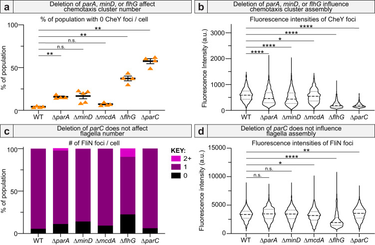
Fig. 6 Deletion of parA, minD, or flhG influences chemotaxis cluster assembly. Deletion of parA, minD, or flhG influences chemotaxis cluster a number (WT: n = 4, ∆parA: n = 4, ∆minD: n = 5, ∆mcdA: n = 4, ∆flhG: n = 4, ∆parC: n = 4 biologically independent samples. Brown-Forsythe and Welch ANOVA test p-values: ∆parA: 0.0013, ∆minD: 0.0642, ∆mcdA: 0.1141, ∆flhG: 0.0043, ∆parC: 0.0012) and b assembly (n = 455 biologically independent cells. Kruskal-Wallis test p-values: ∆parA: <0.0001, ∆minD: <0.0001, ∆mcdA: 0.0171, ∆flhG: <0.0001, ∆parC: <0.0001). Only deletion of flhG substantially influenced flagella c number and d assembly (n = 851 biologically independent cells. Kruskal–Wallis test p-values: ∆parA: 0.1161, ∆minD: 0.4433, ∆mcdA: 0.0184, ∆flhG: <0.0001, ∆parC: 0.0051). Source data are provided as a Source Data file.
Fig. 6 Deletion of parA, minD, or flhG influences chemotaxis cluster assembly. Deletion of parA, minD, or flhG influences chemotaxis cluster a number (WT: n = 4, ∆parA: n = 4, ∆minD: n = 5, ∆mcdA: n = 4, ∆flhG: n = 4, ∆parC: n = 4 biologically independent samples. Brown-Forsythe and Welch ANOVA test p-values: ∆parA: 0.0013, ∆minD: 0.0642, ∆mcdA: 0.1141, ∆flhG: 0.0043, ∆parC: 0.0012) and b assembly (n = 455 biologically independent cells. Kruskal-Wallis test p-values: ∆parA: <0.0001, ∆minD: <0.0001, ∆mcdA: 0.0171, ∆flhG: <0.0001, ∆parC: <0.0001). Only deletion of flhG substantially influenced flagella c number and d assembly (n = 851 biologically independent cells. Kruskal–Wallis test p-values: ∆parA: 0.1161, ∆minD: 0.4433, ∆mcdA: 0.0184, ∆flhG: <0.0001, ∆parC: 0.0051). Source data are provided as a Source Data file.CC BY · PubMed Central
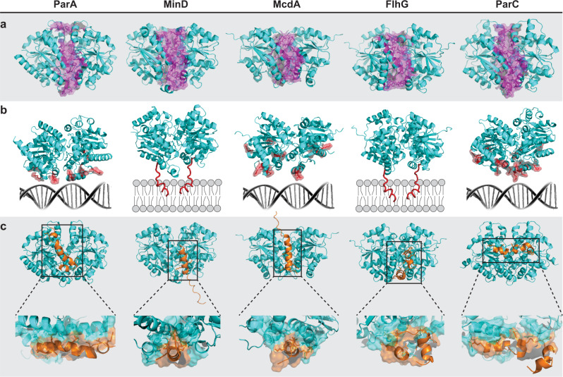
Fig. 7 A/D ATPases have unique interfaces that confer cargo-positioning specificity. a Homodimer structures of the A/D ATPases in H. neapolitanus were generated using AlphaFold2 (cyan). Putative residues for homodimer specificity are highlighted magenta. b Dimers from a are oriented over their positioning matrix—nucleoid DNA or membrane. Putative residues critical for binding nucleoid DNA or membrane are highlighted red. c Dimer structures docked with the N-terminal interacting peptide from putative partner proteins (orange), which confer cargo specificity. Zoomed box: Predicted residues critical for this association are space-filled cyan on the ATPase and orange on the partner protein.
Fig. 7 A/D ATPases have unique interfaces that confer cargo-positioning specificity. a Homodimer structures of the A/D ATPases in H. neapolitanus were generated using AlphaFold2 (cyan). Putative residues for homodimer specificity are highlighted magenta. b Dimers from a are oriented over their positioning matrix—nucleoid DNA or membrane. Putative residues critical for binding nucleoid DNA or membrane are highlighted red. c Dimer structures docked with the N-terminal interacting peptide from putative partner proteins (orange), which confer cargo specificity. Zoomed box: Predicted residues critical for this association are space-filled cyan on the ATPase and orange on the partner protein.CC BY · PubMed Central

Verification

Claims the paper makes

The claims and statistics of this paper haven’t been checked yet.

Run the check — free, no account needed. It reads the open-access text and checks the sources it cites.

A few free checks a day · papers already verified open instantly · results are public

Self-check

Percentages, sums and reported differences re-computed from the paper’s own numbers.

The self-check needs the paper’s full text.

Citations

Cites

Research works printed but not linked: 61

  1. Baxter, J. C. & Funnell, B. E. Plasmid partition mechanisms. Microbiol. Spectr. https://doi.org/10.1128/microbiolspec.PLAS-0023-2014 (2014). · DOI
  2. Jalal, A. S. B. & Le, T. B. K. Bacterial chromosome segregation by the ParABS system. Open Biol. 10, 200097 (2020). · DOI
  3. Lutkenhaus, J. Assembly dynamics of the bacterial MinCDE system and spatial regulation of the Z ring. Annu. Rev. Biochem. 76, 539–562 (2007). · DOI
  4. Maccready, J. S. & Vecchiarelli, A. G. Positioning the model bacterial organelle, the carboxysome. MBio 12, e02519–e02519 (2021). · DOI
  5. Savage, D. F., Afonso, B., Chen, A. H. & Silver, P. A. Spatially ordered dynamics of the bacterial carbon fixation machinery. Science 327, 1258–1261 (2010). · DOI · Paper page
  6. Schuhmacher, J. S. et al. MinD-like ATPase FlhG effects location and number of bacterial flagella during C-ring assembly. Proc. Natl Acad. Sci. USA 112, 3092–3097 (2015). · DOI
  7. Schuhmacher, J. S., Thormann, K. M. & Bange, G. How bacteria maintain location and number of flagella? FEMS Microbiol. Rev. 39, 812–822 (2015). · DOI
  8. Ringgaard, S., Schirner, K., Davis, B. M. & Waldor, M. K. A family of ParA-like ATPases promotes cell pole maturation by facilitating polar localization of chemotaxis proteins. Genes Dev. 25, 1544–1555 (2011). · DOI
  9. Thompson, S. R., Wadhams, G. H. & Armitage, J. P. The positioning of cytoplasmic protein clusters in bacteria. Proc. Natl Acad. Sci. Usa. 103, 8209–8214 (2006). · DOI
  10. Atmakuri, K., Cascales, E., Burton, O. T., Banta, L. M. & Christie, P. J. Agrobacterium ParA/MinD-like VirC1 spatially coordinates early conjugative DNA transfer reactions. EMBO J. 26, 2540–2551 (2007). · DOI
  11. Shan, S. ATPase and GTPase tangos drive intracellular protein transport. Trends Biochem. Sci. 41, 1050–1060 (2016). · DOI
  12. Hester, C. M. & Lutkenhaus, J. Soj (ParA) DNA binding is mediated by conserved arginines and is essential for plasmid segregation. Proc. Natl Acad. Sci. USA 104, 20326–20331 (2007). · DOI
  13. Kiekebusch, D., Michie, K. A., Essen, L.-O., Löwe, J. & Thanbichler, M. Localized dimerization and nucleoid binding drive gradient formation by the bacterial cell division inhibitor MipZ. Mol. Cell 46, 245–259 (2012). · DOI
  14. Szeto, T. H., Rowland, S. L., Rothfield, L. I. & King, G. F. Membrane localization of MinD is mediated by a C-terminal motif that is conserved across eubacteria, archaea, and chloroplasts. Proc. Natl Acad. Sci. USA 99, 15693–15698 (2002). · DOI
  15. Hu, Z. L. & Lutkenhaus, J. A conserved sequence at the C-terminus of MinD is required for binding to the membrane and targeting MinC to the septum. Mol. Microbiol 47, 345–355 (2003). · DOI
  16. Vecchiarelli, A. G. et al. ATP control of dynamic P1 ParA-DNA interactions: a key role for the nucleoid in plasmid partition. Mol. Microbiol. 78, 78–91 (2010). · DOI
  17. Koonin, E. V. A superfamily of ATPases with diverse functions containing either classical or deviant ATP-binding Motif. J. Mol. Biol. 229, 1165–1174 (1993). · DOI
  18. Tilly, K., Checroun, C. & Rosa, P. A. Requirements for Borrelia burgdorferi plasmid maintenance. Plasmid 68, 1–12 (2012). · DOI
  19. Livny, J., Yamaichi, Y. & Waldor, M. K. Distribution of centromere-like parS sites in bacteria: Insights from comparative genomics. J. Bacteriol. 189, 8693–8703 (2007). · DOI
  20. Lutkenhaus, J. The ParA/MinD family puts things in their place. Trends Microbiol. 20, 411–418 (2012). · DOI
  21. Vecchiarelli, A. G., Mizuuchi, K. & Funnell, B. E. Surfing biological surfaces: exploiting the nucleoid for partition and transport in bacteria. Mol. Microbiol. 86, 513–523 (2012). · DOI
  22. Saha, C. K., Sanches Pires, R., Brolin, H., Delannoy, M. & Atkinson, G. C. FlaGs and webFlaGs: discovering novel biology through the analysis of gene neighbourhood conservation. Bioinformatics 37, 1312–1314 (2021). · DOI
  23. Raskin, D. M. & De Boer, P. A. J. Rapid pole-to-pole oscillation of a protein required for directing division to the middle of Escherichia coli. Proc. Natl Acad. Sci. USA 96, 4971–4976 (1999). · DOI
  24. Yu, X. & Margolin, W. FtsZ ring clusters in min and partition mutants: role of both the Min system and the nucleoid in regulating FtsZ ring localization. Mol. Microbiol 32, 315–326 (1999). · DOI
  25. Kerfeld, C. A., Aussignargues, C., Zarzycki, J., Cai, F. & Sutter, M. Bacterial microcompartments. Nat. Rev. Microbiol. 16, 277–290 (2018). · DOI
  26. Turmo, A., Gonzalez-Esquer, C. R. & Kerfeld, C. A. Carboxysomes: metabolic modules for CO2 fixation. FEMS Microbiol. Lett. 364, 18 (2017). · DOI
  27. MacCready, J. S. et al. Protein gradients on the nucleoid position the carbon-fixing organelles of cyanobacteria. Elife 7, e39723 (2018). · DOI
  28. MacCready, J. S., Tran, L., Basalla, J. L., Hakim, P. & Vecchiarelli, A. G. The McdAB system positions α-carboxysomes in proteobacteria. Mol. Microbiol. 00, 1–21 (2021).
  29. Campos-García, J., Nájera, R., Camarena, L. & Soberón-Chávez, G. The Pseudomonas aeruginosa motR gene involved in regulation of bacterial motility. FEMS Microbiol. Lett. 184, 57–62 (2000). · DOI
  30. Dasgupta, N., Arora, S. K. & Ramphal, R. fleN, a gene that regulates flagellar number in Pseudomonas aeruginosa. J. Bacteriol. 182, 357–364 (2000). · DOI
  31. Gao, T., Shi, M., Ju, L. & Gao, H. Investigation into FlhFG reveals distinct features of FlhF in regulating flagellum polarity in Shewanella oneidensis. Mol. Microbiol. 98, 571–585 (2015). · DOI
  32. Kusumoto, A. et al. Collaboration of FlhF and FlhG to regulate polar-flagella number and localization in Vibrio alginolyticus. Microbiol 154, 1390–1399 (2008). · DOI
  33. Kusumoto, A. et al. Regulation of polar flagellar number by the flhF and flhG genes in Vibrio alginolyticus. J. Biochem. 139, 113–121 (2006). · DOI
  34. Arroyo-Pérez, E. E. & Ringgaard, S. Interdependent Polar Localization of FlhF and FlhG and their importance for flagellum formation of vibrio parahaemolyticus. Front. Microbiol. 0, 557 (2021).
  35. Guttenplan, S. B., Shaw, S. & Kearns, D. B. The cell biology of peritrichous flagella in Bacillus subtilis. Mol. Microbiol. 87, 211–229 (2013). · DOI
  36. Balaban, M. & Hendrixson, D. R. Polar flagellar biosynthesis and a regulator of flagellar number influence spatial parameters of cell division in Campylobacter jejuni. PLOS Pathog. 7, e1002420 (2011). · DOI
  37. Gulbronson, C. J. et al. FlhG employs diverse intrinsic domains and influences FlhF GTPase activity to numerically regulate polar flagellar biogenesis in Campylobacter jejuni. Mol. Microbiol. 99, 291–306 (2016). · DOI
  38. Chang, Y. et al. Molecular mechanism for rotational switching of the bacterial flagellar motor. Nat. Struct. Mol. Biol. 27, 1041–1047 (2020). · DOI
  39. Kühn, M. J. et al. Spatial arrangement of several flagellins within bacterial flagella improves motility in different environments. Nat. Commun. 9, 1–12 (2018). · DOI
  40. Ringgaard, S. et al. ParP prevents dissociation of CheA from chemotactic signaling arrays and tethers them to a polar anchor. Proc. Natl Acad. Sci. USA 111, E255–E264 (2014). · DOI
  41. Roberts, M. A. J., Wadhams, G. H., Hadfield, K. A., Tickner, S. & Armitage, J. P. ParA-like protein uses nonspecific chromosomal DNA binding to partition protein complexes. Proc. Natl Acad. Sci. USA 109, 6698–6703 (2012). · DOI
  42. Kentner, D. & Sourjik, V. Dynamic map of protein interactions in the Escherichia coli chemotaxis pathway. Mol. Syst. Biol. 5, 238 (2009). · DOI
  43. Sourjik, V. & Berg, H. C. Localization of components of the chemotaxis machinery of Escherichia coli using fluorescent protein fusions. Mol. Microbiol. 37, 740–751 (2000). · DOI
  44. Briegel, A. et al. Universal architecture of bacterial chemoreceptor arrays. Proc. Natl Acad. Sci. USA 106, 17181–17186 (2009). · DOI
  45. Leonard, T. A., Butler, P. J. & Löwe, J. Bacterial chromosome segregation: structure and DNA binding of the Soj dimer—a conserved biological switch. EMBO J. 24, 270–282 (2005). · DOI
  46. Dunham, T. D., Xu, W., Funnell, B. E. & Schumacher, M. A. Structural basis for ADP‐mediated transcriptional regulation by P1 and P7 ParA. EMBO J. 28, 1792–1802 (2009). · DOI
  47. Wu, W., Park, K.-T., Holyoak, T. & Lutkenhaus, J. Determination of the structure of the MinD-ATP complex reveals the orientation of MinD on the membrane and the relative location of the binding sites for MinE and MinC. Mol. Microbiol. 79, 1515–1528 (2011). · DOI
  48. Schumacher, M. A. et al. Structural mechanism of ATP-induced polymerization of the partition factor ParF. J. Biol. Chem. 287, 26146–26154 (2012). · DOI
  49. Schumacher, D., Harms, A., Bergeler, S., Frey, E. & Søgaard-Andersen, L. Pomx, a para/mind atpase activating protein, is a triple regulator of cell division in myxococcus xanthus. Elife 10, e66160 (2021). · DOI
  50. Park, K.-T., Wu, W., Lovell, S. & Lutkenhaus, J. Mechanism of the asymmetric activation of the MinD ATPase by MinE. Mol. Microbiol. 85, 271–281 (2012). · DOI
  51. Park, K.-T. et al. The min oscillator uses MinD-dependent conformational changes in MinE to spatially regulate cytokinesis. Cell 146, 396–407 (2011). · DOI
  52. Leipe, D. D., Wolf, Y. I., Koonin, E. V. & Aravind, L. Classification and evolution of P-loop GTPases and related ATPases. J. Mol. Biol. 317, 41–72 (2002). · DOI · Paper page
  53. Nußbaum, P. et al. An oscillating MinD protein determines the cellular positioning of the motility machinery in Archaea. Curr. Biol. 30, 4956–4972.e4 (2020). · DOI
  54. Norman, A., Hansen, L. H. & Srensen, S. J. Conjugative plasmids: vessels of the communal gene pool. Philos. Trans. R. Soc. B Biol. Sci. 364, 2275–2289 (2009). · DOI
  55. Schwille, P. & Diez, S. Synthetic biology of minimal systems. Crit. Rev. Biochem. Mol. Biol. 44, 223–242 (2009). · DOI
  56. Schindelin, J. et al. Fiji: an open-source platform for biological-image analysis. Nat. Methods 9, 676–682 (2012). · DOI · Paper page
  57. Ducret, A., Quardokus, E. M. & Brun, Y. V. MicrobeJ, a tool for high throughput bacterial cell detection and quantitative analysis. Nat. Microbiol. 1, 1–7 (2016). · DOI
  58. Mirdita, M. et al. ColabFold—making protein folding accessible to all. Nat. Methods 19, 679–682 (2022). · DOI · Paper page
  59. Raveh, B., London, N., Zimmerman, L. & Schueler-Furman, O. Rosetta FlexPepDockab-initio: Simultaneous folding, docking and refinement of peptides onto their receptors. PLoS One 6, e18934 (2011). · DOI
  60. Li, S. C. & Ng, Y. K. Calibur: A tool for clustering large numbers of protein decoys. BMC Bioinforma. 11, 25 (2010). · DOI
  61. Barlow, K. A. et al. Flex ddG: Rosetta ensemble-based estimation of changes in protein-protein binding affinity upon mutation. J. Phys. Chem. B 122, 5389–5399 (2018). · DOI

Metadata

Authors
Pulianmackal, L. T., Limcaoco, J. M. I., Ravi, K., Yang, S., Zhang, J., Tran, M. K., Ghalmi, M., O’Meara, M. J., & Vecchiarelli, A. G.
Affiliations
University of Michigan (US)
Corresponding author
Vecchiarelli, Anthony G.
Venue
Nature Communications
Published
2023-06-05
Volume
14
Issue
1
Pages
3255-3255
ISSN
2041-1723 (ISSN-L)
OpenAlex
W4379471404
Cited by
24
References
1
Citations by year
2026: 3 · 2025: 14 · 2024: 5 · 2023: 2
FWCI
3.98
Type
journal article
Language
en
Open access
Locations
  • Nature Communications (publishedVersion, cc-by)
  • Research Square (acceptedVersion, cc-by)
  • PubMed Central (publishedVersion, cc-by)
  • PubMed (publishedVersion)
  • DOAJ (DOAJ: Directory of Open Access Journals) (submittedVersion, cc-by-sa)
Funders
National Science Foundation (1817478, 1941966)
Topics
  • Life Sciences › Biochemistry, Genetics and Molecular Biology › Genetics › Bacterial Genetics and Biotechnology
  • Life Sciences › Biochemistry, Genetics and Molecular Biology › Cell Biology › Microtubule and mitosis dynamics
  • Life Sciences › Biochemistry, Genetics and Molecular Biology › Molecular Biology › Genomics and Chromatin Dynamics
Keywords
bacterial chromosome segregation, Halothiobacillus neapolitanus, cell division, spatial regulation
MeSH terms
Adenosine Triphosphatases, Bacteria, Bacterial Proteins, Biological Transport, Cell Division, Chromosome Segregation

About these checks

Registry, retraction, Cites and statistics results are computed, not judged, with one exception: a printed reference that no index lists is read once by a model before it counts as a citation error. Claims are extracted by a model and kept only when their quote appears verbatim in the text; each cited source is then checked, with a model’s help, for whether it says what the claim relies on. Those verdicts can be wrong — read the claim’s quote and the cited source before relying on one.

Statistics and arithmetic are recomputed, not judged.

Confidence bands are shown only for fields calibrated on our annotated test set. None is calibrated yet, so no band is shown.

What counts as a claim ›How we measure confidence ›How we check this ›

Are you an author, or do you see a problem with this page? Request a review