

The DOI matches its Crossref record; 2 of the 2 references it cites were found in a registry or an index.
Discovery of a Ni2+-dependent heterohexameric metformin hydrolase
Open accessPublished version of a preprint
Abstract
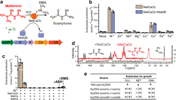
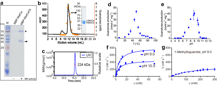
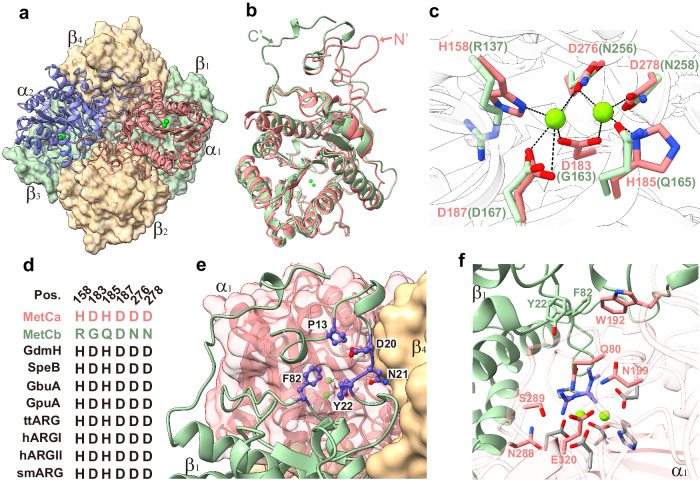
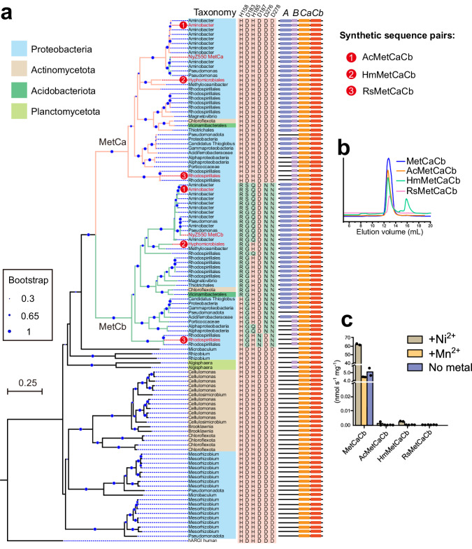
The biguanide drug metformin is a first-line blood glucose-lowering medication for type 2 diabetes, leading to its presence in the global environment. However, little is known about the fate of metformin by microbial catabolism. Here, we characterize a Ni 2+ -dependent heterohexameric enzyme (MetCaCb) from the ureohydrolase superfamily, catalyzing the hydrolysis of metformin into guanylurea and dimethylamine. Either subunit alone is catalytically inactive, but together they work as an active enzyme highly specific for metformin. The crystal structure of the MetCaCb complex shows the coordination of the binuclear metal cluster only in MetCa, with MetCb as a protein binder of its active cognate. An in-silico search and functional assay discover a group of MetCaCb-like protein pairs exhibiting metformin hydrolase activity in the environment. Our findings not only establish the genetic and biochemical foundation for metformin catabolism but also provide additional insights into the adaption of the ancient enzymes toward newly occurred substrate.
Overview
The claims and statistics of this paper haven’t been checked yet.
Run the check — free, no account needed. It reads the open-access text and checks the sources it cites.
Integrity signals
Registry
Cites
Statistics
Figures
Verification
Claims the paper makes
The claims and statistics of this paper haven’t been checked yet.
Run the check — free, no account needed. It reads the open-access text and checks the sources it cites.
Self-check
Percentages, sums and reported differences re-computed from the paper’s own numbers.
The self-check needs the paper’s full text.
Citations
Cites
Highly accurate protein structure prediction with AlphaFold
Jumper, J., Evans, R., Pritzel, A., Green, T., Figurnov, M., Ronneberger, O., Tunyasuvunakool, K., Bates, R., Žídek, A., Potapenko, A., et al. (2021) · Nature · doi:10.1038/s41586-021-03819-2 · Cited by 43,764
McGee, D. J., Zabaleta, J., Viator, R. J., Testerman, T. L., Ochoa, A. C., & Mendz, G. L. (2004) · European Journal of Biochemistry · doi:10.1111/j.1432-1033.2004.04105.x · Cited by 84
Research works printed but not linked: 48
- Dos, S. G. R. et al. Medicines as an emergent contaminant: the review of microbial biodegration potential. Folia Microbiol. (Praha) 67, 157–174 (2022). · DOI
- Wilkinson, J. L. et al. Pharmaceutical pollution of the world’s rivers. Proc. Natl Acad. Sci. USA 119, e2113947119 (2022). · DOI
- Lindell, A. E., Zimmermann-Kogadeeva, M. & Patil, K. R. Multimodal interactions of drugs, natural compounds and pollutants with the gut microbiota. Nat. Rev. Microbiol. 20, 431–443 (2022). · DOI
- Zimmermann, M., Zimmermann-Kogadeeva, M., Wegmann, R. & Goodman, A. L. Mapping human microbiome drug metabolism by gut bacteria and their genes. Nature 570, 462–467 (2019). · DOI · Paper page
- Werner, E. A. & Bell, J. CCXIV.—The preparation of methylguanidine, and of ββ-dimethylguanidine by the interaction of dicyanodiamide, and methylammonium and dimethylammonium chlorides respectively. J. Chem. Soc. Trans. 121, 1790–1794 (1922). · DOI
- Foretz, M., Guigas, B. & Viollet, B. Metformin: update on mechanisms of action and repurposing potential. Nat. Rev. Endocrinol. 19, 460–476 (2023). · DOI
- Balakrishnan, A., Sillanpää, M., Jacob, M. M. & Vo, D. N. Metformin as an emerging concern in wastewater: Occurrence, analysis and treatment methods. Environ. Res. 213, 113613 (2022). · DOI
- Elizalde-Velazquez, G. A. & Gomez-Olivan, L. M. Occurrence, toxic effects and removal of metformin in the aquatic environments in the world: Recent trends and perspectives. Sci. Total Environ. 702, 134924 (2020). · DOI
- Cabreiro, F. et al. Metformin retards aging in C. elegans by altering microbial folate and methionine metabolism. Cell 153, 228–239 (2013). · DOI · Paper page
- Forslund, K. et al. Disentangling type 2 diabetes and metformin treatment signatures in the human gut microbiota. Nature 528, 262–266 (2015). · DOI · Paper page
- He, Y., Zhang, Y. & Ju, F. Metformin contamination in global waters: biotic and abiotic transformation, byproduct generation and toxicity, and evaluation as a pharmaceutical indicator. Environ. Sci. Technol. 56, 13528–13545 (2022). · DOI
- Briones, R. M., Zhuang, W. Q. & Sarmah, A. K. Biodegradation of metformin and guanylurea by aerobic cultures enriched from sludge. Environ. Pollut. 243, 255–262 (2018). · DOI
- Poursat, B. A. J. et al. Biodegradation of metformin and its transformation product, guanylurea, by natural and exposed microbial communities. Ecotox. Environ. Saf. 182, 109414 (2019). · DOI
- Tassoulas, L. J., Robinson, A., Martinez-Vaz, B., Aukema, K. G. & Wackett, L. P. Filling in the gaps in metformin biodegradation: a new enzyme and a metabolic pathway for guanylurea. Appl. Environ. Microbiol. 87, e03003–e03020 (2021). · DOI
- Martinez-Vaz, B. M. et al. Wastewater bacteria remediating the pharmaceutical metformin: Genomes, plasmids and products. Front. Bioeng. Biotechnol. 10, 1086261 (2022). · DOI
- Li, T., Xu, Z.-J. & Zhou, N.-Y. Aerobic Degradation of the antidiabetic drug metformin by Aminobacter sp. strain NyZ550. Environ. Sci. Technol. 57, 1510–1519 (2023). · DOI
- Chaignaud, P. et al. A Methylotrophic bacterium growing with the antidiabetic drug metformin as its sole carbon, nitrogen and energy source. Microorganisms 10, 2302 (2022). · DOI
- Hillmann, K. B. & Niehaus, T. D. Genome sequences of two Pseudomonas isolates that can use metformin as the sole nitrogen source. Microbiol. Resour. Announc. 11, e0063922 (2022). · DOI
- Hernández, V. M., Arteaga, A. & Dunn, M. F. Diversity, properties and functions of bacterial arginases. FEMS Microbiol. Rev. 45, fuab034 (2021). · DOI
- Baggio, R. et al. Inhibition of Mn2+2-arginase by borate leads to the design of a transition state analogue inhibitor, 2(S)-amino-6-boronohexanoic acid. J. Am. Chem. Soc. 119, 8107–8108 (1997). · DOI
- Di Costanzo, L. et al. Crystal structure of human arginase I at 1.29-Å resolution and exploration of inhibition in the immune response. Proc. Natl Acad. Sci. USA 102, 13058–13063 (2005). · DOI
- Velázquez-Libera, J. L., Caballero, J., Tuñón, I., Hernández-Rodríguez, E. W. & Ruiz-Pernía, J. J. On the nature of the enzyme–substrate complex and the reaction mechanism in human arginase I. A combined molecular dynamics and QM/MM study. ACS Catal. 10, 8321–8333 (2020). · DOI
- O’Leary, N. et al. Reference sequence (RefSeq) database at NCBI: current status, taxonomic expansion, and functional annotation. Nucleic Acids Res 44, D733–D745 (2015). · DOI · Paper page
- Tassoulas, L. J., Rankin, J. A., Elias, M. H. & Wackett, L. P. Dinickel enzyme evolved to metabolize the pharmaceuticalmetformin and its implications for wastewater and human microbiomes. Proc. Natl Acad. Sci. USA 121, e2312652121 (2024). · DOI
- Miller, D., Xu, H. & White, R. H. A new subfamily of agmatinases present in Methanogenic Archaea Is Fe(II) dependent. Biochemistry 51, 3067–3078 (2012). · DOI
- Saragadam, T., Kumar, S. & Punekar, N. Characterization of 4-guanidinobutyrase from Aspergillus niger. Microbiology 165, 396–410 (2019). · DOI
- Arakawa, N., Igarashi, M., Kazuoka, T., Oikawa, T. & Soda, K. D-arginase of Arthrobacter sp. KUJ 8602: characterization and its identity with Zn2+-guanidinobutyrase. J. biochem. 133, 33–42 (2003). · DOI
- Funck, D. et al. Discovery of a Ni2+-dependent guanidine hydrolase in bacteria. Nature 603, 515–521 (2022). · DOI
- Romero, N. et al. Mammalian agmatinases constitute unusual members in the family of Mn2+-dependent ureahydrolases. J. Inorg. Biochem. 166, 122–125 (2017). · DOI
- Hai, Y., Kerkhoven, E. J., Barrett, M. P. & Christianson, D. W. Crystal structure of an arginase-like protein from Trypanosoma brucei that evolved without a binuclear manganese cluster. Biochemistry 54, 458–471 (2015). · DOI
- Mothersole, R. G. et al. Sequence divergence in the arginase domain of ornithine decarboxylase/arginase in Fusobacteriacea leads to loss of function in oral associated species. Biochemistry 61, 1378–1391 (2022). · DOI
- Garcia, D., Uribe, E., Salgado, M. & Martínez, M. Mutagenic and kinetic support for an allosteric site in arginase from the extreme thermophile Bacillus caldovelox, which allows activation by arginine. Biochimie. 108, 8–12 (2014). · DOI
- Lobos, M. et al. Insights on the participation of Glu256 and Asp204 in the oligomeric structure and cooperative effects of human arginase type I. J. Struct. Biol. 211, 107533 (2020). · DOI
- Lavulo, L. et al. Subunit-subunit interactions in trimeric arginase. Generation of active monomers by mutation of a single amino acid. J. biol. Chem. 276, 14242–14248 (2001). · DOI
- Adrain, C. & Freeman, M. New lives for old: evolution of pseudoenzyme function illustrated by iRhoms. Nat. rev. Mol. cell biol. 13, 489–498 (2012). · DOI
- Schneider, N. O. et al. Solving the conundrum: widespread proteins annotated for urea metabolism in bacteria are carboxyguanidine deiminases mediating nitrogen assimilation from guanidine. Biochemistry 59, 3258–3270 (2020). · DOI
- Fida, T. T., Palamuru, S., Pandey, G. & Spain, J. C. Aerobic biodegradation of 2,4-dinitroanisole by Nocardioides sp. strain JS1661. Appl. Environ. Microbiol. 80, 7725–7731 (2014). · DOI
- Balaich, J. et al. The human microbiome encodes resistance to the antidiabetic drug acarbose. Nature 600, 110–115 (2021). · DOI
- Tian, J. et al. Inactivation of the antidiabetic drug acarbose by human intestinal microbial-mediated degradation. Nat. Metab. 5, 1–14 (2023). · DOI
- Brown, J. B., Conner, C. & Nichols, G. A. Secondary failure of metformin monotherapy in clinical practice. Diabetes Care 33, 501–506 (2010). · DOI
- Liu, H., Wang, S. J. & Zhou, N. Y. A new isolate of Pseudomonas stutzeri that degrades 2-chloronitrobenzene. Biotechnol. Lett. 27, 275–278 (2005). · DOI
- Kabsch, W. XDS. Acta Crystallogr D. Biol. Crystallogr 66, 125–132 (2010). · DOI · Paper page
- McCoy, A. et al. PHASER crystallographic software. J. appl. Crystallogr. 40, 658–674 (2007). · DOI · Paper page
- Adams, P. et al. The Phenix software for automated determination of macromolecular structures. Methods 55, 94–106 (2011). · DOI
- Emsley, P., Lohkamp, B., Scott, W. G. & Cowtan, K. Features and development of COOT. Acta Crystallogr. Sect. D., Biol. Crystallogr. 66, 486–501 (2010). · DOI · Paper page
- Williams, C. J. et al. MolProbity: More and better reference data for improved all-atom structure validation. Protein Sci. 27, 293–315 (2018). · DOI · Paper page
- Trott, O. & Olson, A. J. AutoDock Vina: improving the speed and accuracy of docking with a new scoring function, efficient optimization, and multithreading. J. Comput. Chem. 31, 455–461 (2010). · DOI · Paper page
- Madeira, F. et al. Search and sequence analysis tools services from EMBL-EBI in 2022. Nucleic acids res 50, W276–W279 (2022). · DOI
Metadata
- Authors
- Li, T., Xu, Z.-J., Zhang, S.-T., Xu, J., Pan, P., & Zhou, N.-Y.
- Affiliations
- Shanghai Jiao Tong University (CN)
- Corresponding author
- Zhou, Ning-Yi
- Venue
- Nature Communications
- Published
- 2024-07-20
- Volume
- 15
- Issue
- 1
- Pages
- 6121-6121
- ISSN
- 2041-1723 (ISSN-L)
- PMC
- PMC11271267
- OpenAlex
- W4400842703
- Cited by
- 13
- References
- 2
- Citations by year
- 2026: 4 · 2025: 5 · 2024: 4
- FWCI
- 1.30
- Type
- journal article
- Language
- en
- Open access
- gold
- doi.org
- Locations
- Nature Communications (publishedVersion, cc-by)
- Research Square (acceptedVersion, cc-by)
- PubMed Central (publishedVersion, cc-by)
- PubMed (publishedVersion)
- RePEc: Research Papers in Economics (submittedVersion)
- DOAJ (DOAJ: Directory of Open Access Journals) (submittedVersion, cc-by-sa)
- Funders
- National Natural Science Foundation of China (32230001)
- Topics
- Physical Sciences › Environmental Science › Pollution › Pharmaceutical and Antibiotic Environmental Impacts
- Life Sciences › Biochemistry, Genetics and Molecular Biology › Structural Biology › Enzyme Structure and Function
- Life Sciences › Biochemistry, Genetics and Molecular Biology › Molecular Biology › Enzyme Catalysis and Immobilization
- Keywords
- metformin, nickel-dependent enzymes, guanylurea, dimethylamine, type 2 diabetes drugs, environmental fate of pharmaceuticals, enzyme evolution, crystal structure
- MeSH terms
- Bacterial Proteins, Hydrolases, Hydrolysis, Metformin, Models, Molecular, Nickel, Substrate Specificity, Crystallography, X-Ray
About these checks
Registry, retraction, Cites and statistics results are computed, not judged, with one exception: a printed reference that no index lists is read once by a model before it counts as a citation error. Claims are extracted by a model and kept only when their quote appears verbatim in the text; each cited source is then checked, with a model’s help, for whether it says what the claim relies on. Those verdicts can be wrong — read the claim’s quote and the cited source before relying on one.
Statistics and arithmetic are recomputed, not judged.
Confidence bands are shown only for fields calibrated on our annotated test set. None is calibrated yet, so no band is shown.
What counts as a claim ›How we measure confidence ›How we check this ›
Are you an author, or do you see a problem with this page? Request a review
