Cited by 15Cancer-related Molecular PathwaysNuclear Structure and FunctionRNA and protein synthesis mechanisms

The DOI matches its Crossref record; 3 of the 3 references it cites were found in a registry or an index.

SURF2 is a MDM2 antagonist in triggering the nucleolar stress response

Open access

Published version of a preprint

Abstract

Cancer cells rely on high ribosome production to sustain their proliferation rate. Many chemotherapies impede ribosome production which is perceived by cells as "nucleolar stress" (NS), triggering p53-dependent and independent pathways leading to cell cycle arrest and/or apoptosis. The 5S ribonucleoprotein (RNP) particle, a sub-ribosomal particle, is instrumental to NS response. Upon ribosome assembly defects, the 5S RNP accumulates as free form. This free form is able to sequester and inhibit MDM2, thus promoting p53 stabilization. To investigate how cancer cells can resist to NS, here we purify free 5S RNP and uncover an interaction partner, SURF2. Functional characterization of SURF2 shows that its depletion increases cellular sensitivity to NS, while its overexpression promotes their resistance to it. Consistently, SURF2 is overexpressed in many cancers and its expression level is an independent marker of prognosis for adrenocortical cancer. Our data demonstrate that SURF2 buffers free 5S RNP particles, and can modulate their activity, paving the way for the research of new molecules that can finely tune the response to nucleolar stress in the framework of cancer therapies.

Overview

The claims and statistics of this paper haven’t been checked yet.

Run the check — free, no account needed. It reads the open-access text and checks the sources it cites.

A few free checks a day · papers already verified open instantly · results are public

Integrity signals

Registry

Crossref · 10.1038/s41467-024-52659-x

Cites

3 of 3 references verified0 citation errors · 0 not checkable

Statistics

Figures

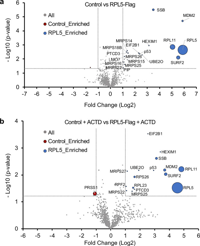
Fig. 1 Label-free quantitative proteomics analysis of RPL5-FLAG co-purified proteins. Nano-liquid chromatography-tandem mass spectrometry (nanoLC-MS/MS) analysis of trypsin digested proteins retained on flag-coated beads issued from U2OS control cells (LEFT quadrant) or cell expressing RPL5-FLAG (RIGHT quadrant). Three independent experimental replicates (n = 3) were performed. Volcano plots showing proteins significantly enriched for control cells (red) versus cells expressing RPL5-FLAG (a) in untreated cells (b) in cells treated with actinomycin D (ACTD, 10 ng/ml) for 24 h. An unpaired two-tailed Student’s t test with equal variance was used. Enrichment significance thresholds are represented by an absolute log2-transformed fold-change (FC) greater than 1 and a -log10-transformed (p-value) greater than 1.3. The iBAQ (intensity-Based Absolute Quantification) values are represented by the diameter of each dot for the proteins that are significantly enriched in order to rank their absolute abundances73. Source data are provided as a Source Data file.
Fig. 1 Label-free quantitative proteomics analysis of RPL5-FLAG co-purified proteins. Nano-liquid chromatography-tandem mass spectrometry (nanoLC-MS/MS) analysis of trypsin digested proteins retained on flag-coated beads issued from U2OS control cells (LEFT quadrant) or cell expressing RPL5-FLAG (RIGHT quadrant). Three independent experimental replicates (n = 3) were performed. Volcano plots showing proteins significantly enriched for control cells (red) versus cells expressing RPL5-FLAG (a) in untreated cells (b) in cells treated with actinomycin D (ACTD, 10 ng/ml) for 24 h. An unpaired two-tailed Student’s t test with equal variance was used. Enrichment significance thresholds are represented by an absolute log2-transformed fold-change (FC) greater than 1 and a -log10-transformed (p-value) greater than 1.3. The iBAQ (intensity-Based Absolute Quantification) values are represented by the diameter of each dot for the proteins that are significantly enriched in order to rank their absolute abundances73. Source data are provided as a Source Data file.CC BY-NC-ND · PubMed Central
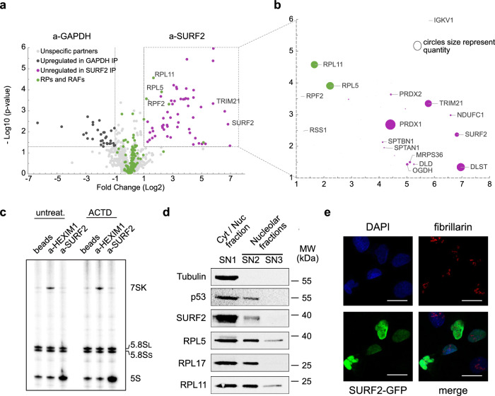
Fig. 2 SURF2 is a partner of free 5S particles and is involved in p53 regulation. a Label-free quantitative proteomics analysis of trypsin-digested proteins retained on beads coated by anti-GAPDH or anti-SURF2 antibodies. Three independent experimental replicates (n = 3) were performed. Volcano plot showing proteins significantly enriched in the GAPDH IP (brown) versus the SURF2 IP (purple). An unpaired two-tailed Student’s t test with equal variance was used. Enrichment significance thresholds are represented by an absolute log2-transformed fold-change (FC) greater than 1 and a -log10-transformed (p-value) greater than 1.3. Proteins involved in ribosome biogenesis and function are indicated in green. b Focus on the iBAQ values, represented by the diameter of each dot, for the proteins significantly enriched in the SURF2 IP. c Detection of RNAs associated with HEXIM1 and SURF2. U2OS RNAs co-immunoprecipitated with endogenous HEXIM1 (a-HEXIM1) or endogenous SURF2 (a-SURF2) were 3’-labeled and separated on a 6% acrylamide gel. Control IP reaction performed without antibodies (beads) is shown (n = 1). d U2OS cell extracts were fractionated using the PSE method. Western-blot analyses showing the contents of different factors (indicated at the left of the gel) in the SN1, SN2, and SN3 fractions obtained with the PSE method (n = 2). e Microscopy analyses of U2OS cells over-expressing SURF2-GFP from TET inducible promoter. Cells were induced with tetracycline to obtain a similar expression as the endogenous copy (tetracycline at 5 ng/mL), and a SURF2-GFP signal was detected (Green). The same cells were also probed for fibrillarin to stain nucleoli. DAPI coloration is used to localize the nuclear compartment. Scale bar 25 μm. Source data are provided as a Source Data file.
Fig. 2 SURF2 is a partner of free 5S particles and is involved in p53 regulation. a Label-free quantitative proteomics analysis of trypsin-digested proteins retained on beads coated by anti-GAPDH or anti-SURF2 antibodies. Three independent experimental replicates (n = 3) were performed. Volcano plot showing proteins significantly enriched in the GAPDH IP (brown) versus the SURF2 IP (purple). An unpaired two-tailed Student’s t test with equal variance was used. Enrichment significance thresholds are represented by an absolute log2-transformed fold-change (FC) greater than 1 and a -log10-transformed (p-value) greater than 1.3. Proteins involved in ribosome biogenesis and function are indicated in green. b Focus on the iBAQ values, represented by the diameter of each dot, for the proteins significantly enriched in the SURF2 IP. c Detection of RNAs associated with HEXIM1 and SURF2. U2OS RNAs co-immunoprecipitated with endogenous HEXIM1 (a-HEXIM1) or endogenous SURF2 (a-SURF2) were 3’-labeled and separated on a 6% acrylamide gel. Control IP reaction performed without antibodies (beads) is shown (n = 1). d U2OS cell extracts were fractionated using the PSE method. Western-blot analyses showing the contents of different factors (indicated at the left of the gel) in the SN1, SN2, and SN3 fractions obtained with the PSE method (n = 2). e Microscopy analyses of U2OS cells over-expressing SURF2-GFP from TET inducible promoter. Cells were induced with tetracycline to obtain a similar expression as the endogenous copy (tetracycline at 5 ng/mL), and a SURF2-GFP signal was detected (Green). The same cells were also probed for fibrillarin to stain nucleoli. DAPI coloration is used to localize the nuclear compartment. Scale bar 25 μm. Source data are provided as a Source Data file.CC BY-NC-ND · PubMed Central
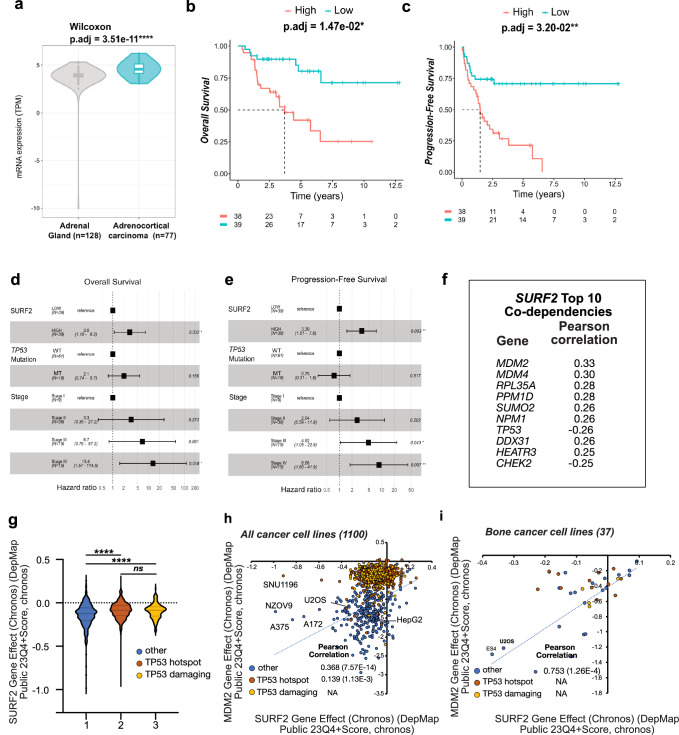
Fig. 3 SURF2 is overexpressed in adrenocortical carcinomas and its knock-out negatively affects cancer cells similarly to MDM2. a Comparison of SURF2 mRNA levels in non-tumoral (left panel, gray values) and tumoral tissues (right panel, blue values). Data were extracted from the standardized Xena database to compare healthy adrenal gland tissues (n = 128, origin GTEx dataset) and adrenocortical carcinoma tissues (n = 77, origin TCGA dataset). Compared to healthy adrenal gland tissues, SURF2 mRNA levels are higher in adrenocortical carcinoma tissue. ****: p < 0.0001 (Wilcoxon Test). Data are presented as a violin plot. It comprises a density plot, the width of which indicates the frequency, and a box plot, where the extreme points reflect the minimum (Q0) and maximum (Q4), the length of the box represents the interquartile range (IQR: percentile Q1 to Q3) and the center represents the median. b, c Association between SURF2 mRNA levels and patient survivals. Using the standardized Xena dataset, associations between SURF2 mRNA levels (median as cut-off value) and overall survival (b) or progression-free survival (c) were determined using Kaplan-Meier curves. A significant association was observed between high levels of SURF2 mRNA levels and poor patient survival. *: p < 0.05; **: p < 0.01 (Log-rank). d, e Association between SURF2 mRNA levels and patient survivals after adjustment on clinical parameters. Using the standardized Xena dataset, multivariate Cox regression models for overall survival (Low n = 39, High n = 38) (d) or progression-free survival (e) were performed on clinical parameters significantly associated with patient survivals (i.e., TP53 mutation status and tumor stage, See Supplementary Table S6). High mRNA levels of SURF2 and stage IV are independent markers of poor survival of patients carrying adrenocortical carcinoma. *: p < 0.05; **: p < 0.01 (Cox Proportional-Hazards Model, two-sided hypothesis test). Error bars show 95%CI (confidence interval) and square HR (hazard ratio), p-value (0.032 and 0.003 respectively), source data are provided as a Source Data file. f Top SURF2 correlated genes in Project Achilles. The Pearson correlation score on the right indicates the strength of co-dependencies between SURF2 and the indicated genes. g SURF2 knock-out chronos effect on different cancer cells classified into three groups depending on their TP53 status: wild-type (blue, n = 386), hot-spot mutations (brown, n = 545), damaging mutations (yellow, n = 169). The data are presented as the mean ± s.d. Statistical analysis by two-way ANOVA with Turkey’s multiple comparisons test. Significant differences are indicated by p-value < 0.0001 (****) (ns = no significance). h Classification of more than 1000 cancer cells based on their associated chronos effect of SURF2 knock-out and MDM2 knock-out. Pearson correlation between the two chronos effects is indicated depending on TP53 status. i Same as in (h) but focusing on bone cancer cell lines. Source data are provided as a Source Data file.
Fig. 3 SURF2 is overexpressed in adrenocortical carcinomas and its knock-out negatively affects cancer cells similarly to MDM2. a Comparison of SURF2 mRNA levels in non-tumoral (left panel, gray values) and tumoral tissues (right panel, blue values). Data were extracted from the standardized Xena database to compare healthy adrenal gland tissues (n = 128, origin GTEx dataset) and adrenocortical carcinoma tissues (n = 77, origin TCGA dataset). Compared to healthy adrenal gland tissues, SURF2 mRNA levels are higher in adrenocortical carcinoma tissue. ****: p < 0.0001 (Wilcoxon Test). Data are presented as a violin plot. It comprises a density plot, the width of which indicates the frequency, and a box plot, where the extreme points reflect the minimum (Q0) and maximum (Q4), the length of the box represents the interquartile range (IQR: percentile Q1 to Q3) and the center represents the median. b, c Association between SURF2 mRNA levels and patient survivals. Using the standardized Xena dataset, associations between SURF2 mRNA levels (median as cut-off value) and overall survival (b) or progression-free survival (c) were determined using Kaplan-Meier curves. A significant association was observed between high levels of SURF2 mRNA levels and poor patient survival. *: p < 0.05; **: p < 0.01 (Log-rank). d, e Association between SURF2 mRNA levels and patient survivals after adjustment on clinical parameters. Using the standardized Xena dataset, multivariate Cox regression models for overall survival (Low n = 39, High n = 38) (d) or progression-free survival (e) were performed on clinical parameters significantly associated with patient survivals (i.e., TP53 mutation status and tumor stage, See Supplementary Table S6). High mRNA levels of SURF2 and stage IV are independent markers of poor survival of patients carrying adrenocortical carcinoma. *: p < 0.05; **: p < 0.01 (Cox Proportional-Hazards Model, two-sided hypothesis test). Error bars show 95%CI (confidence interval) and square HR (hazard ratio), p-value (0.032 and 0.003 respectively), source data are provided as a Source Data file. f Top SURF2 correlated genes in Project Achilles. The Pearson correlation score on the right indicates the strength of co-dependencies between SURF2 and the indicated genes. g SURF2 knock-out chronos effect on different cancer cells classified into three groups depending on their TP53 status: wild-type (blue, n = 386), hot-spot mutations (brown, n = 545), damaging mutations (yellow, n = 169). The data are presented as the mean ± s.d. Statistical analysis by two-way ANOVA with Turkey’s multiple comparisons test. Significant differences are indicated by p-value < 0.0001 (****) (ns = no significance). h Classification of more than 1000 cancer cells based on their associated chronos effect of SURF2 knock-out and MDM2 knock-out. Pearson correlation between the two chronos effects is indicated depending on TP53 status. i Same as in (h) but focusing on bone cancer cell lines. Source data are provided as a Source Data file.CC BY-NC-ND · PubMed Central
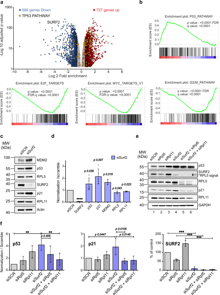
Fig. 4 SURF2 depletion up-regulates the p53 pathway. RNA-seq analyses of SURF2 depletion. U2OS cells were treated by scramble siRNAs or by siRNAs against SURF2 for 96 h. mRNA expression levels were compared between the two conditions, and experiments were repeated (n = 3). a Volcano plot showing significantly differentially expressed genes. Using DESeq2, a comparison of gene expression between the customer-defined groups of samples was performed. The Wald test was used to generate p-values and log2 fold changes. Genes with an adjusted p-value < 0.05 and absolute log2 fold change > 1 were called differentially expressed genes indicated: up-regulated genes (red) and down-regulated (blue) or unaffected (gray). Genes from the TP53 pathway are highlighted yellow. b Gene set enrichment analysis (GSEA) in SURF2 depleted genes or in the control condition (siSCR). ES; enrichment score. GSEA P-values were derived from permutation testing and corrected for multiple testing using the False Discovery Rate (FDR) method. Enrichment score (ES) and FDR value were applied to sort SURF2 depleted and control cells genes-enriched after gene set permutations were performed 1000 times for the analysis. Gene Network Analysis was based on DEGs (log2foldchange > 1). c Western-blot analyses showing the accumulation of different proteins in 20 µg of cellular extracts produced from U2OS cells treated with scrambled siRNAs (siSCR) or with siRNAs directed against SURF2 (siSurf2). d Quantification of signals obtained by western-blot analysis and normalized to actin signals in each condition (c) from independent biological replicates (p53, n = 5; SURF2, n = 5; p21 n = 5; MDM2, n = 3; RPL5, n = 3; RPL11, n = 3). Two-tailed t-test analysis was used for statistics. Data are presented as mean values +/− SD. Significant differences are indicated by stars (p-value ≤0.05*; ≤0.01**;≤0.001*** and ≤0.0001****) or with the precise p-value on the graph (p). e Co-depletion experiment. Western-blot analyses showing the accumulation of different proteins in 20 µg of cellular extracts issued from U2OS cells treated with a combination of different siRNAs: scrambled (siSCR); RPL5 (siRPL5); RPL11 (siRPL11) or SURF2 (siSurf2). f Quantification of signals obtained by western-blot analysis and normalized to GAPDH signals in each condition (h) (n = 3). Data are presented as mean values +/− SD. A one-way ANOVA test was used for statistics. Significant differences are indicated by stars (p-value ≤0.05*; ≤0.01** and ≤0.001***) or with the precise p-value on the graph (p). Source data are provided as a Source Data file.
Fig. 4 SURF2 depletion up-regulates the p53 pathway. RNA-seq analyses of SURF2 depletion. U2OS cells were treated by scramble siRNAs or by siRNAs against SURF2 for 96 h. mRNA expression levels were compared between the two conditions, and experiments were repeated (n = 3). a Volcano plot showing significantly differentially expressed genes. Using DESeq2, a comparison of gene expression between the customer-defined groups of samples was performed. The Wald test was used to generate p-values and log2 fold changes. Genes with an adjusted p-value < 0.05 and absolute log2 fold change > 1 were called differentially expressed genes indicated: up-regulated genes (red) and down-regulated (blue) or unaffected (gray). Genes from the TP53 pathway are highlighted yellow. b Gene set enrichment analysis (GSEA) in SURF2 depleted genes or in the control condition (siSCR). ES; enrichment score. GSEA P-values were derived from permutation testing and corrected for multiple testing using the False Discovery Rate (FDR) method. Enrichment score (ES) and FDR value were applied to sort SURF2 depleted and control cells genes-enriched after gene set permutations were performed 1000 times for the analysis. Gene Network Analysis was based on DEGs (log2foldchange > 1). c Western-blot analyses showing the accumulation of different proteins in 20 µg of cellular extracts produced from U2OS cells treated with scrambled siRNAs (siSCR) or with siRNAs directed against SURF2 (siSurf2). d Quantification of signals obtained by western-blot analysis and normalized to actin signals in each condition (c) from independent biological replicates (p53, n = 5; SURF2, n = 5; p21 n = 5; MDM2, n = 3; RPL5, n = 3; RPL11, n = 3). Two-tailed t-test analysis was used for statistics. Data are presented as mean values +/− SD. Significant differences are indicated by stars (p-value ≤0.05*; ≤0.01**;≤0.001*** and ≤0.0001****) or with the precise p-value on the graph (p). e Co-depletion experiment. Western-blot analyses showing the accumulation of different proteins in 20 µg of cellular extracts issued from U2OS cells treated with a combination of different siRNAs: scrambled (siSCR); RPL5 (siRPL5); RPL11 (siRPL11) or SURF2 (siSurf2). f Quantification of signals obtained by western-blot analysis and normalized to GAPDH signals in each condition (h) (n = 3). Data are presented as mean values +/− SD. A one-way ANOVA test was used for statistics. Significant differences are indicated by stars (p-value ≤0.05*; ≤0.01** and ≤0.001***) or with the precise p-value on the graph (p). Source data are provided as a Source Data file.CC BY-NC-ND · PubMed Central
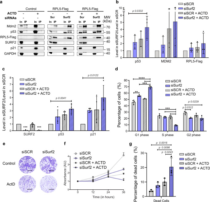
Fig. 5 SURF2 depletion increases free 5S binding to MDM2 and increases cell sensitivity to nucleolar stress. a Detection of proteins associated with RPL5-Flag. Cell extracts produced from U2OS cells overexpressing RPL5-FLAG or not (control) and differentially treated by actinomycin D addition for 24 h (at 10 ng/mL) or not (ACTD + or −) and transfected with scrambled siRNAs (siSCR) or directed against SURF2 (siSurf2) were used to perform immuno-precipitation on beads coated with anti-flag. 10% of the inputs were loaded (In) aside IPs (IP) on a gel to perform western blots using antibodies directed against the indicated proteins to analyze the co-purification efficiency of the different factors. b Quantification of the co-purification efficiency with RPL5-Flag observed (a) for the indicated proteins (n = 3). Results are represented as a comparison of the enrichments observed in cells treated with siRNAs directed against SURF2 (siSurf2) normalized to the ones observed in cells treated with scrambled siRNAs (siSCR). Two-tailed t-test analysis was used for statistics. Data are presented as mean values +/− SD. Significant differences are indicated by stars (p-value ≤0.05*; ≤0.01**;≤0.001*** and ≤0.0001****) or with the precise p-value on the graph (p). c Quantification of the signals corresponding to the indicated proteins in the different inputs of western-blots analyses (a) from independent experiments n = 4 (original data are provided in the source data file). Two-tailed t test analysis was used for statistics. Data are presented as mean values +/− SD. Significant differences are indicated by stars (p-value ≤0.05*; ≤0.01**;≤0.001*** and ≤0.0001****) or with the precise p-value on the graph (p). d DNA content analysis of U2OS cells treated as indicated (siSCR: scrambled siRNAs; siSurf2: siRNAs against SURF2; ACTD: actinomycin D at 10 ng/mL for 24 h) by FACS. Quantification of U2OS cell repartition in the different phases of the cell cycle following different treatments (n = 3). Data are presented as mean values +/− SD. One-way Anova Test was used for statistics. Significant differences are indicated by stars (p-value ≤0.05*; ≤0.01**;≤0.001*** and ≤0.0001****) or with the precise p-value on the graph (p). e, f Proliferation analysis of differentially treated U2OS cells. Cells are treated as indicated (siSCR: scrambled siRNAs; siSurf2: siRNAs against SURF2; ACTD: actinomycin D at 10 ng/mL for 24 h). e Cells are platted on 6 well plate after being transfected with the indicated siRNAs, 24 h before being analyzed, cells were treated with actinomycin D (10 ng/mL) or H2O. Cells were stained with crystal violet to take a picture (n = 3) scale bar (0.5 mm). f Fixed crystal violet was resolubilized and quantified by absorbance for each condition after different time exposure to treatments (n = 3). g Cell apoptosis assays with an annexin-V-FITC and propidium iodide of differentially treated U2OS cells. Both apoptosis and necrosis are regrouped as dead cells (n = 3). Data are presented as mean values +/− SD. The One-way ANOVA test was used for statistics. Significant differences are indicated by stars (p-value ≤0.05*; ≤0.01**;≤0.001*** and ≤0.0001****) or with the precise p-value on the graph (p). Source data are provided as a Source Data file.
Fig. 5 SURF2 depletion increases free 5S binding to MDM2 and increases cell sensitivity to nucleolar stress. a Detection of proteins associated with RPL5-Flag. Cell extracts produced from U2OS cells overexpressing RPL5-FLAG or not (control) and differentially treated by actinomycin D addition for 24 h (at 10 ng/mL) or not (ACTD + or −) and transfected with scrambled siRNAs (siSCR) or directed against SURF2 (siSurf2) were used to perform immuno-precipitation on beads coated with anti-flag. 10% of the inputs were loaded (In) aside IPs (IP) on a gel to perform western blots using antibodies directed against the indicated proteins to analyze the co-purification efficiency of the different factors. b Quantification of the co-purification efficiency with RPL5-Flag observed (a) for the indicated proteins (n = 3). Results are represented as a comparison of the enrichments observed in cells treated with siRNAs directed against SURF2 (siSurf2) normalized to the ones observed in cells treated with scrambled siRNAs (siSCR). Two-tailed t-test analysis was used for statistics. Data are presented as mean values +/− SD. Significant differences are indicated by stars (p-value ≤0.05*; ≤0.01**;≤0.001*** and ≤0.0001****) or with the precise p-value on the graph (p). c Quantification of the signals corresponding to the indicated proteins in the different inputs of western-blots analyses (a) from independent experiments n = 4 (original data are provided in the source data file). Two-tailed t test analysis was used for statistics. Data are presented as mean values +/− SD. Significant differences are indicated by stars (p-value ≤0.05*; ≤0.01**;≤0.001*** and ≤0.0001****) or with the precise p-value on the graph (p). d DNA content analysis of U2OS cells treated as indicated (siSCR: scrambled siRNAs; siSurf2: siRNAs against SURF2; ACTD: actinomycin D at 10 ng/mL for 24 h) by FACS. Quantification of U2OS cell repartition in the different phases of the cell cycle following different treatments (n = 3). Data are presented as mean values +/− SD. One-way Anova Test was used for statistics. Significant differences are indicated by stars (p-value ≤0.05*; ≤0.01**;≤0.001*** and ≤0.0001****) or with the precise p-value on the graph (p). e, f Proliferation analysis of differentially treated U2OS cells. Cells are treated as indicated (siSCR: scrambled siRNAs; siSurf2: siRNAs against SURF2; ACTD: actinomycin D at 10 ng/mL for 24 h). e Cells are platted on 6 well plate after being transfected with the indicated siRNAs, 24 h before being analyzed, cells were treated with actinomycin D (10 ng/mL) or H2O. Cells were stained with crystal violet to take a picture (n = 3) scale bar (0.5 mm). f Fixed crystal violet was resolubilized and quantified by absorbance for each condition after different time exposure to treatments (n = 3). g Cell apoptosis assays with an annexin-V-FITC and propidium iodide of differentially treated U2OS cells. Both apoptosis and necrosis are regrouped as dead cells (n = 3). Data are presented as mean values +/− SD. The One-way ANOVA test was used for statistics. Significant differences are indicated by stars (p-value ≤0.05*; ≤0.01**;≤0.001*** and ≤0.0001****) or with the precise p-value on the graph (p). Source data are provided as a Source Data file.CC BY-NC-ND · PubMed Central
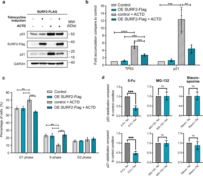
Fig. 6 SURF2 overexpression impedes p53 activation and cell cycle arrest following nucleolar stress. a Western-blot analysis showing the accumulation of different proteins, in 20 µg of cellular extracts produced from control U2OS cells or from U2OS cells that overexpress SURF2-Flag after Tetracycline induction (1 µg/ml) from their T-Rex locus for 24 h. Cells were also treated differentially with or without the addition of actinomycin D (10 mg/ml) for the same period (n = 3). b Quantification of the signals observed in an independent experiment (A) (n = 3). Data are presented as mean values +/− SD. One-way ANOVA tests were used for statistics. Significant differences are indicated by stars (p-value ≤0.05*; ≤0.01** and ≤0.001***). Source data are provided as a Source Data file. c DNA content analysis of U2OS cells treated as indicated (Ctrl: U2OS control cells; OE SURF2-Flag: U2OS cells that overexpress SURF2-Flag; ACTD: Treated with actinomycin D) by FACS. Quantification of different U2OS cell repartition in the different phases of the cell cycle following different treatments is represented as histograms (n = 3). Data are presented as mean values +/− SD. One-way ANOVA tests were used for statistics. Significant differences are indicated by stars (p-value ≤0.05*; ≤0.01** and ≤0.001***). Source data are provided as a Source Data file. d Comparison of p53 and p21 stabilization, assessed by western blots, following drugs treatment (5-Fu: 50 µM; MG-132: 10 µM; staurosporine: 25 nM for 24 h) in U2OS control cells (uninduced) or in cells overexpressing SURF2-Flag (Tetracycline 1 mg/ml). P53 and p21 signals were normalized to 1 in the control experiment, and relative stabilization in cell overexpressing SURF2-Flag was quantified from three independent biological replicates (n = 3). Data are presented as mean values +/− SD. Two-tailed paired T test was used for statistics. Significant differences are indicated by stars (p-value ≤0.05*; ≤0.01** and ≤0.001***). Source data are provided as a Source Data file.
Fig. 6 SURF2 overexpression impedes p53 activation and cell cycle arrest following nucleolar stress. a Western-blot analysis showing the accumulation of different proteins, in 20 µg of cellular extracts produced from control U2OS cells or from U2OS cells that overexpress SURF2-Flag after Tetracycline induction (1 µg/ml) from their T-Rex locus for 24 h. Cells were also treated differentially with or without the addition of actinomycin D (10 mg/ml) for the same period (n = 3). b Quantification of the signals observed in an independent experiment (A) (n = 3). Data are presented as mean values +/− SD. One-way ANOVA tests were used for statistics. Significant differences are indicated by stars (p-value ≤0.05*; ≤0.01** and ≤0.001***). Source data are provided as a Source Data file. c DNA content analysis of U2OS cells treated as indicated (Ctrl: U2OS control cells; OE SURF2-Flag: U2OS cells that overexpress SURF2-Flag; ACTD: Treated with actinomycin D) by FACS. Quantification of different U2OS cell repartition in the different phases of the cell cycle following different treatments is represented as histograms (n = 3). Data are presented as mean values +/− SD. One-way ANOVA tests were used for statistics. Significant differences are indicated by stars (p-value ≤0.05*; ≤0.01** and ≤0.001***). Source data are provided as a Source Data file. d Comparison of p53 and p21 stabilization, assessed by western blots, following drugs treatment (5-Fu: 50 µM; MG-132: 10 µM; staurosporine: 25 nM for 24 h) in U2OS control cells (uninduced) or in cells overexpressing SURF2-Flag (Tetracycline 1 mg/ml). P53 and p21 signals were normalized to 1 in the control experiment, and relative stabilization in cell overexpressing SURF2-Flag was quantified from three independent biological replicates (n = 3). Data are presented as mean values +/− SD. Two-tailed paired T test was used for statistics. Significant differences are indicated by stars (p-value ≤0.05*; ≤0.01** and ≤0.001***). Source data are provided as a Source Data file.CC BY-NC-ND · PubMed Central
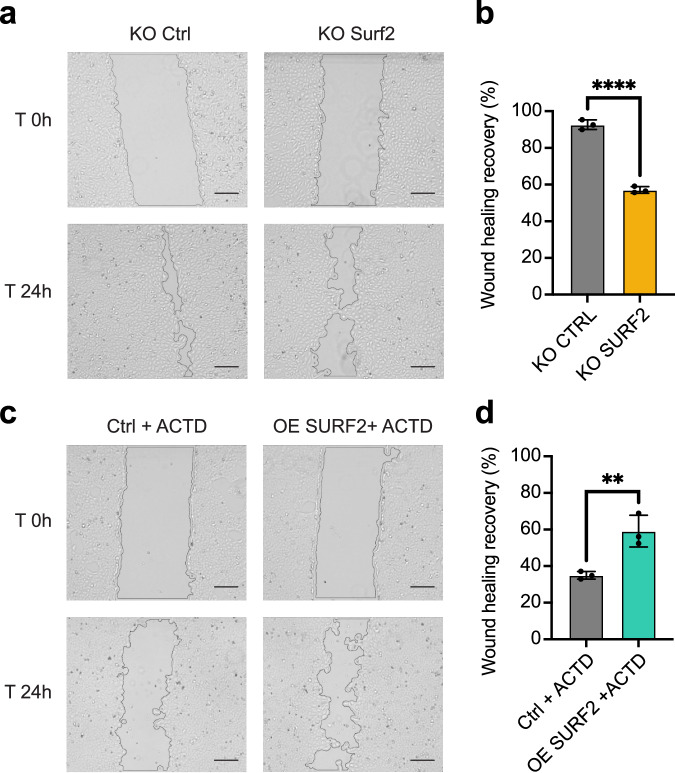
Fig. 7 SURF2 expression level affect U2OS phenotypic traits. Analysis of U2OS KO SURF2 or U2OS overexpressing SURF2-Flag cell migration by wound healing assay. a, c Images were taken with Cell Imaging EVOS (Gx40) at 0 h and 24 h (n = 3) (scale bar = 0.1 mm). b, d Quantification of wound healing by measuring the percentage of persistent scar area after 24 h from three independent biological replicates (n = 3). Use of an unpaired two-tailed t test for statistical tests Data are presented as mean values +/– SD, and significant differences are indicated by stars as follows p-value < 0.05*; 0.01** 0.001*** and 0.0001****) or with the precise p-value on the graph (p). Source data are provided as a Source Data file.
Fig. 7 SURF2 expression level affect U2OS phenotypic traits. Analysis of U2OS KO SURF2 or U2OS overexpressing SURF2-Flag cell migration by wound healing assay. a, c Images were taken with Cell Imaging EVOS (Gx40) at 0 h and 24 h (n = 3) (scale bar = 0.1 mm). b, d Quantification of wound healing by measuring the percentage of persistent scar area after 24 h from three independent biological replicates (n = 3). Use of an unpaired two-tailed t test for statistical tests Data are presented as mean values +/– SD, and significant differences are indicated by stars as follows p-value < 0.05*; 0.01** 0.001*** and 0.0001****) or with the precise p-value on the graph (p). Source data are provided as a Source Data file.CC BY-NC-ND · PubMed Central
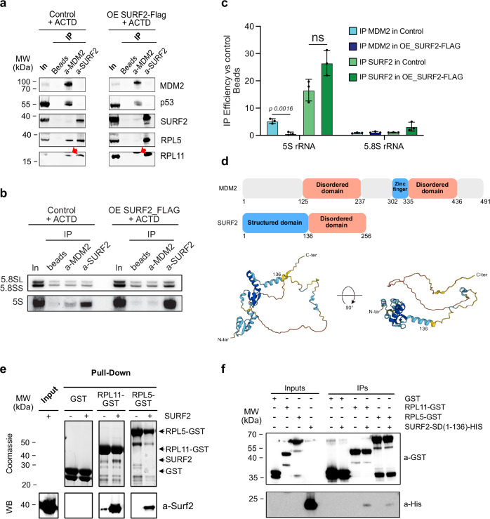
Fig. 8 SURF2 is able to directly interact with both RPL5 and RPL11 and competes for their binding with MDM2 in vivo. a Extracts from U2OS cells that overexpress SURF2 (OE SURF2-Flag) or not (control) and treated by actinomycin D (ACTD) are used to perform IPs using beads only, beads coupled to anti-SURF2 or beads coupled to anti-MDM2. After washes, the remaining proteins are resuspended in loading dye and analyzed by western blots using the indicated antibodies. Red arrows indicate unspecify bands (IgG) (n = 2). b Same as in (a) but using northern blot to test for RNA binding. Probes against 5S and 5.8S were used (n = 3). c Quantification of northern-blot signals to test for specific enrichment of RNA species (n = 3). Use of an unpaired two-tailed t test for statistical tests. Data are presented as mean values +/− SD, and significant differences are indicated by stars as follows p-value < 0.05*; 0.01** 0.001*** and 0.0001****) or with the precise p-value on the graph (p). d Secondary structure of SURF2 and MDM2 proteins. Functional domains are indicated. SURF2 3D structure modelization by Alphafold software is represented. e GST-pulldown assays. Extracts of BL21 that overexpress recombinant SURF2-HIS were mixed with extracts that overexpress either GST alone, GST-RPL5, or GST-RPL11. Proteins specifically retained on glutathione-sepharose beads were analyzed both by Coomassie staining and western-blot analysis (WB). 10% of the extracts were used for inputs (n = 2). f Same experiments as in (e) but replacing extract with SURF2-HIS by extracts that overexpress structural domain of SURF2 (SURF2-SD-HIS) as prey. Proteins specifically retained on glutathione-sepharose beads were analyzed both by western blots with indicated antibodies (n = 2). Source data are provided as a Source Data file.
Fig. 8 SURF2 is able to directly interact with both RPL5 and RPL11 and competes for their binding with MDM2 in vivo. a Extracts from U2OS cells that overexpress SURF2 (OE SURF2-Flag) or not (control) and treated by actinomycin D (ACTD) are used to perform IPs using beads only, beads coupled to anti-SURF2 or beads coupled to anti-MDM2. After washes, the remaining proteins are resuspended in loading dye and analyzed by western blots using the indicated antibodies. Red arrows indicate unspecify bands (IgG) (n = 2). b Same as in (a) but using northern blot to test for RNA binding. Probes against 5S and 5.8S were used (n = 3). c Quantification of northern-blot signals to test for specific enrichment of RNA species (n = 3). Use of an unpaired two-tailed t test for statistical tests. Data are presented as mean values +/− SD, and significant differences are indicated by stars as follows p-value < 0.05*; 0.01** 0.001*** and 0.0001****) or with the precise p-value on the graph (p). d Secondary structure of SURF2 and MDM2 proteins. Functional domains are indicated. SURF2 3D structure modelization by Alphafold software is represented. e GST-pulldown assays. Extracts of BL21 that overexpress recombinant SURF2-HIS were mixed with extracts that overexpress either GST alone, GST-RPL5, or GST-RPL11. Proteins specifically retained on glutathione-sepharose beads were analyzed both by Coomassie staining and western-blot analysis (WB). 10% of the extracts were used for inputs (n = 2). f Same experiments as in (e) but replacing extract with SURF2-HIS by extracts that overexpress structural domain of SURF2 (SURF2-SD-HIS) as prey. Proteins specifically retained on glutathione-sepharose beads were analyzed both by western blots with indicated antibodies (n = 2). Source data are provided as a Source Data file.CC BY-NC-ND · PubMed Central
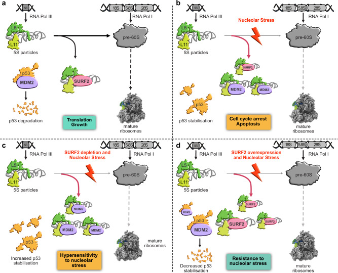
Fig. 9 Model of SURF2 function in free 5S RNP regulation. Schematic representation of free 5S regulation by SURF2 in different conditions. a In normal cells, both 5S and 47S rDNAs are transcribed by RNA polymerase III and I, respectively. Ribosome synthesis is producing pre-60S ribosomes and 5S particles constituted by the association of RPL5 and RPL11 to 5S rRNA are incorporated into these large pre-ribosomes. The remaining overproduced free 5S are bound by SURF2 to avoid MDM2-Free 5S interaction, which would induce p53 stabilization and cell cycle arrest. At the same time, p53 is constantly ubiquitinated by MDM2 to promote its degradation by the proteasome. b After nucleolar stress (drug-induced or caused by genetic mutations/ribosomopathies), ribosome synthesis is impaired, and a larger amount of free 5S particles accumulate in the nucleoplasm. The extra free 5S particles can then be recognized by MDM2, which can no longer ubiquitinylate p53, thereby stabilizing p53 and promoting cell cycle arrest. c In cells lacking SURF2, nucleolar stress still impairs ribosome synthesis, but this time, even more, free 5S RNPs are able to bind to MDM2, inducing stronger stabilization and activation of p53, followed by more cell cycle arrest. d In contrast, in cells overexpressing SURF2, nucleolar stress promotes free 5S RNP accumulation in the nucleoplasm, all of which are recognized by SURF2, which competes with MDM2 for binding. As a result, MDM2 is free and ubiquitinylates p53, conferring to these cells a capacity to resist to nucleolar stress.
Fig. 9 Model of SURF2 function in free 5S RNP regulation. Schematic representation of free 5S regulation by SURF2 in different conditions. a In normal cells, both 5S and 47S rDNAs are transcribed by RNA polymerase III and I, respectively. Ribosome synthesis is producing pre-60S ribosomes and 5S particles constituted by the association of RPL5 and RPL11 to 5S rRNA are incorporated into these large pre-ribosomes. The remaining overproduced free 5S are bound by SURF2 to avoid MDM2-Free 5S interaction, which would induce p53 stabilization and cell cycle arrest. At the same time, p53 is constantly ubiquitinated by MDM2 to promote its degradation by the proteasome. b After nucleolar stress (drug-induced or caused by genetic mutations/ribosomopathies), ribosome synthesis is impaired, and a larger amount of free 5S particles accumulate in the nucleoplasm. The extra free 5S particles can then be recognized by MDM2, which can no longer ubiquitinylate p53, thereby stabilizing p53 and promoting cell cycle arrest. c In cells lacking SURF2, nucleolar stress still impairs ribosome synthesis, but this time, even more, free 5S RNPs are able to bind to MDM2, inducing stronger stabilization and activation of p53, followed by more cell cycle arrest. d In contrast, in cells overexpressing SURF2, nucleolar stress promotes free 5S RNP accumulation in the nucleoplasm, all of which are recognized by SURF2, which competes with MDM2 for binding. As a result, MDM2 is free and ubiquitinylates p53, conferring to these cells a capacity to resist to nucleolar stress.CC BY-NC-ND · PubMed Central

Verification

Claims the paper makes

The claims and statistics of this paper haven’t been checked yet.

Run the check — free, no account needed. It reads the open-access text and checks the sources it cites.

A few free checks a day · papers already verified open instantly · results are public

Self-check

Percentages, sums and reported differences re-computed from the paper’s own numbers.

The self-check needs the paper’s full text.

Citations

Cites

Research works printed but not linked: 70

  1. Zhong, L. et al. Small molecules in targeted cancer therapy: advances, challenges, and future perspectives. Signal Transduct. Target Ther. 6, 201 (2021). · DOI
  2. Sabnis, A. J. & Bivona, T. G. Principles of resistance to targeted cancer therapy: Lessons from basic and translational cancer biology. Trends Mol. Med. 25, 185–197 (2019). · DOI
  3. Montanaro, L., Treré, D. & Derenzini, M. Nucleolus, ribosomes, and cancer. Am. J. Pathol. 173, 301–310 (2008). · DOI
  4. Derenzini, M. et al. Nucleolar size indicates the rapidity of cell proliferation in cancer tissues. J. Pathol. 191, 181–186 (2000). · DOI
  5. Henras, A. K. et al. The post-transcriptional steps of eukaryotic ribosome biogenesis. Cell. Mol. Life Sci. 65, 2334–2359 (2008). · DOI
  6. Albert, B. et al. A ribosome assembly stress response regulates transcription to maintain proteome homeostasis. ELife 8, e45002 (2019). · DOI
  7. Mayer, C. & Grummt, I. Cellular stress and nucleolar function. Cell Cycle 4, 1036–1038 (2005). · DOI · Paper page
  8. Olson, M. O. J. Sensing Cellular Stress: Another New Function for the Nucleolus? Sci. STKE https://doi.org/10.1126/stke.2242004pe10 (2004). · DOI
  9. Nicolas, E. et al. Involvement of human ribosomal proteins in nucleolar structure and p53-dependent nucleolar stress. Nat. Commun. 7, 11390 (2016). · DOI
  10. Golomb, L. Volarevic, S. & Oren, M. p53 and ribosome biogenesis stress: the essentials. FEBS Lett., https://doi.org/10.1016/j.febslet.2014.04.014 (2014). · DOI
  11. Donati, G., Peddigari, S., Mercer, C. A. & Thomas, G. 5S ribosomal RNA is an essential component of a nascent ribosomal precursor complex that regulates the Hdm2-p53 checkpoint. Cell Rep. 4, 87–98 (2013). · DOI
  12. Domostegui, A. et al. Impaired ribosome biogenesis checkpoint activation induces p53-dependent MCL-1 degradation and MYC-driven lymphoma death. Blood 137, 3351–3364 (2021). · DOI
  13. Pelletier, J. et al. Nucleotide depletion reveals the impaired ribosome biogenesis checkpoint as a barrier against DNA damage. EMBO J. 39, e103838 (2020). · DOI
  14. Hannan, K. M. et al. Nuclear stabilization of p53 requires a functional nucleolar surveillance pathway. Cell Rep. 41, 111571 (2022). · DOI
  15. Lindström, M. S., Bartek, J. & Maya-Mendoza, A. p53 at the crossroad of DNA replication and ribosome biogenesis stress pathways. Cell Death Differ. 29, 972–982 (2022). · DOI
  16. Rubbi, C. P. Disruption of the nucleolus mediates stabilization of p53 in response to DNA damage and other stresses. EMBO J. 22, 6068–6077 (2003). · DOI
  17. Zhou, X., Hao, Q., Liao, J., Zhang, Q. & Lu, H. Ribosomal protein S14 unties the MDM2-p53 loop upon ribosomal stress. Oncogene 32, 388–396 (2013). · DOI
  18. Zhou, X., Liao, W.-J., Liao, J.-M., Liao, P. & Lu, H. Ribosomal proteins: functions beyond the ribosome. J. Mol. Cell Biol. 7, 92–104 (2015). · DOI
  19. Yadavilli, S. et al. Ribosomal protein S3: A multi-functional protein that interacts with both p53 and MDM2 through its KH domain. DNA Rep. 8, 1215–1224 (2009). · DOI
  20. Zhang, X. et al. Identification of ribosomal protein S25 (RPS25)-MDM2-p53 regulatory feedback loop. Oncogene 32, 2782–2791 (2013). · DOI
  21. Marechal, V., Elenbaas, B., Piette, J., Nicolas, J. C. & Levine, A. J. The ribosomal L5 protein is associated with mdm-2 and mdm-2-p53 complexes. Mol. Cell. Biol. 14, 7414–7420 (1994).
  22. Sloan, K. E., Bohnsack, M. T. & Watkins, N. J. The 5S RNP couples p53 homeostasis to ribosome biogenesis and nucleolar stress. Cell Rep. 5, 237–247 (2013). · DOI
  23. Nishimura, K. et al. Perturbation of ribosome biogenesis drives cells into senescence through 5S RNP-mediated p53 activation. Cell Rep. 10, 1310–1323 (2015). · DOI
  24. Bursać, S. et al. Mutual protection of ribosomal proteins L5 and L11 from degradation is essential for p53 activation upon ribosomal biogenesis stress. Proc. Natl. Acad. Sci. USA 109, 20467–20472 (2012). · DOI
  25. Cao, P. et al. Genomic gain of RRS1 promotes hepatocellular carcinoma through reducing the RPL11-MDM2-p53 signaling. Sci. Adv. 7, eabf4304 (2021). · DOI
  26. Burger, K. et al. Chemotherapeutic drugs inhibit ribosome biogenesis at various levels. J. Biol. Chem. 285, 12416–12425 (2010). · DOI
  27. Derenzini, E., Rossi, A. & Treré, D. Treating hematological malignancies with drugs inhibiting ribosome biogenesis: when and why. J. Hematol. Oncol. 11, 75 (2018). · DOI
  28. Drygin, D. et al. Targeting RNA polymerase I with an oral small molecule CX-5461 inhibits ribosomal RNA synthesis and solid tumor growth. Cancer Res. 71, 1418–1430 (2011). · DOI
  29. Ferreira, R., Schneekloth, J. S., Panov, K. I., Hannan, K. M. & Hannan, R. D. Targeting the RNA polymerase I transcription for cancer therapy comes of age. Cells 9, 266 (2020). · DOI
  30. Khot, A. et al. First-in-human RNA polymerase I transcription inhibitor CX-5461 in patients with advanced hematologic cancers: Results of a phase I dose-escalation study. Cancer Discov. 9, 1036–1049 (2019). · DOI
  31. Baßler, J. & Hurt, E. Eukaryotic ribosome assembly. Annu. Rev. Biochem. 88, 281–306 (2019). · DOI
  32. Pestov, D. G., Strezoska, Z. & Lau, L. F. Evidence of p53-dependent cross-talk between ribosome biogenesis and the cell cycle: effects of nucleolar protein Bop1 on G(1)/S transition. Mol. Cell. Biol. 21, 4246–4255 (2001). · DOI
  33. Aubert, M. O’Donohue, M.-F. Lebaron, & Gleizes, S. P.-E. Pre-ribosomal RNA processing in human cells: From mechanisms to congenital diseases. Biomolecules https://doi.org/10.3390/biom8040123 (2018). · DOI
  34. Lebaron, S. et al. Functionally impaired RPL8 variants associated with Diamond–Blackfan anemia and a Diamond–Blackfan anemia‐like phenotype. Hum. Mutat. 43, 389–402 (2022). · DOI
  35. Oršolić, I. et al. Cancer-associated mutations in the ribosomal protein L5 gene dysregulate the HDM2/p53-mediated ribosome biogenesis checkpoint. Oncogene 39, 3443–3457 (2020). · DOI
  36. Zheng, J. et al. Structure of human MDM2 complexed with RPL11 reveals the molecular basis of p53 activation. Genes Dev. 29, 1524–1534 (2015). · DOI
  37. Miliani de Marval, P. L. & Zhang, Y. The RP-Mdm2-p53 pathway and tumorigenesis. Oncotarget 2, 234–238 (2011). · DOI
  38. Rinke, J. & Steitz, J. A. Precursor molecules of both human 5S ribosomal RNA and transfer RNAs are bound by a cellular protein reactive with anti-La lupus antibodies. Cell 29, 149–159 (1982). · DOI
  39. Steitz, J. A. et al. A 5S rRNA/L5 complex is a precursor to ribosome assembly in mammalian cells. J. Cell Biol. 106, 545–556 (1988). · DOI
  40. Kressler, D. et al. Synchronizing nuclear import of ribosomal proteins with ribosome assembly. Science 338, 666–671 (2012). · DOI
  41. O’Donohue, M.-F. et al. HEATR3 variants impair nuclear import of uL18 (RPL5) and drive Diamond-Blackfan anemia. Blood https://doi.org/10.1182/blood.2021011846 (2022). · DOI
  42. Castillo Duque de Estrada, N. M. et al. Structure of nascent 5S RNPs at the crossroad between ribosome assembly and MDM2-p53 pathways. Nat. Struct. Mol. Biol. 30, 1119–1131 (2023). · DOI
  43. Vassilev, L. T. et al. In vivo activation of the p53 pathway by small-molecule antagonists of MDM2. Science 303, 844–848 (2004). · DOI · Paper page
  44. Duhig, T., Ruhrberg, C., Mor, O. & Fried, M. The human Surfeit locus. Genomics 52, 72–78 (1998). · DOI
  45. Vernon, E. G. & Gaston, K. Myc and YY1 mediate activation of the Surf-1 promoter in response to serum growth factors. Biochim. Biophys. Acta 1492, 172–179 (2000). · DOI
  46. Mellacheruvu, D. et al. The CRAPome: a contaminant repository for affinity purification-mass spectrometry data. Nat. Methods 10, 730–736 (2013). · DOI · Paper page
  47. Madru, C. et al. Chaperoning 5S RNA assembly. Genes Dev. 29, 1432–1446 (2015). · DOI
  48. Asano, N. et al. Structural and functional analysis of the Rpf2-Rrs1 complex in ribosome biogenesis. Nucleic Acids Res. https://doi.org/10.1093/nar/gkv305 (2015). · DOI
  49. Kharde, S. Calvino, F. R. Gumiero, A. Wild, K. & Sinning, I. The structure of Rpf2-Rrs1 explains its role in ribosome biogenesis. Nucleic Acids Res. https://doi.org/10.1093/nar/gkv640 (2015). · DOI
  50. Nieto, B. et al. Efficient fractionation and analysis of ribosome assembly intermediates in human cells. RNA Biol. 18, 182–197 (2021). · DOI
  51. Wang, M., Anikin, L. & Pestov, D. G. Two orthogonal cleavages separate subunit RNAs in mouse ribosome biogenesis. Nucleic Acids Res. 42, 11180–11191 (2014). · DOI
  52. Lindström, M. S., Jin, A., Deisenroth, C., Wolf, G. White & Zhang, Y. Cancer-associated mutations in the MDM2 zinc finger domain disrupt ribosomal protein interaction and attenuate MDM2-induced p53 degradation. Mol. Cell. Biol. 27, 1056–1068 (2007). · DOI
  53. Russo, A. & Russo, G. Ribosomal proteins control or bypass p53 during Nucleolar Stress. Int. J. Mol. Sci. 18, 140 (2017). · DOI
  54. Dai, M.-S., Arnold, H., Sun, X.-X., Sears, R. & Lu, H. Inhibition of c-Myc activity by ribosomal protein L11. EMBO J. 26, 3332–3345 (2007). · DOI
  55. Carotenuto, P., Pecoraro, A., Palma, G., Russo, G. & Russo, A. Therapeutic approaches targeting nucleolus in cancer. Cells 8, 1090 (2019). · DOI
  56. Howard, G. C. et al. Ribosome subunit attrition and activation of the p53–MDM4 axis dominate the response of MLL-rearranged cancer cells to WDR5 WIN site inhibition. ELife 12, RP90683 (2024). · DOI
  57. Jansen, J. Bohnsack, K. E. Böhlken-Fascher, S. Bohnsack, M. T. & Dobbelstein, M. The ribosomal protein L22 binds the MDM4 pre-mRNA and promotes exon skipping to activate p53 upon nucleolar stress. Cell Rep 43, 114610 (2023). · DOI
  58. Ghosh, C., Hu, J. & Kebebew, E. Advances in translational research of the rare cancer type adrenocortical carcinoma. Nat. Rev. Cancer 23, 805–824 (2023). · DOI
  59. Yin, M. et al. Identification of key genes and pathways in adrenocortical carcinoma: evidence from bioinformatic analysis. Front. Endocrinol. 14, 1250033 (2023). · DOI
  60. Aspesi, A. et al. Lymphoblastoid cell lines from Diamond Blackfan anaemia patients exhibit a full ribosomal stress phenotype that is rescued by gene therapy. Sci. Rep. 7, 12010 (2017). · DOI
  61. Le Goff, S. et al. p53 activation during ribosome biogenesis regulates normal erythroid differentiation. Blood 137, 89–102 (2021). · DOI
  62. Jones, N. C. et al. Prevention of the neurocristopathy Treacher Collins syndrome through inhibition of p53 function. Nat. Med. 14, 125–133 (2008). · DOI
  63. Vlachos, A., Rosenberg, P. S., Atsidaftos, E., Alter, B. P. & Lipton, J. M. Incidence of neoplasia in Diamond Blackfan anemia: a report from the Diamond Blackfan Anemia Registry. Blood 119, 3815–3819 (2012). · DOI · Paper page
  64. Kennedy, A. L. et al. Distinct genetic pathways define pre-malignant versus compensatory clonal hematopoiesis in Shwachman-Diamond syndrome. Nat. Commun. 12, 1334 (2021). · DOI
  65. Tan, S. et al. Somatic genetic rescue of a germline ribosome assembly defect. Nat. Commun. 12, 5044 (2021). · DOI
  66. Reilly, C. R. & Shimamura, A. Predisposition to myeloid malignancies in Shwachman-Diamond syndrome: biological insights and clinical advances. Blood 141, 1513–1523 (2023). · DOI
  67. HaileMariam, M. et al. S-Trap, an ultrafast sample-preparation approach for shotgun proteomics. J. Proteome Res. 17, 2917–2924 (2018). · DOI
  68. Bouyssié, D. et al. mzDB: A file format using multiple indexing strategies for the efficient analysis of large LC-MS/MS and SWATH-MS data sets ∗. Mol. Cell. Proteomics 14, 771–781 (2015). · DOI
  69. Bouyssié, D. et al. Proline: an efficient and user-friendly software suite for large-scale proteomics. Bioinformatics 36, 3148–3155 (2020). · DOI · Paper page
  70. Lebaron, S. et al. The ATPase and helicase activities of Prp43p are stimulated by the G-patch protein Pfa1p during yeast ribosome biogenesis. EMBO J. 28, 3808–3819 (2009). · DOI

Metadata

Authors
Tagnères, S., Santo, P. E., Radermecker, J., Rinaldi, D., Froment, C., Provost, Q., Bongers, M., Capeille, S., Watkins, N., Marcoux, J., Gleizes, P.-E., Marcel, V., Plisson-Chastang, C., & Lebaron, S.
Affiliations
  • Centre National de la Recherche Scientifique (FR)
  • Lyon 1 Université (FR)
  • Newcastle University (GB)
Corresponding author
Lebaron, Simon
Venue
Nature Communications
Published
2024-09-27
Volume
15
Issue
1
Pages
8404-8404
ISSN
2041-1723 (ISSN-L)
OpenAlex
W4402903566
Cited by
15
References
3
Citations by year
2026: 6 · 2025: 9
FWCI
2.57
Type
journal article
Language
en
Open access
Locations
  • Nature Communications (publishedVersion, cc-by-nc-nd)
  • bioRxiv (Cold Spring Harbor Laboratory) (acceptedVersion, other-oa)
  • PubMed Central (publishedVersion, cc-by-nc-nd)
  • PubMed (publishedVersion)
  • HAL (submittedVersion, cc-by-nc-nd)
  • RePEc: Research Papers in Economics (submittedVersion)
  • DOAJ (DOAJ: Directory of Open Access Journals) (submittedVersion, cc-by-sa)
Funders
  • Ligue Contre le Cancer (62339, Labelisation)
  • Institut National Du Cancer (PLBIO022-065)
Topics
  • Health Sciences › Medicine › Oncology › Cancer-related Molecular Pathways
  • Life Sciences › Biochemistry, Genetics and Molecular Biology › Molecular Biology › Nuclear Structure and Function
  • Life Sciences › Biochemistry, Genetics and Molecular Biology › Molecular Biology › RNA and protein synthesis mechanisms
Keywords
nucleolar stress response, ribosome biogenesis, p53 stabilization, tumor resistance, adrenocortical carcinoma, prognostic factors, chemoresistance
MeSH terms
RNA-Seq, Antineoplastic Agents, Cell Nucleolus, Cell Survival, Humans, Neoplasms, Proteins, Ribonucleoproteins, Ribosomes, Stress, Physiological, Gene Expression Regulation, Neoplastic, Tumor Suppressor Protein p53, Apoptosis, Cell Line, Tumor, Cell Proliferation, Proto-Oncogene Proteins c-mdm2, Protein Stability

About these checks

Registry, retraction, Cites and statistics results are computed, not judged, with one exception: a printed reference that no index lists is read once by a model before it counts as a citation error. Claims are extracted by a model and kept only when their quote appears verbatim in the text; each cited source is then checked, with a model’s help, for whether it says what the claim relies on. Those verdicts can be wrong — read the claim’s quote and the cited source before relying on one.

Statistics and arithmetic are recomputed, not judged.

Confidence bands are shown only for fields calibrated on our annotated test set. None is calibrated yet, so no band is shown.

What counts as a claim ›How we measure confidence ›How we check this ›

Are you an author, or do you see a problem with this page? Request a review