Cited by 1Protein Structure and DynamicsMachine Learning in BioinformaticsEnzyme Structure and Function

The DOI matches its Crossref record; 2 of the 2 references it cites were found in a registry or an index.

A fast approach for structural and evolutionary analysis based on energetic profile protein comparison

Open access

Published version of a preprint

Abstract

In structural bioinformatics, the efficiency of predicting protein similarity, function, and evolutionary relationships is crucial. Our approach proposed herein leverages protein energy profiles derived from a knowledge-based potential, deviating from traditional methods relying on structural alignment or atomic distances. This method assigns unique energy profiles to individual proteins, facilitating rapid comparative analysis for both structural similarities and evolutionary relationships across various hierarchical levels. Our study demonstrates that energy profiles contain substantial information about protein structure at class, fold, superfamily, and family levels. Notably, these profiles accurately distinguish proteins across species, illustrated by the classification of coronavirus spike glycoproteins and bacteriocin proteins. Introducing a separation measure based on energy profile similarity, our method shows significant correlation with a network-based approach, emphasizing the potential of energy profiles as efficient predictors for drug combinations with faster computational requirements. Our key insight is that the sequence-based energy profile strongly correlates with structure-derived energy, enabling rapid and efficient protein comparisons based solely on sequences.

Overview

The claims and statistics of this paper haven’t been checked yet.

Run the check — free, no account needed. It reads the open-access text and checks the sources it cites.

A few free checks a day · papers already verified open instantly · results are public

Integrity signals

Registry

Crossref · 10.1038/s41467-025-57374-9

Cites

2 of 2 references verified0 citation errors · 0 not checkable

Statistics

Figures

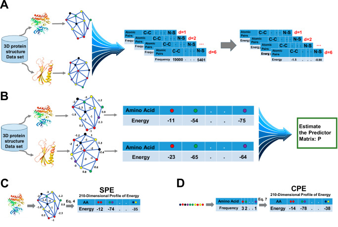
Fig. 1 Development of knowledge-based potential function and profile of energy. A Construction of the knowledge-based potential function. B Estimation of the predictor matrix P. C Construction of the structural profile of energy (SPE) based on protein structure. D Construction of the compositional profile of energy (CPE) based on protein sequence.
Fig. 1 Development of knowledge-based potential function and profile of energy. A Construction of the knowledge-based potential function. B Estimation of the predictor matrix P. C Construction of the structural profile of energy (SPE) based on protein structure. D Construction of the compositional profile of energy (CPE) based on protein sequence.CC BY · PubMed Central
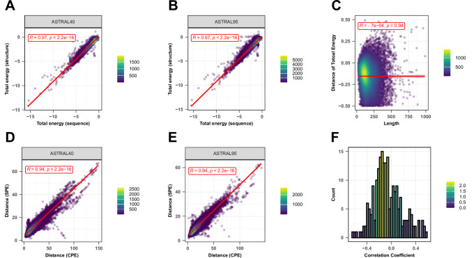
Fig. 2 Sequence-Structure relationship. Two-sided Pearson correlation comparing total energy estimates derived from protein sequence (X-axis) and protein structure (Y-axis) for protein domains in the (A) ASTRAL40 data set and (B) ASTRAL95 dataset. C Two-sided Pearson correlation between the difference in total energy from sequence and structure (Y-axis) and protein length (X-axis). The red lines represent the least squares regression line, and the gray shaded area represents the 95% confidence intervals around the regression line. Each point in (A–C) represents a protein domain. Two-sided Pearson correlation comparing the distances of profile of energy derived from sequence (X-axis) and structure (Y-axis) for all pairs of domains in D) ASTRAL40 and E) ASTRAL95 datasets, respectively. Each point in (D, E) represents a pair of protein domain. In plots (A–D), R indicates the correlation coefficient, and p shows the p-value. The exact p-value is less than 10e-16, which is below the precision threshold of standard statistical computations. F Histogram showing the distribution of correlation coefficients between the difference in energy estimates (from sequence and structure) and protein length across all 210 pairwise interactions. Source data are provided as a Source Data file.
Fig. 2 Sequence-Structure relationship. Two-sided Pearson correlation comparing total energy estimates derived from protein sequence (X-axis) and protein structure (Y-axis) for protein domains in the (A) ASTRAL40 data set and (B) ASTRAL95 dataset. C Two-sided Pearson correlation between the difference in total energy from sequence and structure (Y-axis) and protein length (X-axis). The red lines represent the least squares regression line, and the gray shaded area represents the 95% confidence intervals around the regression line. Each point in (A–C) represents a protein domain. Two-sided Pearson correlation comparing the distances of profile of energy derived from sequence (X-axis) and structure (Y-axis) for all pairs of domains in D) ASTRAL40 and E) ASTRAL95 datasets, respectively. Each point in (D, E) represents a pair of protein domain. In plots (A–D), R indicates the correlation coefficient, and p shows the p-value. The exact p-value is less than 10e-16, which is below the precision threshold of standard statistical computations. F Histogram showing the distribution of correlation coefficients between the difference in energy estimates (from sequence and structure) and protein length across all 210 pairwise interactions. Source data are provided as a Source Data file.CC BY · PubMed Central
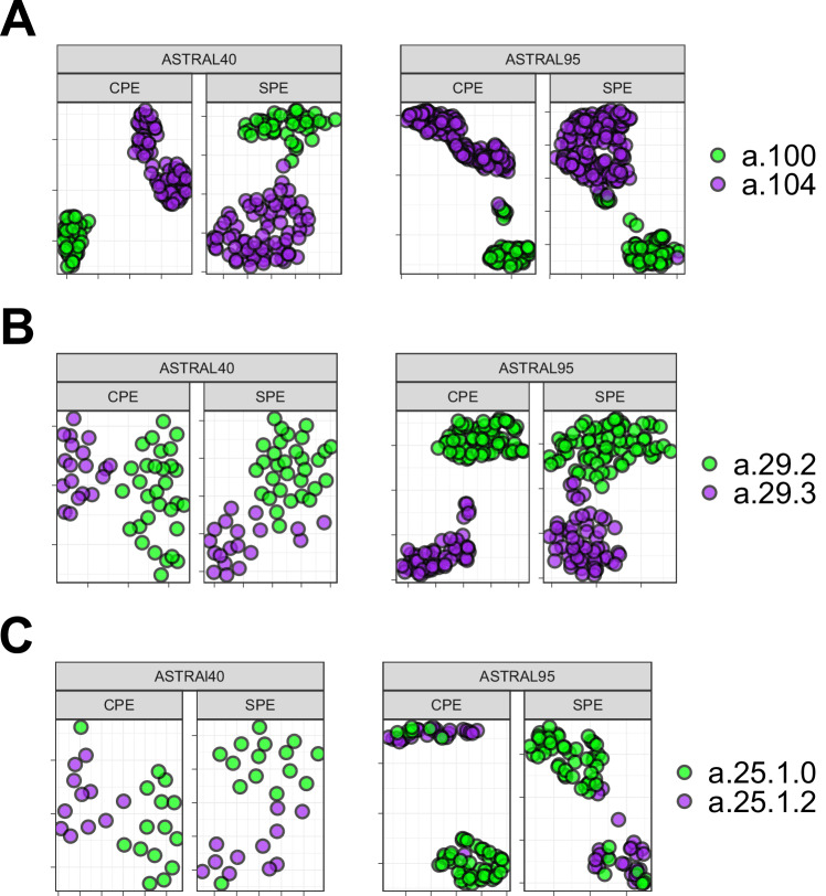
Fig. 3 UMAP Visualization of Energy Profiles. The UMAP projection of structural profile of energy (SPE) and Compositional Energy Profiles (CPE) of protein domains from ASTRAL40 and ASTRAL95 represents the structural information embedded in energy profiles across hierarchical levels of SCOP; each panel includes two figures, one generated by CPE (left panel) and the other by SPE (right panel), revealing that protein domains sharing the same (A) fold, (B) superfamily, and (C) family exhibit comparable energy profile patterns. The folds a.100 and a.104, superfamilies a.29.2 and a.29.3, as well as families a.25.1.0 and a.25.1.2, are randomly selected for analysis, and the UMAP plots were generated using parameters n_neighbors = 30 and min_dist = 0.1. Source data are provided as a Source Data file.
Fig. 3 UMAP Visualization of Energy Profiles. The UMAP projection of structural profile of energy (SPE) and Compositional Energy Profiles (CPE) of protein domains from ASTRAL40 and ASTRAL95 represents the structural information embedded in energy profiles across hierarchical levels of SCOP; each panel includes two figures, one generated by CPE (left panel) and the other by SPE (right panel), revealing that protein domains sharing the same (A) fold, (B) superfamily, and (C) family exhibit comparable energy profile patterns. The folds a.100 and a.104, superfamilies a.29.2 and a.29.3, as well as families a.25.1.0 and a.25.1.2, are randomly selected for analysis, and the UMAP plots were generated using parameters n_neighbors = 30 and min_dist = 0.1. Source data are provided as a Source Data file.CC BY · PubMed Central
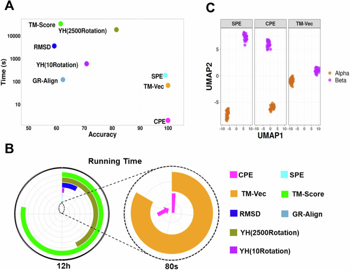
Fig. 4 Performance and Computational Efficiency of Protein Dissimilarity Measures. A Time versus accuracy for the 1-NN classifier using GR-Align, RMSD, TM-score, Yau-Hausdorff distance, TM-Vec, and the distance between energy profiles SPE and CPE as measures of protein dissimilarity. B Running times of the evaluated methods, scaled to 12 h, with an inset zooming in on the region indicated by the dashed circle. The entire circle represents 80 s. Each method is represented by different colors as indicated in the figure legend. C The UMAP projection of α and β globins from the hemoglobin biological unit using CPE, SPE, and TM-Vec representations. n_neighbors = 13, min_dist = 0.1. Source data are provided as a Source Data file.
Fig. 4 Performance and Computational Efficiency of Protein Dissimilarity Measures. A Time versus accuracy for the 1-NN classifier using GR-Align, RMSD, TM-score, Yau-Hausdorff distance, TM-Vec, and the distance between energy profiles SPE and CPE as measures of protein dissimilarity. B Running times of the evaluated methods, scaled to 12 h, with an inset zooming in on the region indicated by the dashed circle. The entire circle represents 80 s. Each method is represented by different colors as indicated in the figure legend. C The UMAP projection of α and β globins from the hemoglobin biological unit using CPE, SPE, and TM-Vec representations. n_neighbors = 13, min_dist = 0.1. Source data are provided as a Source Data file.CC BY · PubMed Central
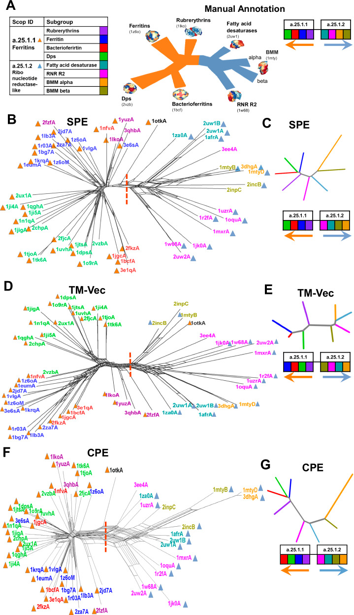
Fig. 5 Phylogenetic network reconstruction of the ferritin-like superfamily. A Schematic representation of the relationships among major ferritin-like protein families, with each subgroup shown in a distinct color. B Phylogenetic network reconstructed using SPE. C Neighbor-joining tree generated based on the average distances between subgroups using SPE. D Phylogenetic network reconstructed using TM-Vec. E Neighbor-joining tree generated using the average distances between subgroups with TM-Vec. F Phylogenetic network reconstructed using CPE. G Neighbor-joining tree based on the average distances between subgroups using CPE. The red dotted line highlights the separation between two SCOP families: ferritins (SCOP ID a.25.1.1), which includes the Bacterioferritin, Ferritins, Dps, and Rubrerythrin subgroups, and the Ribonucleotide Reductase-like family (SCOP ID a.25.1.2), which includes the BMM-alpha, BMM-beta, Fatty_acid, and RNRR2 subgroups. The inferred arrangement of subfamilies using each method is shown to the right of (C, E, G). Source data are provided as a Source Data file.
Fig. 5 Phylogenetic network reconstruction of the ferritin-like superfamily. A Schematic representation of the relationships among major ferritin-like protein families, with each subgroup shown in a distinct color. B Phylogenetic network reconstructed using SPE. C Neighbor-joining tree generated based on the average distances between subgroups using SPE. D Phylogenetic network reconstructed using TM-Vec. E Neighbor-joining tree generated using the average distances between subgroups with TM-Vec. F Phylogenetic network reconstructed using CPE. G Neighbor-joining tree based on the average distances between subgroups using CPE. The red dotted line highlights the separation between two SCOP families: ferritins (SCOP ID a.25.1.1), which includes the Bacterioferritin, Ferritins, Dps, and Rubrerythrin subgroups, and the Ribonucleotide Reductase-like family (SCOP ID a.25.1.2), which includes the BMM-alpha, BMM-beta, Fatty_acid, and RNRR2 subgroups. The inferred arrangement of subfamilies using each method is shown to the right of (C, E, G). Source data are provided as a Source Data file.CC BY · PubMed Central
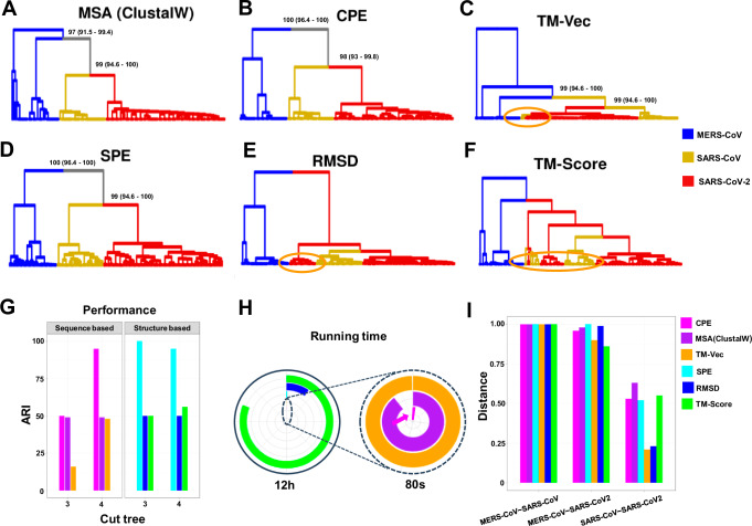
Fig. 6 Clustering analysis of spike glycoprotein structures from SARS-CoV, SARS-CoV-2, and MERS-CoV. The dendrograms depict the clustering of spike glycoprotein structures from the three viruses: SARS-CoV, SARS-CoV-2, and MERS-CoV. The clustering is based on pairwise distances calculated from different methods: (A) protein sequence, (B) CPE, (C) TM-Vec, (D) SPE, (E) RMSD, and (F) TM-Score. The leaves of each tree are color-coded to indicate the originating virus for each spike glycoprotein structure. G Displays the ARI values for each method,(H) shows the running time associated with each method scaled to 12 h, with an inset zooming in on the region indicated by the dashed circle. The entire circle represents 80 s, and (I) presents the average distance between the three virus groups as calculated by each method. Source data are provided as a Source Data file.
Fig. 6 Clustering analysis of spike glycoprotein structures from SARS-CoV, SARS-CoV-2, and MERS-CoV. The dendrograms depict the clustering of spike glycoprotein structures from the three viruses: SARS-CoV, SARS-CoV-2, and MERS-CoV. The clustering is based on pairwise distances calculated from different methods: (A) protein sequence, (B) CPE, (C) TM-Vec, (D) SPE, (E) RMSD, and (F) TM-Score. The leaves of each tree are color-coded to indicate the originating virus for each spike glycoprotein structure. G Displays the ARI values for each method,(H) shows the running time associated with each method scaled to 12 h, with an inset zooming in on the region indicated by the dashed circle. The entire circle represents 80 s, and (I) presents the average distance between the three virus groups as calculated by each method. Source data are provided as a Source Data file.CC BY · PubMed Central
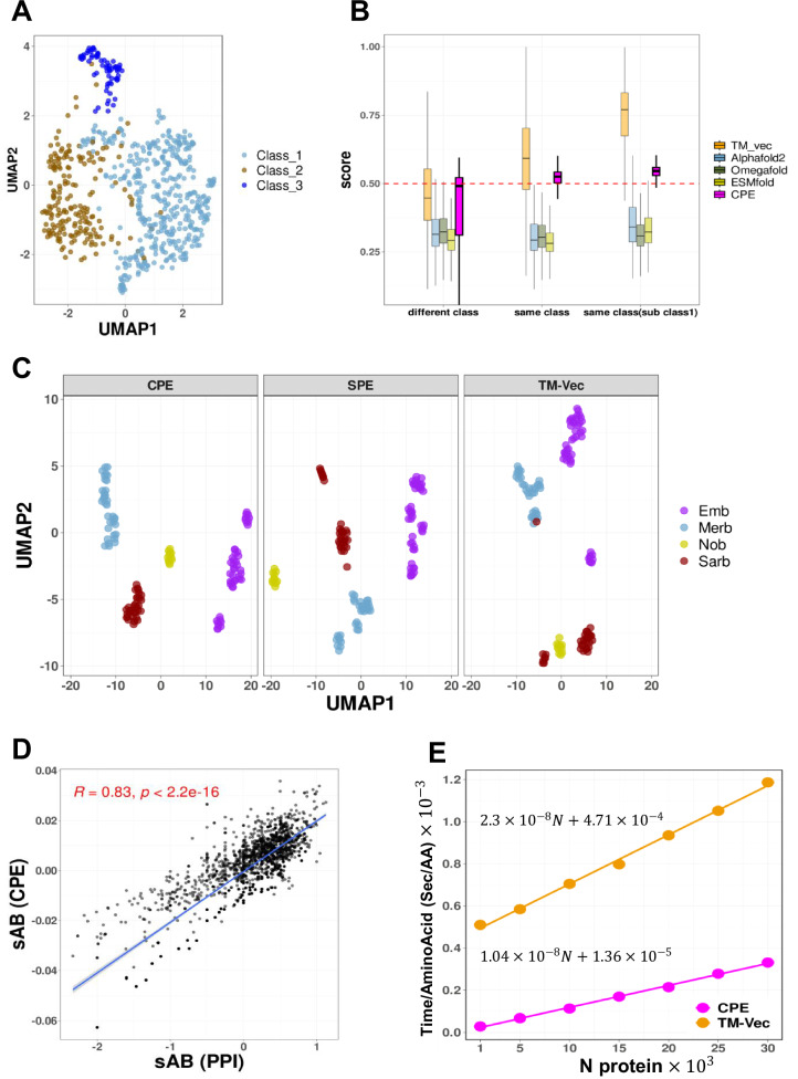
Fig. 7 UMAP Projection of Energy Profiles for Bacteriocins and Betacoronavirus Domains, with Distance Comparisons and Scalability Analysis. A UMAP projection of Compositional Energy Profiles (CPE) for 690 peptides, representing three different classes of bacteriocins. B Comparison of CPE distances with the TM-scores produced by running TM-align on structures predicted by AlphaFold2, OmegaFold and ESMFold, and TM-Vec for all pairs of bacteriocins, pairs at different classes (n = 125869), pairs at the same class (n = 111349), pairs from the same class from subclass1 (n = 13431). Statistics assessed by two-tailed Student’s t-test. Boxplots display the median (center line), the 25th and 75th percentiles (bounds of the box), and the minimum and maximum values (whiskers) excluding outliers. Comparisons of CPE distances revealed statistically significant differences across the groups (different class, same class, and subclass1 within the same class), with p-values for all three tests being <10e−16. The exact p-value is less than 10e-16, which is below the precision threshold of standard statistical computations. CPE distances are normalized by min-max normalization. C Papain-like Protease (PLPro) domains across Betacoronavirus subgenera. The UMAP projection shows clustering of PLPro domains from Sarbecovirus (n = 31), Nobecovirus (n = 11), Merbecovirus (n = 34), and Embecovirus (n = 45) using SPE, CPE, and TMVec representations. n_neighbors = 13, min_dist = 0.5. D Two-sided Pearson correlation comparing Protein-Protein Interaction Network Distances (X-axis) with Energy Profile Distances (Y-axis). The blue line illustrates the least squares regression line, and the gray shaded area represents the 95% confidence intervals around the regression line. Each point corresponds to a drug combination. R indicates the correlation coefficient, and p shows the p-value. E Scalability of CPE and TM-Vec. Processing time per amino acid for subsets from the ASTRAL95 dataset, ranging in size from 1000 to 30,000 proteins (at intervals of 5000). Source data are provided as a Source Data file.
Fig. 7 UMAP Projection of Energy Profiles for Bacteriocins and Betacoronavirus Domains, with Distance Comparisons and Scalability Analysis. A UMAP projection of Compositional Energy Profiles (CPE) for 690 peptides, representing three different classes of bacteriocins. B Comparison of CPE distances with the TM-scores produced by running TM-align on structures predicted by AlphaFold2, OmegaFold and ESMFold, and TM-Vec for all pairs of bacteriocins, pairs at different classes (n = 125869), pairs at the same class (n = 111349), pairs from the same class from subclass1 (n = 13431). Statistics assessed by two-tailed Student’s t-test. Boxplots display the median (center line), the 25th and 75th percentiles (bounds of the box), and the minimum and maximum values (whiskers) excluding outliers. Comparisons of CPE distances revealed statistically significant differences across the groups (different class, same class, and subclass1 within the same class), with p-values for all three tests being <10e−16. The exact p-value is less than 10e-16, which is below the precision threshold of standard statistical computations. CPE distances are normalized by min-max normalization. C Papain-like Protease (PLPro) domains across Betacoronavirus subgenera. The UMAP projection shows clustering of PLPro domains from Sarbecovirus (n = 31), Nobecovirus (n = 11), Merbecovirus (n = 34), and Embecovirus (n = 45) using SPE, CPE, and TMVec representations. n_neighbors = 13, min_dist = 0.5. D Two-sided Pearson correlation comparing Protein-Protein Interaction Network Distances (X-axis) with Energy Profile Distances (Y-axis). The blue line illustrates the least squares regression line, and the gray shaded area represents the 95% confidence intervals around the regression line. Each point corresponds to a drug combination. R indicates the correlation coefficient, and p shows the p-value. E Scalability of CPE and TM-Vec. Processing time per amino acid for subsets from the ASTRAL95 dataset, ranging in size from 1000 to 30,000 proteins (at intervals of 5000). Source data are provided as a Source Data file.CC BY · PubMed Central

Verification

Claims the paper makes

The claims and statistics of this paper haven’t been checked yet.

Run the check — free, no account needed. It reads the open-access text and checks the sources it cites.

A few free checks a day · papers already verified open instantly · results are public

Self-check

Percentages, sums and reported differences re-computed from the paper’s own numbers.

The self-check needs the paper’s full text.

Citations

Cites

Research works printed but not linked: 48

  1. Sayers, E. W. et al. Database resources of the national center for biotechnology information. Nucleic acids Res. 49, D10 (2021). · DOI
  2. Altschul, S. F. et al. Gapped BLAST and PSI-BLAST: a new generation of protein database search programs. Nucleic acids Res. 25, 3389–3402 (1997). · DOI · Paper page
  3. Kilinc, M., Jia, K. & Jernigan, R. L. Improved global protein homolog detection with major gains in function identification. Proc. Natl Acad. Sci. 120, e2211823120 (2023). · DOI
  4. Quan, Y. et al. Evolution-strengthened knowledge graph enables predicting the targetability and druggability of genes. PNAS nexus 2, pgad147 (2023). · DOI
  5. Du, Z. et al. pLM4ACE: A protein language model based predictor for antihypertensive peptide screening. Food Chem. 431, 137162 (2024). · DOI
  6. Wang, Y. et al. RNAincoder: a deep learning-based encoder for RNA and RNA-associated interaction. Nucleic Acids Res. 51, gkad404 (2023). · DOI
  7. Sippl, M. J. Boltzmann’s principle, knowledge-based mean fields and protein folding. An approach to the computational determination of protein structures. J. computer-aided Mol. Des. 7, 473–501 (1993). · DOI
  8. Mirzaie, M., Eslahchi, C., Pezeshk, H. & Sadeghi, M. A distance‐dependent atomic knowledge‐based potential and force for discrimination of native structures from decoys. Proteins: Struct., Funct., Bioinforma. 77, 454–463 (2009). · DOI
  9. Mirzaie, M. Identification of native protein structures captured by principal interactions. BMC Bioinforma. 20, 1–10 (2019). · DOI
  10. Mirzaie, M. Discrimination power of knowledge-based potential dictated by the dominant energies in native protein structures. Amino acids 51, 1029–1038 (2019). · DOI
  11. Mirzaie, M. Hydrophobic residues can identify native protein structures. Proteins: Struct., Funct., Bioinforma. 86, 467–474 (2018). · DOI
  12. Dosztanyi, Z., Csizmok, V., Tompa, P. & Simon, I. The pairwise energy content estimated from amino acid composition discriminates between folded and intrinsically unstructured proteins. J. Mol. Biol. 347, 827–839 (2005). · DOI
  13. Bowie, J. U., Lüthy, R. & Eisenberg, D. A method to identify protein sequences that fold into a known three-dimensional structure. Science 253, 164–170 (1991). · DOI · Paper page
  14. Goldstein, R. A., Luthey-Schulten, Z. A. & Wolynes, P. G. Protein tertiary structure recognition using optimized Hamiltonians with local interactions. Proc. Natl Acad. Sci. 89, 9029–9033 (1992). · DOI
  15. Sillitoe, I. et al. CATH: increased structural coverage of functional space. Nucleic acids Res. 49, D266–D273 (2021). · DOI · Paper page
  16. Lo Conte, L. et al. SCOP: a structural classification of proteins database. Nucleic acids Res. 28, 257–259 (2000). · DOI
  17. Fox, N. K., Brenner, S. E. & Chandonia, J.-M. SCOPe: Structural Classification of Proteins—extended, integrating SCOP and ASTRAL data and classification of new structures. Nucleic acids Res. 42, D304–D309 (2014). · DOI · Paper page
  18. Lundin, D., Poole, A. M., Sjberg, B.-M. & Hgbom, M. Use of structural phylogenetic networks for classification of the ferritin-like superfamily. J. Biol. Chem. 287, 20565–20575 (2012). · DOI
  19. Gowthaman, R. et al. CoV3D: a database of high resolution coronavirus protein structures. Nucleic acids Res. 49, D282–D287 (2021). · DOI
  20. van Heel, A. J., de Jong, A., Montalbán-López, M., Kok, J. & Kuipers, O. P. BAGEL3: automated identification of genes encoding bacteriocins and (non-)bactericidal posttranslationally modified peptides. Nucleic Acids Res. 41, W448–W453 (2013). · DOI
  21. Cheng, F., Kovács, I. A. & Barabási, A.-L. Network-based prediction of drug combinations. Nat. Commun. 10, 1197 (2019). · DOI
  22. Freiberger, M. I. et al. Local energetic frustration conservation in protein families and superfamilies. Nat. Commun. 14, 8379 (2023). · DOI
  23. Maiorov, V. N. & Crippen, G. M. Significance of root-mean-square deviation in comparing three-dimensional structures of globular proteins. J. Mol. Biol. 235, 625–634 (1994). · DOI
  24. Zhang, Y. Skolnick J. TM-align: a protein structure alignment algorithm based on the TM-score. Nucleic acids Res. 33, 2302–2309 (2005). · DOI · Paper page
  25. Hamamsy, T. et al. Protein remote homology detection and structural alignment using deep learning. Nat. Biotechnol. 42, 975–985 (2024). · DOI
  26. Malod-Dognin, N. & Pržulj, N. GR-Align: fast and flexible alignment of protein 3D structures using graphlet degree similarity. Bioinformatics 30, 1259–1265 (2014). · DOI
  27. Tian, K, Zhao, X., Zhang, Y. & Yau, S. Comparing protein structures and inferring functions with a novel three-dimensional Yau–Hausdorff method. J. Biomol. Struct. Dyn. 37, 4151–4160 (2019). · DOI
  28. Wintjens, R. T., Rooman, M. J. & Wodak, S. J. Automatic classification and analysis of αα-turn motifs in proteins. J. Mol. Biol. 255, 235–253 (1996). · DOI
  29. Malik, A. J., Poole, A. M. & Allison, J. R. Structural phylogenetics with confidence. Mol. Biol. Evol. 37, 2711–2726 (2020). · DOI
  30. Puente-Lelievre, C. et al. Tertiary-interaction characters enable fast, model-based structural phylogenetics beyond the twilight zone. bioRxiv, 2023.2012. 2012.571181 (2023). · DOI
  31. Bryant, D. Moulton V. Neighbor-net: an agglomerative method for the construction of phylogenetic networks. Mol. Biol. evolution 21, 255–265 (2004). · DOI
  32. Schliep, K. P. phangorn: phylogenetic analysis in R. Bioinformatics 27, 592–593 (2011). · DOI
  33. Huson, D. H. & Bryant, D. Application of Phylogenetic Networks in Evolutionary Studies. Mol. Biol. Evolution 23, 254–267 (2005). · DOI
  34. Bodenhofer, U., Bonatesta, E., Horejš-Kainrath, C. & Hochreiter, S. msa: an R package for multiple sequence alignment. Bioinformatics 31, 3997–3999 (2015). · DOI
  35. Charif, D., Lobry, J. R. SeqinR 1.0-2: a contributed package to the R project for statistical computing devoted to biological sequences retrieval and analysis. In: Structural approaches to sequence evolution: Molecules, networks, populations). Springer (2007). · DOI
  36. Wu, R. et al. High-resolution de novo structure prediction from primary sequence. bioRxiv 2022. · DOI · Paper page
  37. Mirzaie, M. et al. Designing patient-oriented combination therapies for acute myeloid leukemia based on efficacy/toxicity integration and bipartite network modeling. Oncogenesis 13, 11 (2024). · DOI
  38. Jafari, M. et al. Bipartite network models to design combination therapies in acute myeloid leukaemia. Nat. Commun. 13, 2128 (2022). · DOI
  39. Gordon, D. E. et al. A SARS-CoV-2 protein interaction map reveals targets for drug repurposing. Nature 583, 459–468 (2020). · DOI · Paper page
  40. Katoh, K., Misawa, K., Ki, K. & Miyata, T. MAFFT: a novel method for rapid multiple sequence alignment based on fast Fourier transform. Nucleic acids Res. 30, 3059–3066 (2002). · DOI · Paper page
  41. Li, W., Jaroszewski, L. & Godzik, A. Clustering of highly homologous sequences to reduce the size of large protein databases. Bioinformatics 17, 282–283 (2001). · DOI
  42. Rausell, A., Juan, D., Pazos, F. & Valencia, A. Protein interactions and ligand binding: from protein subfamilies to functional specificity. Proc. Natl Acad. Sci. 107, 1995–2000 (2010). · DOI
  43. Wang, G. & Dunbrack, Jr. R. L. PISCES: a protein sequence culling server. Bioinformatics 19, 1589–1591 (2003). · DOI
  44. Mirzaie, M. & Sadeghi, M. Delaunay‐based nonlocal interactions are sufficient and accurate in protein fold recognition. Proteins: Struct., Funct., Bioinforma. 82, 415–423 (2014). · DOI
  45. Mirzaie, M. & Sadeghi, M. Distance‐dependent atomic knowledge‐based force in protein fold recognition. Proteins: Struct., Funct., Bioinforma. 80, 683–690 (2012). · DOI
  46. Barber, C. B., Dobkin, D. P. & Huhdanpaa, H. The quickhull algorithm for convex hulls. ACM Trans. Math. Softw. (TOMS) 22, 469–483 (1996). · DOI
  47. Grant, B. J., Rodrigues, A. P., ElSawy, K. M., McCammon, J. A. & Caves, L. S. Bio3d: an R package for the comparative analysis of protein structures. Bioinformatics 22, 2695–2696 (2006). · DOI
  48. Wickham, H. ggplot2. Wiley Interdiscip. Rev.: computational Stat. 3, 180–185 (2011). · DOI

Metadata

Authors
Choopanian, P., Andressoo, J.-O., & Mirzaie, M.
Affiliations
University of Helsinki (FI)
Corresponding author
  • Andressoo, Jaan-Olle
  • Mirzaie, Mehdi
Venue
Nature Communications
Published
2025-03-06
Volume
16
Issue
1
Pages
2231-2231
ISSN
2041-1723 (ISSN-L)
OpenAlex
W4408198396
Cited by
1
References
2
Citations by year
2026: 1
FWCI
0.30
Type
journal article
Language
en
Open access
Locations
  • Nature Communications (publishedVersion, cc-by)
  • bioRxiv (Cold Spring Harbor Laboratory) (acceptedVersion)
  • PubMed Central (publishedVersion, cc-by)
  • PubMed (publishedVersion)
  • RePEc: Research Papers in Economics (submittedVersion)
  • DOAJ (DOAJ: Directory of Open Access Journals) (submittedVersion, cc-by-sa)
  • Työväentutkimus Vuosikirja (submittedVersion)
Funders
Academy of Finland (297727 and 350678)
Topics
  • Life Sciences › Biochemistry, Genetics and Molecular Biology › Molecular Biology › Protein Structure and Dynamics
  • Life Sciences › Biochemistry, Genetics and Molecular Biology › Molecular Biology › Machine Learning in Bioinformatics
  • Life Sciences › Biochemistry, Genetics and Molecular Biology › Structural Biology › Enzyme Structure and Function
Keywords
knowledge-based potentials, protein structural similarity, protein family classification, protein fold classification, drug combination prediction
MeSH terms
Bacteriocins, Models, Molecular, Protein Conformation, Proteins, Thermodynamics, Evolution, Molecular, Computational Biology, Spike Glycoprotein, Coronavirus

About these checks

Registry, retraction, Cites and statistics results are computed, not judged, with one exception: a printed reference that no index lists is read once by a model before it counts as a citation error. Claims are extracted by a model and kept only when their quote appears verbatim in the text; each cited source is then checked, with a model’s help, for whether it says what the claim relies on. Those verdicts can be wrong — read the claim’s quote and the cited source before relying on one.

Statistics and arithmetic are recomputed, not judged.

Confidence bands are shown only for fields calibrated on our annotated test set. None is calibrated yet, so no band is shown.

What counts as a claim ›How we measure confidence ›How we check this ›

Are you an author, or do you see a problem with this page? Request a review