Cited by 6Wnt/β-catenin signaling in development and cancerComputational Drug Discovery MethodsReceptor Mechanisms and Signaling

The DOI matches its Crossref record; 1 of the 1 references it cites were found in a registry or an index.

In silico docking yields small molecule negative allosteric modulators targeting the core of Frizzled 7

Open access
Scharf, Magdalena M., Kinsolving, Julia, Grätz, Lukas, Voss, Jan Hendrik, Carrasco-Busturia, David, Forsberg, Björn, Kolb, Peter, Schulte, Gunnar·2025·Nature Communications

Published version of a preprint

Abstract

Targeting the Frizzled family (FZD 1-10 ) of WNT receptors pharmacologically has, despite substantial therapeutic potential, proven difficult. Given an almost complete lack of validated, effective small molecules targeting FZDs, no putative ligand binding site has so far been identified. In order to target FZD 7 , a potential target for the treatment of intestinal tumors, we combine an approach of adapted docking setups and large molecular library docking screens, identifying compound C407. Applying pharmacological assays, genetically-encoded biosensors, site-directed mutagenesis, cryo-electron microscopy and molecular dynamics simulations, the compound binding site in the core of the seven transmembrane bundle is validated and C407 is confirmed as a negative allosteric modulator of WNT-induced and FZD-mediated WNT/ β -catenin signaling. In summary, we provide here the proof-of-principle that targeting FZDs with small molecule compounds is possible and effective. Future hit optimization and functional validation in disease-relevant in vitro and in vivo models will pave the way towards clinical exploration.

Overview

The claims and statistics of this paper haven’t been checked yet.

Run the check — free, no account needed. It reads the open-access text and checks the sources it cites.

A few free checks a day · papers already verified open instantly · results are public

Integrity signals

Registry

Crossref · 10.1038/s41467-025-67147-z

Cites

1 of 1 references verified0 citation errors · 0 not checkable

Statistics

Figures

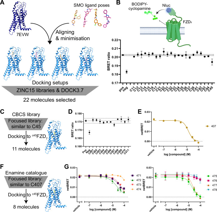
Fig. 1 Initial compound screens. A Scheme summarizing the preparation of docking setups and the large library screen. B Representative measurement of all compounds selected from the primary screen in the BRET-based competition binding assay (Nluc-FZD7 and BODIPY-cyclopamine) at a single concentration of 10 μM. The scheme above the graph illustrates the assay principle. A full concentration response curve for the binding of the potential hit compound C45 can be found in Supplementary Fig. 2. C Scheme of the in silico follow-up screen based on initial hit C45 using the CBCS library. D Representative measurement of all compounds selected from the screen in (C) in a BRET-based competition binding assay (HiBiT-FZD7 and BODIPY-cyclopamine) at a single concentration of 10 μM. E Concentration-response curve for C407 from a BRET-based competition binding assay (Nluc-FZD7 and BODIPY-cyclopamine). F Scheme of the follow-up screen based on hit molecule C407 and using the Enamine catalog. G Concentration-response curves for the compounds selected in (F) from BRET-based competition binding assays (Nluc-FZD7 and BODIPY-cyclopamine). B, D Data points are mean ± SD of technical triplicates from a representative experiment of n = 3 independent experiments. Dotted lines represent mean ± 3 ⋅ SD of the positive control as a measure of significance. Pos = positive control: DMSO and BODIPY-cyclopamine; neg = negative control: buffer only (with DMSO; no BODIPY-cyclopamine). E, G Data points represent mean ± SEM of n = 3–5 independent experiments. B Scheme created in BioRender. Kinsolving, J. (2025), https://BioRender.com/8drrhgy. Source data are provided as a Source Data file.
Fig. 1 Initial compound screens. A Scheme summarizing the preparation of docking setups and the large library screen. B Representative measurement of all compounds selected from the primary screen in the BRET-based competition binding assay (Nluc-FZD7 and BODIPY-cyclopamine) at a single concentration of 10 μM. The scheme above the graph illustrates the assay principle. A full concentration response curve for the binding of the potential hit compound C45 can be found in Supplementary Fig. 2. C Scheme of the in silico follow-up screen based on initial hit C45 using the CBCS library. D Representative measurement of all compounds selected from the screen in (C) in a BRET-based competition binding assay (HiBiT-FZD7 and BODIPY-cyclopamine) at a single concentration of 10 μM. E Concentration-response curve for C407 from a BRET-based competition binding assay (Nluc-FZD7 and BODIPY-cyclopamine). F Scheme of the follow-up screen based on hit molecule C407 and using the Enamine catalog. G Concentration-response curves for the compounds selected in (F) from BRET-based competition binding assays (Nluc-FZD7 and BODIPY-cyclopamine). B, D Data points are mean ± SD of technical triplicates from a representative experiment of n = 3 independent experiments. Dotted lines represent mean ± 3 ⋅ SD of the positive control as a measure of significance. Pos = positive control: DMSO and BODIPY-cyclopamine; neg = negative control: buffer only (with DMSO; no BODIPY-cyclopamine). E, G Data points represent mean ± SEM of n = 3–5 independent experiments. B Scheme created in BioRender. Kinsolving, J. (2025), https://BioRender.com/8drrhgy. Source data are provided as a Source Data file.CC BY · PubMed Central
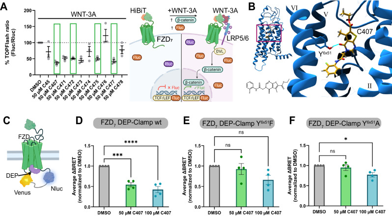
Fig. 2 Pharmacological characterization of compounds. A Impact of compounds on WNT-3A-induced (300 ng/mL) TOPFlash at HiBiT-FZD7 normalized to WNT-3A response in absence of test compound (DMSO). A scheme illustrating the assay principle is shown on the right. To determine true hits (C407, C472, C475, and C477; green boxes), control assays (Supplementary Fig. 5) and cell toxicity (Supplementary Fig. 6) have to be taken into account. Data (lines) represent mean ± SEM of three independent experiments. B Predicted binding pose of C407 (goldenrod) to FZD7 (blue) from the docking calculations. The compound is predicted to bind deep within the 7TMD (inset: side-view of the FZD7-C407 complex) and forms polar interactions to residue Y4896x51 (bright blue line). The chemical structure of C407 is given for reference. C Scheme of the FZD-DEP-Clamp sensor. Upon WNT-stimulation a rearrangement of the complex results in a change in BRET. D: FZD7-DEP-Clamp measurements determining the WNT-3A-induced (1 μg/mL) sensor rearrangement in the absence (DMSO) and presence of different concentrations of C407. E, F Same measurements as in (D), but after introducing Y4896x51F (E) or Y4896x51A (F) point mutations into the FZD7-DEP-Clamp. D–F Data (lines) represent mean ± SEM of four independent experiments. Time-courses can be found in Supplementary Fig. 7. Statistical analysis was performed with a one-way analysis of variance (ANOVA) followed by Dunnett’s post-hoc test comparing all other means with the means of the DMSO control. *p = 0.026, ***p = 0.0004, ****p < 0.0001. A Scheme created in BioRender. Kinsolving, J. (2025) https://BioRender.com/8drrhgy. C Scheme created in BioRender. Grätz, L. (2025) https://BioRender.com/mvf137p. Source data are provided as a Source Data file.
Fig. 2 Pharmacological characterization of compounds. A Impact of compounds on WNT-3A-induced (300 ng/mL) TOPFlash at HiBiT-FZD7 normalized to WNT-3A response in absence of test compound (DMSO). A scheme illustrating the assay principle is shown on the right. To determine true hits (C407, C472, C475, and C477; green boxes), control assays (Supplementary Fig. 5) and cell toxicity (Supplementary Fig. 6) have to be taken into account. Data (lines) represent mean ± SEM of three independent experiments. B Predicted binding pose of C407 (goldenrod) to FZD7 (blue) from the docking calculations. The compound is predicted to bind deep within the 7TMD (inset: side-view of the FZD7-C407 complex) and forms polar interactions to residue Y4896x51 (bright blue line). The chemical structure of C407 is given for reference. C Scheme of the FZD-DEP-Clamp sensor. Upon WNT-stimulation a rearrangement of the complex results in a change in BRET. D: FZD7-DEP-Clamp measurements determining the WNT-3A-induced (1 μg/mL) sensor rearrangement in the absence (DMSO) and presence of different concentrations of C407. E, F Same measurements as in (D), but after introducing Y4896x51F (E) or Y4896x51A (F) point mutations into the FZD7-DEP-Clamp. D–F Data (lines) represent mean ± SEM of four independent experiments. Time-courses can be found in Supplementary Fig. 7. Statistical analysis was performed with a one-way analysis of variance (ANOVA) followed by Dunnett’s post-hoc test comparing all other means with the means of the DMSO control. *p = 0.026, ***p = 0.0004, ****p < 0.0001. A Scheme created in BioRender. Kinsolving, J. (2025) https://BioRender.com/8drrhgy. C Scheme created in BioRender. Grätz, L. (2025) https://BioRender.com/mvf137p. Source data are provided as a Source Data file.CC BY · PubMed Central
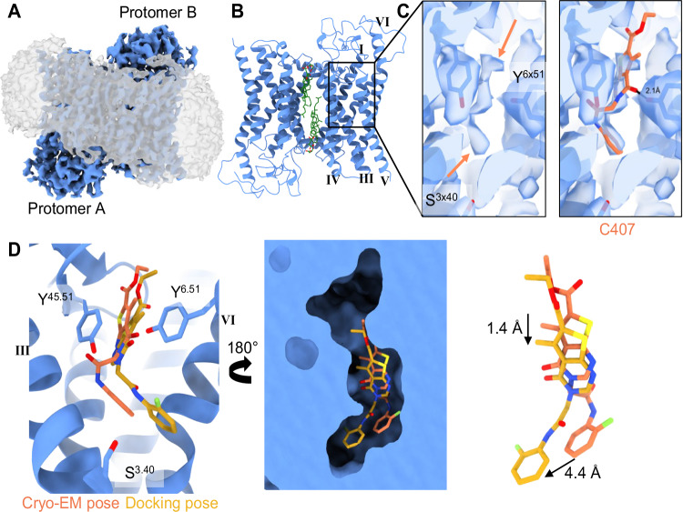
Fig. 3 Cryo-EM structure of the FZD7-dimer containing a density potentially corresponding to C407. A Cryo-EM map of the antiparallel FZD7-dimer (blue) solubilized in LMNG detergent (micelle shown in gray). B Model of the FZD7-dimer after focused masking. The lipidic interface consisting of conserved cholesterol binding sites (green) mediates interactions between the protomers at the dimer interface. C Reconstruction and zoom in on protomer B showing the tentative placement of C407 in a weak density observed in the 7TMD cavity. The density (highlighted with orange arrows; left) is consistent with low-occupancy ligand binding, but does not allow unambiguous assignment. The predicted binding pose of C407 is modeled in this density (orange molecule; right), D Model (left) and surface representation (middle) of protomer B of the FZD7-dimer depicting the cavity within the 7TMD and comparing the cryo-EM pose (orange) and docking pose (yellow) of C407 that can be accommodated in the pocket. Docking pose and cryo-EM pose are slightly shifted (right).
Fig. 3 Cryo-EM structure of the FZD7-dimer containing a density potentially corresponding to C407. A Cryo-EM map of the antiparallel FZD7-dimer (blue) solubilized in LMNG detergent (micelle shown in gray). B Model of the FZD7-dimer after focused masking. The lipidic interface consisting of conserved cholesterol binding sites (green) mediates interactions between the protomers at the dimer interface. C Reconstruction and zoom in on protomer B showing the tentative placement of C407 in a weak density observed in the 7TMD cavity. The density (highlighted with orange arrows; left) is consistent with low-occupancy ligand binding, but does not allow unambiguous assignment. The predicted binding pose of C407 is modeled in this density (orange molecule; right), D Model (left) and surface representation (middle) of protomer B of the FZD7-dimer depicting the cavity within the 7TMD and comparing the cryo-EM pose (orange) and docking pose (yellow) of C407 that can be accommodated in the pocket. Docking pose and cryo-EM pose are slightly shifted (right).CC BY · PubMed Central
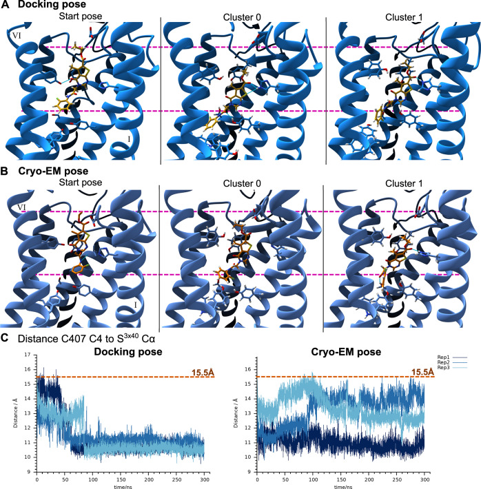
Fig. 4 Movement of C407 throughout the MD trajectories to a binding pose located deeper within the 7TMD core. A MD simulations starting from the docking pose of C407 (yellow sticks). Left: Start pose as predicted by the docking calculations. Middle and right image represent the most frequent C407 binding poses throughout the trajectory with 51% of frames (cluster 0) and 35% of frames (cluster 1) (Supplementary Data 4). Pink dashed lines represent the highest and lowest reaching parts of C407 in the starting pose. TM7 is hidden for improved clarity and numbering of the TMs is indicated in roman numerals. Full side-views of the receptor can be found in Supplementary Fig. 16. B Same as (A), but for the MD simulations starting from the pose of C407 (orange sticks) as modeled based on the structural cryo-EM data. The most frequent binding poses of C407 throughout the trajectory were found in 56% (cluster 0) and 30% (cluster 1) of the frames (Supplemenatry Data 4). C Distance between atom C4 of C407 and the S3513x40 Cα atom of FZD7 for the MD simulations starting from the docking pose (left) or the cryo-EM pose (right). The initial distance as measured in the starting model is indicated by the orange dashed line. Refer to Supplementary Fig. 16 for location of residue and atoms used for distance measurements and to Supplementary Fig. 14 for complementary distance measurements. Data for the three independent replica are shown in shades of blue.
Fig. 4 Movement of C407 throughout the MD trajectories to a binding pose located deeper within the 7TMD core. A MD simulations starting from the docking pose of C407 (yellow sticks). Left: Start pose as predicted by the docking calculations. Middle and right image represent the most frequent C407 binding poses throughout the trajectory with 51% of frames (cluster 0) and 35% of frames (cluster 1) (Supplementary Data 4). Pink dashed lines represent the highest and lowest reaching parts of C407 in the starting pose. TM7 is hidden for improved clarity and numbering of the TMs is indicated in roman numerals. Full side-views of the receptor can be found in Supplementary Fig. 16. B Same as (A), but for the MD simulations starting from the pose of C407 (orange sticks) as modeled based on the structural cryo-EM data. The most frequent binding poses of C407 throughout the trajectory were found in 56% (cluster 0) and 30% (cluster 1) of the frames (Supplemenatry Data 4). C Distance between atom C4 of C407 and the S3513x40 Cα atom of FZD7 for the MD simulations starting from the docking pose (left) or the cryo-EM pose (right). The initial distance as measured in the starting model is indicated by the orange dashed line. Refer to Supplementary Fig. 16 for location of residue and atoms used for distance measurements and to Supplementary Fig. 14 for complementary distance measurements. Data for the three independent replica are shown in shades of blue.CC BY · PubMed Central
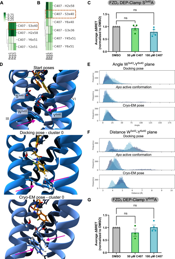
Fig. 5 Interactions and potential mechanism of action of C407 as derived from MD simulations. Most frequent hydrogen bonds between C407 and FZD7 throughout the MD simulations starting from the docking pose (A) or the cryo-EM pose (B). The interaction of C407 with S3513x40 is highlighted with an orange box. C FZD7-DEP-Clamp measurements determining the WNT-3A-induced (1 μg/mL) sensor rearrangement in absence (DMSO) and presence of different concentrations of C407 after introduction of point mutation S3513x40A. D Closeup view of the microswitch consisting of residues W3543x43 and Y4786x40 in the starting models (top) and in reference to the most frequent poses of C407 throughout the MD simulations starting from the docking pose (middle; C407 in yellow) and the cryo-EM pose (bottom; C407 in orange). Side-chains of the two residues are highlighted with magenta arrows. TM5 is hidden in these images for improved clarity. Numbering of the TMs is indicated in roman numerals. E Frequency distributions of observed angles between the vectors perpendicular to the aromatic planes of W3543x43 and Y4786x40. F Frequency distributions of observed distances between the center of mass of the aromatic planes of W3543x43 and Y4786x40. E, F Distributions for the MD simulations starting from the docking pose (top) or the cryo-EM pose (bottom) are compared with those of the apo FZD7 structure as used for the docking calculations (middle). Data are shown for three independent replicas in different shades of blue. Corresponding time-courses can be found in Supplementary Fig. 18. G FZD7-DEP-Clamp measurements determining the WNT-3A-induced (1 μg/mL) sensor rearrangement in absence (DMSO) and presence of different concentrations of C407 after introduction of point mutation Y4786x40A. C, G Data (lines) represent mean ± SEM of four independent experiments. Statistical analysis was performed with a one-way analysis of variance (ANOVA) followed by Dunnett’s post-hoc test comparing all other means with the means of the DMSO control. Corresponding time-course measurements can be found in Supplementary Fig. 7. Source data are provided as a Source Data file.
Fig. 5 Interactions and potential mechanism of action of C407 as derived from MD simulations. Most frequent hydrogen bonds between C407 and FZD7 throughout the MD simulations starting from the docking pose (A) or the cryo-EM pose (B). The interaction of C407 with S3513x40 is highlighted with an orange box. C FZD7-DEP-Clamp measurements determining the WNT-3A-induced (1 μg/mL) sensor rearrangement in absence (DMSO) and presence of different concentrations of C407 after introduction of point mutation S3513x40A. D Closeup view of the microswitch consisting of residues W3543x43 and Y4786x40 in the starting models (top) and in reference to the most frequent poses of C407 throughout the MD simulations starting from the docking pose (middle; C407 in yellow) and the cryo-EM pose (bottom; C407 in orange). Side-chains of the two residues are highlighted with magenta arrows. TM5 is hidden in these images for improved clarity. Numbering of the TMs is indicated in roman numerals. E Frequency distributions of observed angles between the vectors perpendicular to the aromatic planes of W3543x43 and Y4786x40. F Frequency distributions of observed distances between the center of mass of the aromatic planes of W3543x43 and Y4786x40. E, F Distributions for the MD simulations starting from the docking pose (top) or the cryo-EM pose (bottom) are compared with those of the apo FZD7 structure as used for the docking calculations (middle). Data are shown for three independent replicas in different shades of blue. Corresponding time-courses can be found in Supplementary Fig. 18. G FZD7-DEP-Clamp measurements determining the WNT-3A-induced (1 μg/mL) sensor rearrangement in absence (DMSO) and presence of different concentrations of C407 after introduction of point mutation Y4786x40A. C, G Data (lines) represent mean ± SEM of four independent experiments. Statistical analysis was performed with a one-way analysis of variance (ANOVA) followed by Dunnett’s post-hoc test comparing all other means with the means of the DMSO control. Corresponding time-course measurements can be found in Supplementary Fig. 7. Source data are provided as a Source Data file.CC BY · PubMed Central

Verification

Claims the paper makes

The claims and statistics of this paper haven’t been checked yet.

Run the check — free, no account needed. It reads the open-access text and checks the sources it cites.

A few free checks a day · papers already verified open instantly · results are public

Self-check

Percentages, sums and reported differences re-computed from the paper’s own numbers.

The self-check needs the paper’s full text.

Citations

Cites

Research works printed but not linked: 57

  1. Lorente, J. S. et al. GPCR drug discovery: new agents, targets and indications. Nat. Rev. Drug Discov. 24, 458–479 (2025). · DOI
  2. Schulte, G. International Union of Basic and Clinical Pharmacology. CXV: the class F of G protein-coupled receptors. Pharmacol. Rev. 76, 1009–1037 (2024). · DOI
  3. Nusse, R. & Clevers, H. Wnt/β-catenin signaling, disease, and emerging therapeutic modalities. Cell 169, 985–999 (2017). · DOI · Paper page
  4. Maltzahn, J. V., Bentzinger, C. F. & Rudnicki, M. A. Wnt7a-Fzd7 signalling directly activates the Akt/mTOR anabolic growth pathway in skeletal muscle. Nat. Cell Biol. 14, 186–191 (2012). · DOI
  5. Fernandez, A. et al. The WNT receptor FZD7 is required for maintenance of the pluripotent state in human embryonic stem cells. Proc. Natl. Acad. Sci. USA 111, 1409–1414 (2014). · DOI
  6. Xu, L. et al. Cryo-EM structure of constitutively active human Frizzled 7 in complex with heterotrimeric Gs. Cell Res. 31, 1311–1314 (2021). · DOI
  7. Wright, S. C. et al. A conserved molecular switch in Class F receptors regulates receptor activation and pathway selection. Nat. Commun. 10, 667 (2019). · DOI
  8. Flanagan, D. J. et al. Frizzled-7 is required for Wnt signaling in gastric tumors with and without APC mutations. Cancer Res. 79, 970–981 (2019). · DOI
  9. Larasati, Y., Boudou, C., Koval, A. & Katanaev, V. L. Unlocking the Wnt pathway: therapeutic potential of selective targeting FZD7 in cancer. Drug Discov. Today 27, 777–792 (2022). · DOI
  10. Tao, L. et al. Frizzled proteins are colonic epithelial receptors for C. difficile toxin B. Nature 538, 350–355 (2016). · DOI
  11. Kinsolving, J. et al. Structural and functional insight into the interaction of Clostridioides difficile toxin B and FZD7. Cell Rep. 43, 113727 (2024). · DOI
  12. Nile, A. H. et al. A selective peptide inhibitor of Frizzled 7 receptors disrupts intestinal stem cells. Nat. Chem. Biol. 14, 582–590 (2018). · DOI
  13. Hansen, S., Nile, A. H., Mehta, S. C., Fuhrmann, J. & Hannoush, R. N. Lead optimization yields high affinity Frizzled 7-targeting peptides that modulate Clostridium difficile toxin B pathogenicity in epithelial cells. J. Med. Chem. 62, 7739–7750 (2019). · DOI
  14. Koval, A., Boudou, C. & Katanaev, V. L. Challenging reported frizzled-targeting compounds in selective assays reveals lack of functional inhibition and claimed profiles. ACS Pharmacol. Transl. Sci. 7, 4144–4154 (2024). · DOI
  15. Kinsolving, J. et al. A putative Frizzled 7-targeting compound acts as a firefly luciferase inhibitor. J. Med. Chem. 67, 22332–22341 (2024). · DOI
  16. Bous, J. et al. Structural basis of frizzled 7 activation and allosteric regulation. Nat. Commun. 15, 7422 (2024). · DOI
  17. Grätz, L. et al. Pathway selectivity in Frizzleds is achieved by conserved micro-switches defining pathway-determining, active conformations. Nat. Commun. 14, 4573 (2023). · DOI
  18. Turku, A., Schihada, H., Kozielewicz, P., Bowin, C. F. & Schulte, G. Residue 6.43 defines receptor function in class F GPCRs. Nat. Commun. 12, 3919 (2021). · DOI
  19. Kozielewicz, P., Turku, A. & Schulte, G. Molecular pharmacology of class F receptor activation. Mol. Pharmacol. 97, 62–71 (2020). · DOI
  20. Qian, Y. et al. Structural basis of Frizzled 4 in recognition of Dishevelled 2 unveils mechanism of WNT signaling activation. Nat. Commun. 15, 7644 (2024). · DOI
  21. Grätz, L., Voss, J. H. & Schulte, G. Class-wide analysis of Frizzled-Dishevelled interactions using BRET biosensors reveals functional differences among receptor paralogs. ACS Sens. 9, 4626–4636 (2024). · DOI
  22. Janda, C. Y., Waghray, D., Levin, A. M., Thomas, C. & Garcia, K. C. Structural basis of Wnt recognition by Frizzled. Science 337, 59–64 (2012). · DOI
  23. Grätz, L. et al. SAG1.3-derived Frizzled-targeting small molecule compounds. J. Biol. Chem. 301, 110751 (2025). · DOI
  24. Kozielewicz, P. et al. Structural insight into small molecule action on Frizzleds. Nat. Commun. 11, 414 (2020). · DOI
  25. Isberg, V. et al. Generic GPCR residue numbers—aligning topology maps while minding the gaps. Trends Pharmacol. Sci. 36, 22–31 (2015). · DOI
  26. Bowin, C. F. et al. WNT stimulation induces dynamic conformational changes in the Frizzled–Dishevelled interaction. Sci. Signal. 16, eabo4974 (2023). · DOI
  27. Berkeley, R. F., Cook, B. D. & Herzik, M. A. Machine learning approaches to cryoEM density modification differentially affect biomacromolecule and ligand density quality. Front. Mol. Biosci. 11, 1404885 (2024). · DOI
  28. Latorraca, N. R., Venkatakrishnan, A. J. & Dror, R. O. GPCR dynamics: structures in motion. Chem. Rev. 117, 139–155 (2017). · DOI
  29. Schulte, G. et al. Frizzleds act as dynamic pharmacological entities. Trends Pharmacol. Sci. 45, 419–429 (2024). · DOI
  30. Maurice, M. M. & Angers, S. Mechanistic insights into Wnt–catenin pathway activation and signal transduction. Nat. Rev. Mol. Cell Biol. 26, 371–388 (2025). · DOI
  31. Shapovalov, M. V. & Dunbrack, R. L. A smoothed backbone-dependent rotamer library for proteins derived from adaptive kernel density estimates and regressions. Structure 19, 844–858 (2011). · DOI · Paper page
  32. Pettersen, E. F. et al. UCSF–Chimera — A visualization system for exploratory research and analysis. J. Comput. Chem. 25, 1605–1612 (2004). · DOI
  33. Momany, F. A. & Rone, R. Validation of the general purpose QUANTA 3.2/CHARMm force field. J. Comput. Chem. 13, 888–900 (1992). · DOI
  34. Moriarty, N. W., Liebschner, D., Tronrud, D. E. & Adams, P. D. Arginine off-kilter: guanidinium is not as planar as restraints denote. Acta Crystallogr. Sect. D 76, 1159–1166 (2020). · DOI
  35. Qi, X., Friedberg, L., Bose-Boyd, R. D., Long, T. & Li, X. Sterols in an intramolecular channel of Smoothened mediate Hedgehog signaling. Nat. Chem. Biol. 16, 1368–1375 (2020). · DOI
  36. Weierstall, U. et al. Lipidic cubic phase injector facilitates membrane protein serial femtosecond crystallography. Nat. Commun. 5, 3309 (2014). · DOI
  37. Byrne, E. F. et al. Structural basis of Smoothened regulation by its extracellular domains. Nature 535, 517–522 (2016). · DOI
  38. Wang, C. et al. Structural basis for Smoothened receptor modulation and chemoresistance to anticancer drugs. Nat. Commun. 5, 4355 (2014). · DOI
  39. Sterling, T. & Irwin, J. J. ZINC15 – Ligand discovery for everyone. J. Chem. Inf. Model. 55, 2324–2337 (2015). · DOI · Paper page
  40. Coleman, R. G., Carchia, M., Sterling, T., Irwin, J. J. & Shoichet, B. K. Ligand pose and orientational sampling in molecular docking. PLoS ONE 8, e75992 (2013). · DOI
  41. Varadi, M. et al. AlphaFold Protein Structure Database: massively expanding the structural coverage of protein-sequence space with high-accuracy models. Nucleic Acids Res. 50, D439–D444 (2022). · DOI · Paper page
  42. He, X., Man, V. H., Yang, W., Lee, T.-S. & Wang, J. A fast and high-quality charge model for the next generation general AMBER force field. J. Chem. Phys. 153, 114502 (2020). · DOI
  43. Wang, J., Wolf, R. M., Caldwell, J. W., Kollman, P. A. & Case, D. A. Development and testing of a general amber force field. J. Comput. Chem. 25, 1157–1174 (2004). · DOI
  44. Shao, Y. et al. Advances in molecular quantum chemistry contained in the Q-Chem 4 program package. Mol. Phys. 113, 184–215 (2015). · DOI
  45. Sousa da Silva, A. W. & Vranken, W. F. ACPYPE – AnteChamber PYthon Parser interfacE. BMC Res. Notes 5, 367 (2012). · DOI
  46. Wang, J., Wang, W., Kollman, P. A. & Case, D. A. Automatic atom type and bond type perception in molecular mechanical calculations. J. Mol. Graph 25, 247–260 (2006). · DOI
  47. Jo, S., Kim, T., Iyer, V. G. & Im, W. CHARMM-GUI : a web-based graphical user interface for CHARMM. J. Comput. Chem. 29, 1859–1865 (2008). · DOI · Paper page
  48. Jo, S., Lim, J. B., Klauda, J. B. & Im, W. CHARMM-GUI membrane builder for mixed bilayers and its application to yeast membranes. Biophys. J. 97, 50–58 (2009). · DOI
  49. Lomize, M. A., Pogozheva, I. D., Joo, H., Mosberg, H. I. & Lomize, A. L. OPM database and PPM web server: resources for positioning of proteins in membranes. Nucleic Acids Res. 40, D370–D376 (2011). · DOI
  50. Van Der Spoel, D. et al. GROMACS: fast, flexible, and free. J. Comput. Chem. 26, 1701–1718 (2005). · DOI
  51. Tian, C. et al. ff19SB: amino-acid-specific protein backbone parameters trained against quantum mechanics energy surfaces in solution. J. Chem. Theory Comput. 16, 528–552 (2020). · DOI
  52. Dickson, C. J., Walker, R. C. & Gould, I. R. Lipid21: complex lipid membrane simulations with AMBER. J. Chem. Theory Comput. 18, 1726–1736 (2022). · DOI
  53. Hess, B., Bekker, H., Berendsen, H. J. C. & Fraaije, J. G. E. M. LINCS: a linear constraint solver for molecular simulations. J. Comput. Chem. 18, 1463–1472 (1997). · DOI
  54. Essmann, U. et al. A smooth particle mesh Ewald method. J. Chem. Phys. 103, 8577–8593 (1995). · DOI
  55. Roe, D. R. & Cheatham III, T. E. PTRAJ and CPPTRAJ: software for processing and analysis of molecular dynamics trajectory data. J. Chem. Theory Comput. 9, 3084–3095 (2013). · DOI
  56. Rodriguez-Espigares, I. et al. GPCRmd uncovers the dynamics of the 3D-GPCRome. Nat. Methods 17, 777–787 (2020). · DOI
  57. Goddard, T. D. et al. UCSF ChimeraX: meeting modern challenges in visualization and analysis. Protein Sci. 27, 14–25 (2018). · DOI · Paper page

Metadata

Authors
Scharf, M. M., Kinsolving, J., Grätz, L., Voss, J. H., Carrasco-Busturia, D., Forsberg, B., Kolb, P., & Schulte, G.
Affiliations
  • Karolinska Institutet (SE)
  • University of Bonn (DE)
  • Linköping University (SE)
  • Philipps University of Marburg (DE)
Corresponding author
Schulte, Gunnar
Venue
Nature Communications
Published
2025-12-14
Volume
16
Issue
1
Pages
11138-11138
ISSN
2041-1723 (ISSN-L)
OpenAlex
W4417303264
Cited by
6
References
1
Citations by year
2026: 6
FWCI
1.80
Type
journal article
Language
en
Open access
Locations
  • Nature Communications (publishedVersion, cc-by)
  • Research Square (acceptedVersion, cc-by)
  • Europe PMC (PubMed Central) (publishedVersion, cc-by)
  • PubMed (publishedVersion)
  • PubMed Central (publishedVersion, cc-by)
  • KTH Publication Database DiVA (KTH Royal Institute of Technology) (submittedVersion, other-oa)
  • DOAJ (DOAJ: Directory of Open Access Journals) (submittedVersion, cc-by-sa)
Funders
  • European Commission (101199012, 2020 ‘)
  • Deutsche Forschungsgemeinschaft (520506488, 470002134, 504098926)
  • Cancerfonden (NNF22OC0078104, 23 2825 Pj, 20 1102 PjF, 2017/561, 2019-, CAN2017/561)
  • Svenska Sällskapet för Medicinsk Forskning (PG-22-0379)
  • Karolinska Institutet
  • Knut och Alice Wallenbergs Stiftelse (KAW 2020.0239)
  • Novo Nordisk
  • Vetenskapsrådet (2019-01190, 01190, 2017-04676, 2024-02515, 2022-06725)
  • Science for Life Laboratory (KAW 2020.0239)
  • Novo Nordisk Fonden (NNF22OC0078104, NNF21OC0070008)
  • HORIZON EUROPE Framework Programme
Topics
  • Life Sciences › Biochemistry, Genetics and Molecular Biology › Molecular Biology › Wnt/β-catenin signaling in development and cancer
  • Life Sciences › Pharmacology, Toxicology and Pharmaceutics › Drug Discovery › Computational Drug Discovery Methods
  • Life Sciences › Biochemistry, Genetics and Molecular Biology › Molecular Biology › Receptor Mechanisms and Signaling
Keywords
Frizzled-7, Wnt receptors, negative allosteric modulators, small-molecule ligands, Wnt/β-catenin signaling, ligand binding sites, intestinal tumors, genetically encoded biosensors, molecular docking
MeSH terms
Allosteric Regulation, Binding Sites, Humans, Ligands, Mutagenesis, Site-Directed, Cryoelectron Microscopy, Frizzled Receptors, beta Catenin, Small Molecule Libraries, Molecular Dynamics Simulation, HEK293 Cells, Wnt Signaling Pathway, Molecular Docking Simulation

About these checks

Registry, retraction, Cites and statistics results are computed, not judged, with one exception: a printed reference that no index lists is read once by a model before it counts as a citation error. Claims are extracted by a model and kept only when their quote appears verbatim in the text; each cited source is then checked, with a model’s help, for whether it says what the claim relies on. Those verdicts can be wrong — read the claim’s quote and the cited source before relying on one.

Statistics and arithmetic are recomputed, not judged.

Confidence bands are shown only for fields calibrated on our annotated test set. None is calibrated yet, so no band is shown.

What counts as a claim ›How we measure confidence ›How we check this ›

Are you an author, or do you see a problem with this page? Request a review