

The DOI matches its Crossref record; 97 of the 97 references it cites were found in a registry or an index.
Phase separation and ageing of glycine-rich protein from tick adhesive
Open accessPublished version of a preprint
Abstract
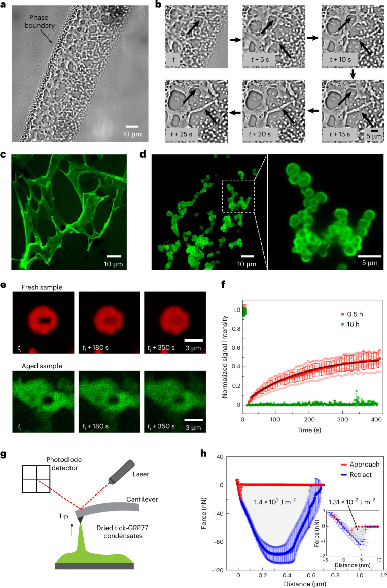
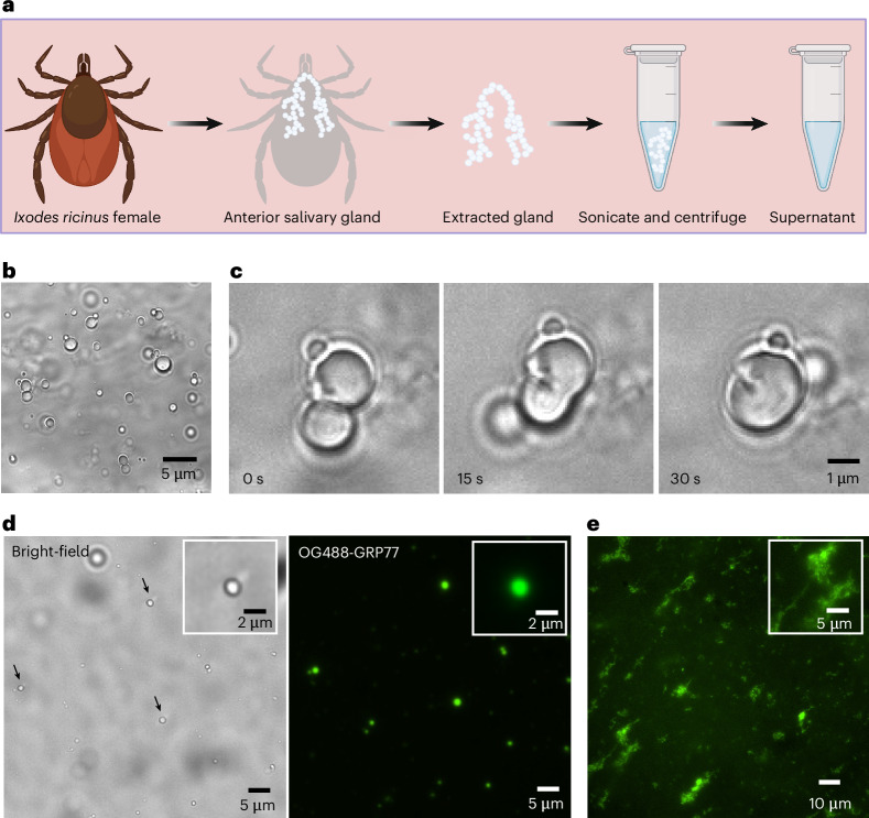
Hard ticks feed on their host for multiple days. To ensure firm attachment, they secrete a protein-rich saliva that eventually forms a solid cement cone. The underlying mechanism of this liquid-to-solid transition is currently not understood. This study focuses on the phase transitions of a disordered glycine-rich protein (GRP) found in tick saliva. We show that GRP undergoes liquid–liquid phase separation via simple coacervation to form biomolecular condensates in salty environments. Cation– π and π – π interactions mediated by periodically placed arginine and aromatic amino-acid residues are the primary driving forces that promote phase separation. Interestingly, GRP condensates exhibit ageing by undergoing liquid-to-gel transition over time and exhibit adhesive properties, similar to the naturally occurring cement cone. Finally, we provide evidence for protein-rich condensates in natural tick saliva. Our findings provide a starting point to gain further insights into the bioadhesion of ticks, to develop novel tick control strategies, and towards achieving biomedical applications such as tissue sealants.
Overview
The claims and statistics of this paper haven’t been checked yet.
Run the check — free, no account needed. It reads the open-access text and checks the sources it cites.
Integrity signals
Registry
Cites
Statistics
Figures
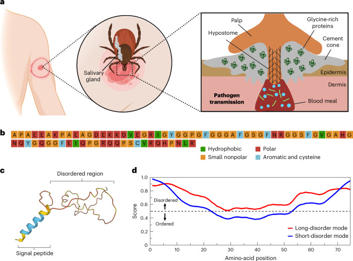
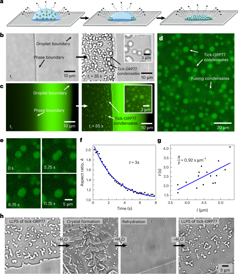
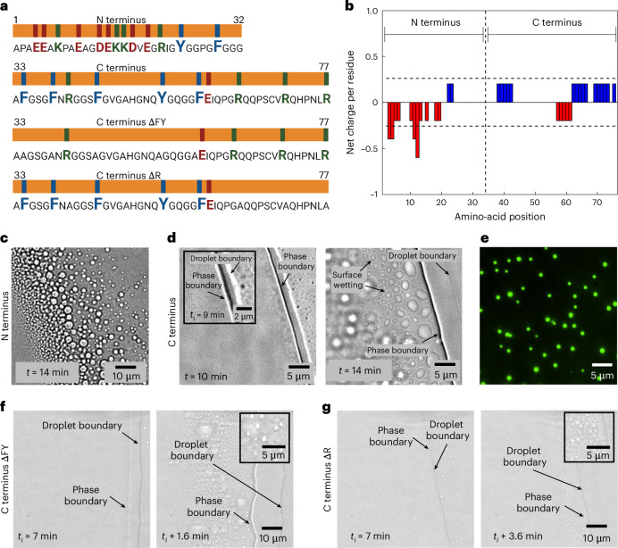
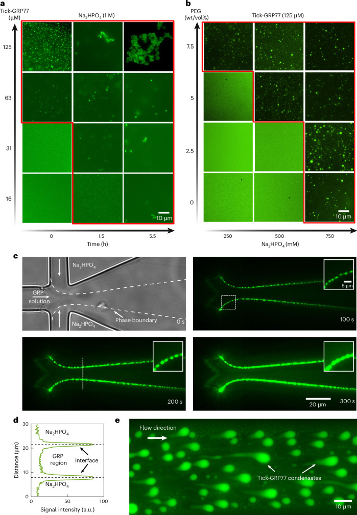
Verification
Claims the paper makes
The claims and statistics of this paper haven’t been checked yet.
Run the check — free, no account needed. It reads the open-access text and checks the sources it cites.
Self-check
Percentages, sums and reported differences re-computed from the paper’s own numbers.
The self-check needs the paper’s full text.
Citations
Cites
Highly accurate protein structure prediction with AlphaFold
Jumper, J., Evans, R., Pritzel, A., Green, T., Figurnov, M., Ronneberger, O., Tunyasuvunakool, K., Bates, R., Žídek, A., Potapenko, A., et al. (2021) · Nature · doi:10.1038/s41586-021-03819-2 · Cited by 43,764
Capillary flow as the cause of ring stains from dried liquid drops
Deegan, R. D., Bakajin, O., Dupont, T. F., Huber, G., Nagel, S. R., & Witten, T. A. (1997) · Nature · doi:10.1038/39827 · Cited by 6,453
Liquid phase condensation in cell physiology and disease
Shin, Y., & Brangwynne, C. P. (2017) · Science · doi:10.1126/science.aaf4382 · Cited by 4,213
Liquid-Liquid Phase Separation in Biology
Hyman, A. A., Weber, C. A., & Jülicher, F. (2014) · Annual Review of Cell and Developmental Biology · doi:10.1146/annurev-cellbio-100913-013325 · Cited by 3,155
Alberti, S., Gladfelter, A., & Mittag, T. (2019) · Cell · doi:10.1016/j.cell.2018.12.035 · Cited by 2,856
Interactions between macromolecules and ions: the Hofmeister series
Zhang, Y., & Cremer, P. S. (2006) · Current Opinion in Chemical Biology · doi:10.1016/j.cbpa.2006.09.020 · Cited by 2,100
The global importance of ticks
JONGEJAN, F., & UILENBERG, G. (2004) · Parasitology · doi:10.1017/s0031182004005967 · Cited by 2,091
Wang, J., Choi, J.-M., Holehouse, A. S., Lee, H. O., Zhang, X., Jahnel, M., Maharana, S., Lemaitre, R., Pozniakovsky, A., Drechsel, D., et al. (2018) · Cell · doi:10.1016/j.cell.2018.06.006 · Cited by 2,076
Nott, T. J., Petsalaki, E., Farber, P., Jervis, D., Fussner, E., Plochowietz, A., Craggs, T. D., Bazett-Jones, D. P., Pawson, T., Forman-Kay, J. D., et al. (2015) · Molecular Cell · doi:10.1016/j.molcel.2015.01.013 · Cited by 1,803
Mussel-Inspired Adhesives and Coatings
Lee, B. P., Messersmith, P. B., Israelachvili, J. N., & Waite, J. H. (2011) · Annual Review of Materials Research · doi:10.1146/annurev-matsci-062910-100429 · Cited by 1,668
Polymer physics of intracellular phase transitions
Brangwynne, C. P., Tompa, P., & Pappu, R. V. (2015) · Nature Physics · doi:10.1038/nphys3532 · Cited by 1,571
Mészáros, B., Erdős, G., & Dosztányi, Z. (2018) · Nucleic Acids Research · doi:10.1093/nar/gky384 · Cited by 1,454
The PSIPRED Protein Analysis Workbench: 20 years on
Buchan, D. W. A., & Jones, D. T. (2019) · Nucleic Acids Research · doi:10.1093/nar/gkz297 · Cited by 1,351
Elbaum-Garfinkle, S., Kim, Y., Szczepaniak, K., Chen, C. C.-H., Eckmann, C. R., Myong, S., & Brangwynne, C. P. (2015) · Proceedings of the National Academy of Sciences · doi:10.1073/pnas.1504822112 · Cited by 1,278
Self-citationCation−π Interaction: Its Role and Relevance in Chemistry, Biology, and Material Science
Mahadevi, A. S., & Sastry, G. N. (2013) · Chemical Reviews · doi:10.1021/cr300222d · Cited by 1,149
Valence and patterning of aromatic residues determine the phase behavior of prion-like domains
Martin, E. W., Holehouse, A. S., Peran, I., Farag, M., Incicco, J. J., Bremer, A., Grace, C. R., Soranno, A., Pappu, R. V., & Mittag, T. (2020) · Science · doi:10.1126/science.aaw8653 · Cited by 1,147
Hofmeister Phenomena: An Update on Ion Specificity in Biology
Nostro, P. L., & Ninham, B. W. (2012) · Chemical Reviews · doi:10.1021/cr200271j · Cited by 1,029
Overview: Ticks as vectors of pathogens that cause disease in humans and animals
Fuente, J. D. L. (2008) · Frontiers in Bioscience · doi:10.2741/3200 · Cited by 934
Mussel adhesion – essential footwork
Waite, J. H. (2017) · Journal of Experimental Biology · doi:10.1242/jeb.134056 · Cited by 675
Biomolecular Phase Separation: From Molecular Driving Forces to Macroscopic Properties
Dignon, G. L., Best, R. B., & Mittal, J. (2020) · Annual Review of Physical Chemistry · doi:10.1146/annurev-physchem-071819-113553 · Cited by 657
Bioinspired underwater adhesives by using the supramolecular toolbox
Hofman, A. H., van Hees, I. A., Yang, J., & Kamperman, M. (2018) · Advanced Materials · doi:10.1002/adma.201704640 · Cited by 652
Convery, N., & Gadegaard, N. (2019) · Micro and Nano Engineering · doi:10.1016/j.mne.2019.01.003 · Cited by 648
Erdős, G., Pajkos, M., & Dosztányi, Z. (2021) · Nucleic Acids Research · doi:10.1093/nar/gkab408 · Cited by 608
The role of saliva in tick feeding
Ivo, M. B. F. (2009) · Frontiers in bioscience · doi:10.2741/3363 · Cited by 598
Mecozzi, S., West, A. P., & Dougherty, D. A. (1996) · Proceedings of the National Academy of Sciences · doi:10.1073/pnas.93.20.10566 · Cited by 592
Estimating the Frequency of Lyme Disease Diagnoses, United States, 2010–2018
Kugeler, K. J., Schwartz, A. M., Delorey, M. J., Mead, P. S., & Hinckley, A. F. (2021) · Emerging infectious diseases · doi:10.3201/eid2702.202731 · Cited by 592
CIDER: Resources to Analyze Sequence-Ensemble Relationships of Intrinsically Disordered Proteins
Holehouse, A. S., Das, R. K., Ahad, J., Richardson, M. O. G., & Pappu, R. V. (2017) · Biophysical Journal · doi:10.1016/j.bpj.2016.11.3200 · Cited by 551
Peptide-based coacervates as biomimetic protocells
Abbas, M., Lipiński, W. P., Wang, J., & Spruijt, E. (2021) · Chemical Society Reviews · doi:10.1039/d0cs00307g · Cited by 497
Direct Visual Observation of Thermal Capillary Waves
Aarts, D. G. A. L., Schmidt, M., & LEKKERKERKER, H. N. W. (2004) · Science · doi:10.1126/science.1097116 · Cited by 478
Bakker, E. S., Gill, J. L., Johnson, C. N., Vera, F. W. M., Sandom, C. J., Asner, G. P., & Svenning, J.-C. (2016) · Proceedings of the National Academy of Sciences · doi:10.1073/pnas.1502545112 · Cited by 368
An annotated catalog of salivary gland transcripts from Ixodes scapularis ticks
Ribeiro, J. M. C., Alarcon-Chaidez, F., B. Francischetti, I. M., Mans, B. J., Mather, T. N., Valenzuela, J. G., & Wikel, S. K. (2006) · Insect Biochemistry and Molecular Biology · doi:10.1016/j.ibmb.2005.11.005 · Cited by 366
Complex coacervates as a foundation for synthetic underwater adhesives
Stewart, R. J., Wang, C. S., & Shao, H. (2010) · Advances in Colloid and Interface Science · doi:10.1016/j.cis.2010.10.009 · Cited by 356
A short peptide synthon for liquid–liquid phase separation
Abbas, M., Lipiński, W. P., Nakashima, K. K., Huck, W. T. S., & Spruijt, E. (2021) · Nature Chemistry · doi:10.1038/s41557-021-00788-x · Cited by 355
Wikel, S. K. (1996) · Annual Review of Entomology · doi:10.1146/annurev.en.41.010196.000245 · Cited by 353
The Essential Role of Tick Salivary Glands and Saliva in Tick Feeding and Pathogen Transmission
Šimo, L., Kazimírová, M., Richardson, J. P., & Bonnet, S. I. (2017) · Frontiers in Cellular and Infection Microbiology · doi:10.3389/fcimb.2017.00281 · Cited by 341
Investigating the Hydrogen-Bonding Model of Urea Denaturation
Sagle, L. B., Zhang, Y., Litosh, V. A., Chen, X., Cho, Y., & Cremer, P. S. (2009) · Journal of the American Chemical Society · doi:10.1021/ja9016057 · Cited by 306
Poudyal, R. R., Guth-Metzler, R., Veenis, A. J., Frankel, E. A., Keating, C. D., & Bevilacqua, P. C. (2019) · Nature Communications · doi:10.1038/s41467-019-08353-4 · Cited by 305
Complexation and coacervation of like-charged polyelectrolytes inspired by mussels
Kim, S., Huang, J. S., Lee, Y., Dutta, S., Yoo, H. Y., Jung, Y. M., Jho, Y., Zeng, H., & Hwang, D. S. (2016) · Proceedings of the National Academy of Sciences · doi:10.1073/pnas.1521521113 · Cited by 248
Yang, X., Goldberg, M. S., Popova, T. G., Schoeler, G. B., Wikel, S. K., Hagman, K. E., & Norgard, M. V. (2000) · Molecular Microbiology · doi:10.1046/j.1365-2958.2000.02104.x · Cited by 245
HullRad: Fast Calculations of Folded and Disordered Protein and Nucleic Acid Hydrodynamic Properties
Fleming, P. J., & Fleming, K. G. (2018) · Biophysical Journal · doi:10.1016/j.bpj.2018.01.002 · Cited by 231
Tick Salivary Gland Physiology
Sauer, J. R., McSwain, J. L., Bowman, A. S., & Essenberg, R. C. (1995) · Annual Review of Entomology · doi:10.1146/annurev.en.40.010195.001333 · Cited by 224
Pathogen transmission in relation to duration of attachment by Ixodes scapularis ticks
Eisen, L. (2018) · Ticks and Tick-borne Diseases · doi:10.1016/j.ttbdis.2018.01.002 · Cited by 199
Berthier, J., & Brakke, K. A. (2012) · Wiley eBooks · doi:10.1002/9781118401323 · Cited by 190
The role of coacervation and phase transitions in the sandcastle worm adhesive system
Stewart, R. J., Wang, C. S., Song, I. T., & Jones, J. P. (2016) · Advances in Colloid and Interface Science · doi:10.1016/j.cis.2016.06.008 · Cited by 188
Biomolecular condensates undergo a generic shear-mediated liquid-to-solid transition
Shen, Y., Ruggeri, F. S., Vigolo, D., Kamada, A., Qamar, S., Levin, A., Iserman, C., Alberti, S., George‐Hyslop, P. S., & Knowles, T. P. J. (2020) · Nature Nanotechnology · doi:10.1038/s41565-020-0731-4 · Cited by 184
Hong, Y., Najafi, S., Casey, T. M., Shea, J., Han, S., & Hwang, D. S. (2022) · Nature Communications · doi:10.1038/s41467-022-35001-1 · Cited by 181
Tick salivary glands: function, physiology and future
Bowman, A. S., & Sauer, J. R. (2004) · Parasitology · doi:10.1017/s0031182004006468 · Cited by 178
Evaporation-driven liquid flow in sessile droplets
Gelderblom, H., Diddens, C., & Marin, A. (2022) · Soft Matter · doi:10.1039/d2sm00931e · Cited by 175
KEMP, D. H., Stone, B. F., & Binnington, K. C. (1982) · Elsevier eBooks · doi:10.1016/b978-0-08-024937-7.50009-3 · Cited by 173
The adhesive properties of coacervated recombinant hybrid mussel adhesive proteins
Lim, S., Choi, Y. S., Kang, D. G., Song, Y. H., & Chi, H. J. (2010) · Biomaterials · doi:10.1016/j.biomaterials.2010.01.063 · Cited by 171
Hydrogen-bonds mediate liquid-liquid phase separation of mussel derived adhesive peptides
Guo, Q., Zou, G., Qian, X., Chen, S., Gao, H., & Yu, J. (2022) · Nature Communications · doi:10.1038/s41467-022-33545-w · Cited by 158
On-chip microfluidic production of cell-sized liposomes
Deshpande, S., & Dekker, C. (2018) · Nature Protocols · doi:10.1038/nprot.2017.160 · Cited by 157
Self-citationLin, Y., Fichou, Y., Longhini, A. P., Llanes, L. C., Yin, P., Bazan, G. C., Kosik, K. S., & Han, S. (2020) · Journal of Molecular Biology · doi:10.1016/j.jmb.2020.166731 · Cited by 153
A cross-reactive tick cement antigen is a candidate broad-spectrum tick vaccine
Trimnell, A. R., Davies, G. M., Lissina, O., Hails, R. S., & Nuttall, P. A. (2005) · Vaccine · doi:10.1016/j.vaccine.2005.03.041 · Cited by 142
Coacervate delivery systems for proteins and small molecule drugs
Johnson, N. R., & Wang, Y. (2014) · Expert Opinion on Drug Delivery · doi:10.1517/17425247.2014.941355 · Cited by 140
Smith, A. M., & Callow, J. A. (2006) · Springer eBooks · doi:10.1007/978-3-540-31049-5 · Cited by 137
Peptide-based liquid droplets as emerging delivery vehicles
Liu, J., Spruijt, E., Miserez, A., & Langer, R. (2023) · Nature Reviews Materials · doi:10.1038/s41578-022-00528-8 · Cited by 130
Molecular biology of spider silk
Winkler, S., & Kaplan, D. L. (2000) · Reviews in Molecular Biotechnology · doi:10.1016/s1389-0352(00)00005-2 · Cited by 113
Nuttall, P. A. (2018) · Ticks and Tick-borne Diseases · doi:10.1016/j.ttbdis.2018.11.005 · Cited by 112
Dual action ectoparasite vaccine targeting ‘exposed’ and ‘concealed’ antigens
Trimnell, A. R., Hails, R. S., & Nuttall, P. A. (2002) · Vaccine · doi:10.1016/s0264-410x(02)00334-1 · Cited by 110
Bishop, R. P., Lambson, B. E., Wells, C. W., Pandit, P., Osaso, J., Nkonge, C., Morzaria, S., Musoke, A. J., & Nene, V. M. (2002) · International Journal for Parasitology · doi:10.1016/s0020-7519(02)00027-9 · Cited by 106
Controlling ticks and tick-borne diseases…looking forward
de la Fuente, J. (2018) · Ticks and Tick-borne Diseases · doi:10.1016/j.ttbdis.2018.04.001 · Cited by 106
Hennebert, E., Maldonado, B., Ladurner, P., Flammang, P., & Santos, R. (2014) · Interface Focus · doi:10.1098/rsfs.2014.0064 · Cited by 101
Methods for characterizing the material properties of biomolecular condensates
Alshareedah, I., Kaur, T., & Banerjee, P. R. (2020) · Methods in enzymology on CD-ROM/Methods in enzymology · doi:10.1016/bs.mie.2020.06.009 · Cited by 93
Phase separation by the SARS-CoV-2 nucleocapsid protein: Consensus and open questions
Cascarina, S. M., & Ross, E. D. (2022) · Journal of Biological Chemistry · doi:10.1016/j.jbc.2022.101677 · Cited by 90
Walker, A. R., Fletcher, J. D., & Gill, H. S. (1985) · International Journal for Parasitology · doi:10.1016/0020-7519(85)90106-7 · Cited by 88
Ribeiro, J. M. C., & Mans, B. J. (2020) · Frontiers in Cellular and Infection Microbiology · doi:10.3389/fcimb.2020.00374 · Cited by 86
Tick attachment cement – reviewing the mysteries of a biological skin plug system
Suppan, J., Engel, B., Marchetti‐Deschmann, M., & Nürnberger, S. (2017) · Biological reviews/Biological reviews of the Cambridge Philosophical Society · doi:10.1111/brv.12384 · Cited by 82
Liquid–liquid phase separation of the Golgi matrix protein GM130
Rebane, A. A., Ziltener, P., LaMonica, L. C., Bauer, A. H., Zheng, H., López‐Montero, I., Pincet, F., Rothman, J. E., & Ernst, A. M. (2019) · FEBS Letters · doi:10.1002/1873-3468.13715 · Cited by 79
Salomon, J., Hamer, S. A., & Swei, A. (2020) · Journal of Insect Science · doi:10.1093/jisesa/ieaa073 · Cited by 74
Zhou, J., Gong, H., Zhou, Y., Xuan, X., & Fujisaki, K. (2006) · Parasitology Research · doi:10.1007/s00436-006-0243-7 · Cited by 73
Saliva, Salivary Gland, and Hemolymph Collection from Ixodes Scapularis Ticks
Patton, T. G., Dietrich, G., Brandt, K. S., Dolan, M. C., Piesman, J. F., & Gilmore, R. D. (2012) · Journal of Visualized Experiments · doi:10.3791/3894 · Cited by 70
GREGSON, J. D. (1967) · Parasitology · doi:10.1017/s0031182000071845 · Cited by 64
Maruyama, S. R., Anatriello, E., Anderson, J. M., Ribeiro, J. M. C., Brandão, L. G., Valenzuela, J. G., Ferreira, B. R., Garcia, G. R., Szabó, M. P. J., Henson, S., et al. (2010) · BMC Genomics · doi:10.1186/1471-2164-11-363 · Cited by 62
Controllable coacervation of recombinantly produced spider silk protein using kosmotropic salts
Mohammadi, P., Jonkergouw, C., Beaune, G., Engelhardt, P., Kamada, A., Timonen, J. V. I., Knowles, T. P. J., Penttilä, M. E., & Linder, M. B. (2019) · Journal of Colloid and Interface Science · doi:10.1016/j.jcis.2019.10.058 · Cited by 59
Bullard, R. L., Allen, P. E., Chao, C., Douglas, J. R., Das, P., Morgan, S. E., Ching, W., & Karim, S. (2016) · Ticks and Tick-borne Diseases · doi:10.1016/j.ttbdis.2016.04.006 · Cited by 58
Spontaneous Switching among Conformational Ensembles in Intrinsically Disordered Proteins
Choi, U. B., Sanabria, H., Smirnova, T. I., Bowen, M. E., & Weninger, K. (2019) · Biomolecules · doi:10.3390/biom9030114 · Cited by 55
Bhowmick, B., & Han, Q. (2020) · Frontiers in Veterinary Science · doi:10.3389/fvets.2020.00319 · Cited by 54
Lim, J., Kumar, A., Low, K. J. Y., Verma, C. S., Mu, Y., Miserez, A., & Pervushin, K. V. (2021) · The Journal of Physical Chemistry B · doi:10.1021/acs.jpcb.0c11476 · Cited by 51
Harnessing disorder: onychophorans use highly unstructured proteins, not silks, for prey capture
Haritos, V. S., Niranjane, A., Weisman, S., Trueman, H. E., Sriskantha, A., & Sutherland, T. D. (2010) · Proceedings of the Royal Society B Biological Sciences · doi:10.1098/rspb.2010.0604 · Cited by 47
Kar, M., Posey, A. E., Dar, F., Hyman, A. A., & Pappu, R. V. (2021) · Biochemistry · doi:10.1021/acs.biochem.1c00501 · Cited by 41
Identification and characterization of proteins in the Amblyomma americanum tick cement cone
Hollmann, T., Kim, T. K., Tirloni, L., Radulović, Ž. M., Pinto, A. F. M., Diedrich, J. K., Yates, J. R., Vaz, I. D. S., & Mulenga, A. (2017) · International Journal for Parasitology · doi:10.1016/j.ijpara.2017.08.018 · Cited by 39
Villar, M., Pacheco, I., Mateos‐Hernández, L., Cabezas‐Cruz, A., Tabor, A. E., Rodriguez-Valle, M., Mulenga, A., Kocan, K. M., Blouin, E. F., & de la Fuente, J. (2021) · Expert Review of Proteomics · doi:10.1080/14789450.2021.2018305 · Cited by 37
Studying phase separation in confinement
Deshpande, S., & Dekker, C. (2021) · Current Opinion in Colloid & Interface Science · doi:10.1016/j.cocis.2021.101419 · Cited by 34
Self-citationWang, H., Zhang, X., Wang, X., Zhang, B., Wang, M., Yang, X., Han, X., Wang, R., Ren, S., Hu, Y., et al. (2019) · Frontiers in Physiology · doi:10.3389/fphys.2018.01943 · Cited by 30
Actinosomes: Condensate-Templated Containers for Engineering Synthetic Cells
Ganar, K. A., Leijten, L., & Deshpande, S. (2022) · ACS Synthetic Biology · doi:10.1021/acssynbio.2c00290 · Cited by 28
Self-citationModulating liquid–liquid phase separation of FUS: mechanisms and strategies
Ji, Y., Li, F., & Qiao, Y. (2022) · Journal of Materials Chemistry B · doi:10.1039/d2tb01688e · Cited by 28
Nuijens, T., Toplak, A., de Meulenreek, M. B. A. C. V., Schmidt, M., Goldbach, M., & Quaedflieg, P. J. L. M. (2016) · Tetrahedron Letters · doi:10.1016/j.tetlet.2016.06.132 · Cited by 24
Translational biotechnology for the control of ticks and tick-borne diseases
de la Fuente, J. (2021) · Ticks and Tick-borne Diseases · doi:10.1016/j.ttbdis.2021.101738 · Cited by 23
Lu, Y., Sharma, B., Soon, W. L., Shi, X., Zhao, T., Lim, Y. T., Sobota, R. M., Hoon, S., Pilloni, G., Usadi, A., et al. (2022) · Advanced Science · doi:10.1002/advs.202201444 · Cited by 23
Bouzetos, E., Ganar, K. A., Mastrobattista, E., Deshpande, S., & John van der, O. O. S. T. (2021) · Trends in biotechnology · doi:10.1016/j.tibtech.2021.04.009 · Cited by 22
Self-citationFibers on the Fly: Multiscale Mechanisms of Fiber Formation in the Capture Slime of Velvet Worms
Baer, A., Schmidt, S., Mayer, G., & Harrington, M. J. (2019) · Integrative and Comparative Biology · doi:10.1093/icb/icz048 · Cited by 18
Bullard, R. L., Sharma, S. R., Das, P. K., Morgan, S. E., & Karim, S. (2019) · Frontiers in Physiology · doi:10.3389/fphys.2019.00744 · Cited by 18
Immunization of guinea pigs with cement extract induces resistance against Ixodes scapularis ticks
Lynn, G. E., Černý, J., Kurokawa, C., Diktaş, H., Matias, J., Sajid, A., Arora, G., DePonte, K., Narasimhan, S., & Fikrig, E. (2022) · Ticks and Tick-borne Diseases · doi:10.1016/j.ttbdis.2022.102017 · Cited by 11
Agwunobi, D. O., Wang, N., Huang, L., Zhang, Y., Chang, G., Wang, K., Li, M., Wang, H., & Liu, J. (2022) · Frontiers in Cellular and Infection Microbiology · doi:10.3389/fcimb.2021.769026 · Cited by 8
On-Chip Octanol-Assisted Liposome Assembly for Bioengineering
Chen, C., Ganar, K. A., & Deshpande, S. (2023) · Journal of Visualized Experiments · doi:10.3791/65032 · Cited by 8
Self-citationPhase Separation and Ageing of Glycine-Rich Protein from Tick Adhesive
Ganar, K. A., Nandy, M., Turbina, P., Chen, C., Suylen, D. P. L., Nihoul, E., Pascoe, E. L., Beelen, S. V. D., Plaum, M., Bos, L. V. D., et al. (2024) · Figshare · doi:10.6084/m9.figshare.26490541 · Cited by 1
Self-citation
Research works printed but not linked: 1
- Sonenshine, D. E. & Roe, R. M. Biology of Ticks Vol. 2 (Oxford Univ. Press, 2014).
Metadata
- Authors
- Ganar, K. A., Nandy, M., Turbina, P., Chen, C., Suylen, D., Nihoul, E., Pascoe, E. L., van der Beelen, S., Plaum, M., van den Bos, L., Koenraadt, C. J. M., Dijkgraaf, I., & Deshpande, S.
- Affiliations
- Wageningen University & Research (NL)
- Maastricht University (NL)
- Fondazione Edmund Mach (IT)
- Corresponding author
- Dijkgraaf, Ingrid
- Deshpande, Siddharth
- ORCID
- Venue
- Nature Chemistry
- Published
- 2024-11-29
- Volume
- 17
- Issue
- 2
- Pages
- 186-197
- ISSN
- 1755-4330 (ISSN-L)
- 1755-4349
- PMC
- PMC11794139
- OpenAlex
- W4404860401
- Cited by
- 42
- References
- 97
- Citations by year
- 2026: 26 · 2025: 14 · 2024: 1
- FWCI
- 3.86
- Type
- journal article
- Language
- en
- Open access
- hybrid
- doi.org
- Licence
- cc-by
- creativecommons.org/licenses/by/4.0 (tdm)
- creativecommons.org/licenses/by/4.0 (vor)
- Locations
- Nature Chemistry (publishedVersion, cc-by)
- bioRxiv (Cold Spring Harbor Laboratory) (acceptedVersion, cc-by-nc-nd)
- PubMed Central (publishedVersion, cc-by)
- PubMed (publishedVersion)
- Socio-Environmental Systems Modeling (submittedVersion, cc-by)
- Research Publications (Maastricht University) (submittedVersion, cc-by)
- Funders
- Nederlandse Organisatie voor Wetenschappelijk Onderzoek (OCENW.KLEIN.465)
- Topics
- Physical Sciences › Materials Science › Biomaterials › Supramolecular Self-Assembly in Materials
- Life Sciences › Biochemistry, Genetics and Molecular Biology › Molecular Biology › Protein Structure and Dynamics
- Life Sciences › Biochemistry, Genetics and Molecular Biology › Molecular Biology › RNA Research and Splicing
- Keywords
- glycine-rich proteins, tick saliva, liquid-liquid phase separation, coacervation, biomolecular condensates, cation-π interactions, π-π stacking, bioadhesion, tick control, tissue sealants
- MeSH terms
- Phase Separation, Adhesives, Animals, Glycine, Saliva, Salivary Proteins and Peptides, Ticks, Phase Transition
About these checks
Registry, retraction, Cites and statistics results are computed, not judged, with one exception: a printed reference that no index lists is read once by a model before it counts as a citation error. Claims are extracted by a model and kept only when their quote appears verbatim in the text; each cited source is then checked, with a model’s help, for whether it says what the claim relies on. Those verdicts can be wrong — read the claim’s quote and the cited source before relying on one.
Statistics and arithmetic are recomputed, not judged.
Confidence bands are shown only for fields calibrated on our annotated test set. None is calibrated yet, so no band is shown.
What counts as a claim ›How we measure confidence ›How we check this ›
Are you an author, or do you see a problem with this page? Request a review
Data sources
Bibliographic data: Semantic Scholar, OpenAlex, Crossref.
Contains information from the Semantic Scholar Academic Graph Datasets, made available under the ODC Attribution License (ODC-BY 1.0).
Semantic Scholar’s data is described in: Kinney, R., et al. (2023). The Semantic Scholar Open Data Platform. arXiv:2301.10140.
