

The DOI matches its Crossref record; 86 of the 86 references it cites were found in a registry or an index.
Deep learning in rheumatological image interpretation
Open accessAbstract
Artificial intelligence techniques, specifically deep learning, have already affected daily life in a wide range of areas. Likewise, initial applications have been explored in rheumatology. Deep learning might not easily surpass the accuracy of classic techniques when performing classification or regression on low-dimensional numerical data. With images as input, however, deep learning has become so successful that it has already outperformed the majority of conventional image-processing techniques developed during the past 50 years. As with any new imaging technology, rheumatologists and radiologists need to consider adapting their arsenal of diagnostic, prognostic and monitoring tools, and even their clinical role and collaborations. This adaptation requires a basic understanding of the technical background of deep learning, to efficiently utilize its benefits but also to recognize its drawbacks and pitfalls, as blindly relying on deep learning might be at odds with its capabilities. To facilitate such an understanding, it is necessary to provide an overview of deep-learning techniques for automatic image analysis in detecting, quantifying, predicting and monitoring rheumatic diseases, and of currently published deep-learning applications in radiological imaging for rheumatology, with critical assessment of possible limitations, errors and confounders, and conceivable consequences for rheumatologists and radiologists in clinical practice. Deep learning is a powerful technique with great potential for the analysis and interpretation of rheumatological images. To successfully use deep learning, rheumatologists should understand the tasks involved in image processing and the potential confounders and limitations that can affect the analysis of clinical data.
Overview
The claims and statistics of this paper haven’t been checked yet.
Free: it reads the open-access text and checks the first 5 claims that cite a source we can check. Create a free account to check every claim.
Integrity signals
Registry
Cites
Statistics
Figures
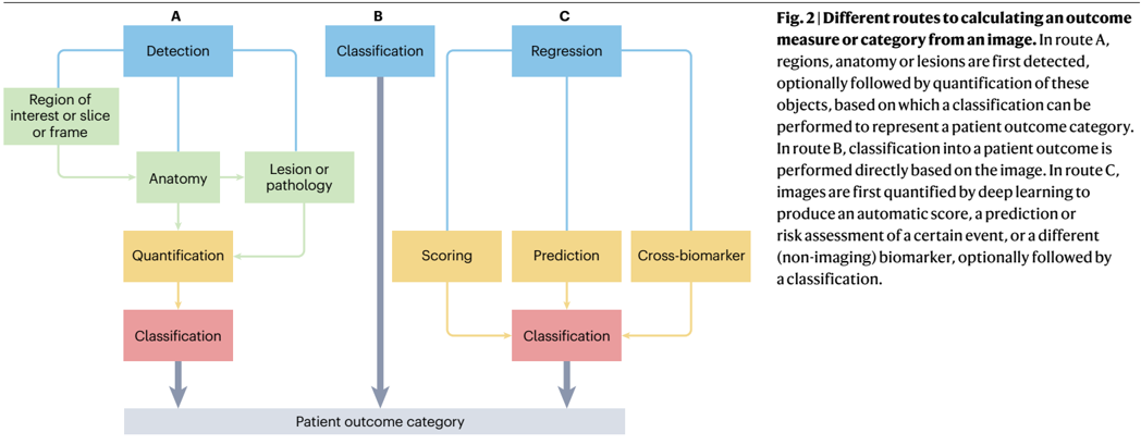
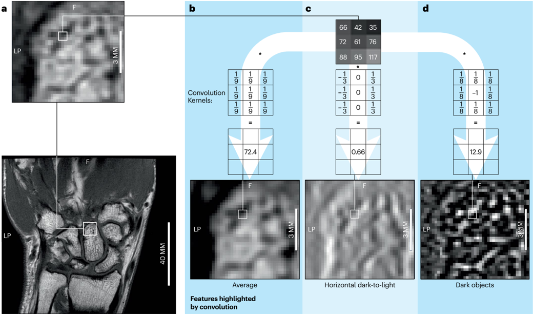
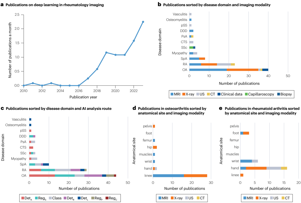
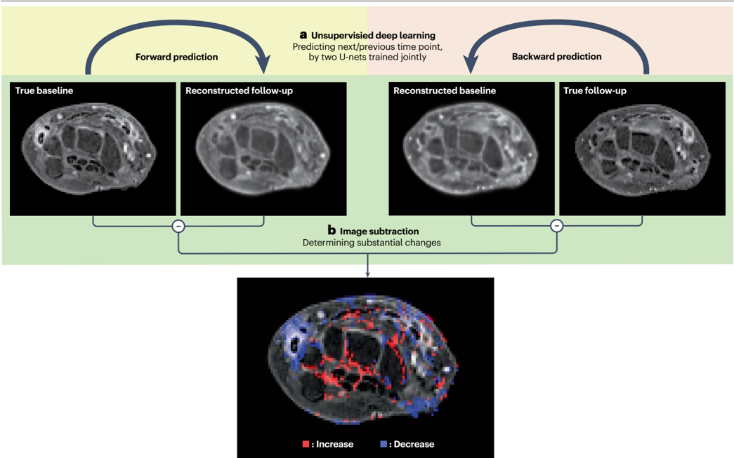
Verification
Claims the paper makes
The claims and statistics of this paper haven’t been checked yet.
Free: it reads the open-access text and checks the first 5 claims that cite a source we can check. Create a free account to check every claim.
Claims other papers make about this one
Each is checked against this paper’s own text: does it say what the citing paper says it does?
The source backs part of the claim, not all of it — or backs it for a different population, condition or comparison.
In the citing paper
“The fundamental criterion for evaluating a medical AI agent is its precision in generating diagnostic and treatment recommendations.80,100,118,119,120”
From A foundational architecture for AI agents in healthcare (2025) · cites this paper as [80]
Experimental — accuracy not yet measuredThe source backs part of the claim, not all of it — or backs it for a different population, condition or comparison.
In the citing paper
“AI-driven medical imaging analysis has emerged as a pivotal tool in modern healthcare, significantly improving the accuracy and efficiency of diagnostic processes.66,74,76,77,78,79,80,81,82,83”
From A foundational architecture for AI agents in healthcare (2025) · cites this paper as [80]
Experimental — accuracy not yet measured
Self-check
Percentages, sums and reported differences re-computed from the paper’s own numbers.
The self-check needs the paper’s full text.
Citations
Cites
Shortcut learning in deep neural networks
Geirhos, R., Jacobsen, J.-H., Michaelis, C., Zemel, R., Brendel, W., Bethge, M., & Wichmann, F. A. (2020) · Nature Machine Intelligence · doi:10.1038/s42256-020-00257-z · Cited by 2,224 · cited 1×
“In this example, any sign in the image that indicates which imaging system was used can inform the disease classification 78 , leading to a phenomenon called shortcut learning 79 .”
Christodoulou, E., Ma, J., Collins, G. S., Steyerberg, E. W., Verbakel, J. Y., & Van Calster, B. (2019) · Journal of Clinical Epidemiology · doi:10.1016/j.jclinepi.2019.02.004 · Cited by 2,119 · cited 1×
“For analysis of low-dimensional clinical data, such as a series of single blood biomarkers, or demographic data, deep learning might not easily surpass the accuracy of statistical models 2 .”
Zech, J. R., Badgeley, M. A., Liu, M., Costa, A. B., Titano, J. J., & Oermann, E. K. (2018) · PLOS Medicine · doi:10.1371/journal.pmed.1002683 · Cited by 1,719 · cited 1×
“In this example, any sign in the image that indicates which imaging system was used can inform the disease classification 78 , leading to a phenomenon called shortcut learning 79 .”
Checklist for Artificial Intelligence in Medical Imaging (CLAIM): a guide for authors and reviewers
Mongan, J., Moy, L., & Kahn, C. E. (2020) · Radiology: Artificial Intelligence · doi:10.1148/ryai.2020200029 · Cited by 1,443
Sanity Checks for Saliency Maps
Adebayo, J., Gilmer, J., Muelly, M., Goodfellow, I., Hardt, M., & Kim, B. (2018) · arXiv · doi:10.48550/arxiv.1810.03292 · Cited by 1,294 · cited 1×
“We should be careful not to only accept the quality of a saliency map if it meets our expectations or to select a mapping method only based on its visual appeal 75 .”
Deep Feature Learning for Knee Cartilage Segmentation Using a Triplanar Convolutional Neural Network
Prasoon, A., Petersen, K., Igel, C., Lauze, F., Dam, E., & Nielsen, M. (2013) · Lecture Notes in Computer Science · doi:10.1007/978-3-642-40763-5_31 · Cited by 582 · cited 2×
“Initial deep-learning research was done on solely the detection of cartilage and/or (cortical) bone 5,24–27 , as this segmentation is a challenging task in itself.”
Pesapane, F., Volonté, C., Codari, M., & Sardanelli, F. (2018) · Insights into Imaging · doi:10.1007/s13244-018-0645-y · Cited by 479 · cited 1×
“Additionally, regulations apply for safety, the ethical use of sensitive image information and protection of privacy, and these regulations vary across jurisdictions 83 , which represents a challenge for software companies’ business models.”
Norman, B., Pedoia, V., & Majumdar, S. (2018) · Radiology · doi:10.1148/radiol.2018172322 · Cited by 419 · cited 1×
“Subsequently, these MRI segmentation results were available to enable the use of morphology (such as cartilage thickness or bone flattening) or relaxometry (T2 mapping) to quantify knee OA severity 28–31 .”
Ambellan, F., Tack, A., Ehlke, M., & Zachow, S. (2019) · Medical Image Analysis · doi:10.1016/j.media.2018.11.009 · Cited by 368 · cited 1×
“Initial deep-learning research was done on solely the detection of cartilage and/or (cortical) bone 5,24–27 , as this segmentation is a challenging task in itself.”
Liu, F., Zhou, Z., Jang, H., Samsonov, A., Zhao, G., & Kijowski, R. (2018) · Magnetic Resonance in Medicine · doi:10.1002/mrm.26841 · Cited by 319 · cited 1×
“Initial deep-learning research was done on solely the detection of cartilage and/or (cortical) bone 5,24–27 , as this segmentation is a challenging task in itself.”
Research works printed but not linked: 5
- Banerjee, S., Bhunia, S. & Schaefer, G. Osteophyte detection for hand osteoarthritis identification in X-ray images using CNNs. Conf. Proc. IEEE Eng. Med. Biol. Soc. 2011, 6196–6199 (2011).
- Ras, G., Xie, N., Gerven, M. V. & Doran, D. Explainable deep learning: a field guide for the uninitiated. J. Artif. Int. Res. 73, 68 (2022).
- Tiulpin, A., Thevenot, J., Rahtu, E., Lehenkari, P. & Saarakkala, S. Automatic knee osteoarthritis diagnosis from plain radiographs: a deep learning-based approach. Sci. Rep. 8, 1727 (2018). · DOI
- Shamonin, D. P. et al. POS0920 quantification of tenosynovitis from wrist MRIs, based on deep learning. Ann. Rheum. Dis. 82, 770–771 (2023).
- Hassanzadeh, T. et al. A deep learning model to locate inflammatory changes in rheumatoid arthritis. Ann. Rheum. Dis. 82, 298–299 (2023).
Cited by
A foundational architecture for AI agents in healthcare
Liu, F., Niu, Y., Zhang, Q., Wang, K., Dong, Z., Wong, I. N., Cheng, L., Li, T., Duan, L., Li, K., et al. (2025) · Cell Reports Medicine · doi:10.1016/j.xcrm.2025.102374 · Cited by 41
Bılgın, E. (2025) · Therapeutic Advances in Musculoskeletal Disease · doi:10.1177/1759720x251343579 · Cited by 22
“12,13 Radiomics Radiomics is a technique responsible for extracting and analyzing complex features from medical images, such as X-rays, computed tomography (CT), and magnetic resonance imaging (MRI) scans.”
Lu, Y. J., Zhang, J., Zhao, Y. H., Zhang, J. W., Zhang, Z., Jiang, R. Z., Liang, J. C., Li, H. D., Dai, J. Y., Cui, T. J., et al. (2024) · Engineering · doi:10.1016/j.eng.2024.11.028 · Cited by 19
AI-Driven Innovations in Pediatric Dentistry: Enhancing Care and Improving Outcome
AlHarbi, N. A. H., & Alharbi, A. S. (2024) · Cureus · doi:10.7759/cureus.69250 · Cited by 33
“The "deep" in DL refers to the multiple layers through which data is processed, allowing the system to learn complex patterns and features from large datasets [15].”
BackgroundArtificial intelligence in rheumatoid arthritis
Sun, Y., Lin, J., & Chen, W. (2025) · Rheumatology & autoimmunity · doi:10.1002/rai2.12171 · Cited by 17
Metadata
- Authors
- Stoel, B. C., Staring, M., Reijnierse, M., & van der Helm-van Mil, A. H. M.
- Affiliations
- Leiden University Medical Center (NL)
- Corresponding author
- Stoel, Berend C.
- Venue
- Nature Reviews Rheumatology
- Published
- 2024-02-08
- Volume
- 20
- Issue
- 3
- Pages
- 182-195
- ISSN
- 1759-4790 (ISSN-L)
- 1759-4804
- OpenAlex
- W4391642764
- Cited by
- 25
- References
- 86
- Citations by year
- 2026: 7 · 2025: 14 · 2024: 4
- FWCI
- 0.57
- Type
- journal article
- Language
- en
- Open access
- hybrid
- doi.org
- Licence
- cc-by
- www.springernature.com/gp/researchers/text-and-data-mining (tdm)
- www.springernature.com/gp/researchers/text-and-data-mining (vor)
- Locations
- Nature Reviews Rheumatology (publishedVersion, cc-by)
- PubMed (publishedVersion)
- Leiden Repository (Leiden University) (submittedVersion, cc-by)
- Funders
- Nederlandse Organisatie voor Wetenschappelijk Onderzoek (17970)
- Topics
- Health Sciences › Medicine › Health Informatics › Artificial Intelligence in Healthcare and Education
- Health Sciences › Medicine › Radiology, Nuclear Medicine and Imaging › Radiomics and Machine Learning in Medical Imaging
- Physical Sciences › Engineering › Biomedical Engineering › Medical Imaging and Analysis
- Keywords
- deep learning, radiological imaging, rheumatic diseases, automatic image analysis, disease detection, disease quantification, disease surveillance, clinical decision support
- MeSH terms
- Deep Learning, Artificial Intelligence, Diagnostic Imaging, Humans, Rheumatic Diseases, Rheumatology
About these checks
Registry, retraction, Cites and statistics results are computed, not judged, with one exception: a printed reference that no index lists is read once by a model before it counts as a citation error. Claims are extracted by a model and kept only when their quote appears verbatim in the text; each cited source is then checked, with a model’s help, for whether it says what the claim relies on. Those verdicts can be wrong — read the claim’s quote and the cited source before relying on one.
Statistics and arithmetic are recomputed, not judged.
Confidence bands are shown only for fields calibrated on our annotated test set. None is calibrated yet, so no band is shown.
What counts as a claim ›How we measure confidence ›How we check this ›
Are you an author, or do you see a problem with this page? Request a review
How to cite
Cite CoChat for the checks on this page, and the paper for its own findings.
- This page
- CoChat. (2026). Citation check: Deep learning in rheumatological image interpretation. https://app.cochat.ai/papers/10.1038/s41584-023-01074-5
- The paper
- Stoel, B. C., Staring, M., Reijnierse, M., & van der Helm-van Mil, A. H. M. (2024). Deep learning in rheumatological image interpretation. Nature Reviews Rheumatology. https://doi.org/10.1038/s41584-023-01074-5
Data sources
Bibliographic data: Semantic Scholar, OpenAlex, Crossref.
Contains information from the Semantic Scholar Academic Graph Datasets, made available under the ODC Attribution License (ODC-BY 1.0).
Semantic Scholar’s data is described in: Kinney, R., et al. (2023). The Semantic Scholar Open Data Platform. arXiv:2301.10140.
