Student or educator? Sign up with your school email and get 6 months of Pro free.
Cited by 30Genetics and Neurodevelopmental DisordersNeuroscience and Neuropharmacology ResearchGenomic variations and chromosomal abnormalities

The DOI matches its Crossref record; 80 of the 80 references it cites were found in a registry or an index; none of the 54 sources its claims cite is checked for support yet.

Phenotypic complexities of rare heterozygous neurexin-1 deletions

Open access

Published version of a preprint

Abstract

Given the large number of genes significantly associated with risk for neuropsychiatric disorders, a critical unanswered question is the extent to which diverse mutations-sometimes affecting the same gene-will require tailored therapeutic strategies. Here we consider this in the context of rare neuropsychiatric disorder-associated copy number variants (2p16.3) resulting in heterozygous deletions in NRXN1, which encodes a presynaptic cell-adhesion protein that serves as a critical synaptic organizer in the brain. Complex patterns of NRXN1 alternative splicing are fundamental to establishing diverse neurocircuitry, vary between the cell types of the brain and are differentially affected by unique (non-recurrent) deletions 1. We contrast the cell-type-specific effect of patient-specific mutations in NRXN1 using human-induced pluripotent stem cells, finding that perturbations in NRXN1 splicing result in divergent cell-type-specific synaptic outcomes. Through distinct loss-of-function (LOF) and gain-of-function (GOF) mechanisms, NRXN1+/- deletions cause decreased synaptic activity in glutamatergic neurons, yet increased synaptic activity in GABAergic neurons. Reciprocal isogenic manipulations causally demonstrate that aberrant splicing drives these changes in synaptic activity. For NRXN1 deletions, and perhaps more broadly, precision medicine will require stratifying patients based on whether their gene mutations act through LOF or GOF mechanisms, to achieve individualized restoration of NRXN1 isoform repertoires by increasing wild-type and/or ablating mutant isoforms. Given the increasing number of mutations predicted to engender both LOF and GOF mechanisms in brain disorders, our findings add nuance to future considerations of precision medicine.

Overview

CoChat Summary

TL;DR

Patient-specific heterozygous NRXN1 deletions act through distinct loss-of-function (5′-Del) or gain-of-function (3′-Del) splicing mechanisms, producing opposing synaptic phenotypes in human glutamatergic versus GABAergic neurons. The authors propose that precision medicine for NRXN1 deletions will require stratifying patients by LOF/GOF mechanism rather than by clinical diagnosis alone.

Key findings

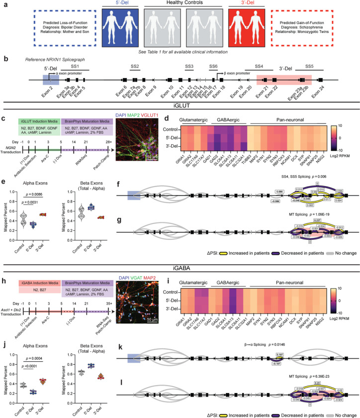
  • NRXN1+/− 5′-Del cases (≈115-kb deletion affecting the alpha-transcript promoter) showed reduced β→α splicing, consistent with LOF, while 3′-Del cases (≈136-kb deletion impacting SS4/SS5 across exons 21–23) expressed mutant exon 20–24 splice junctions, consistent with GOF.
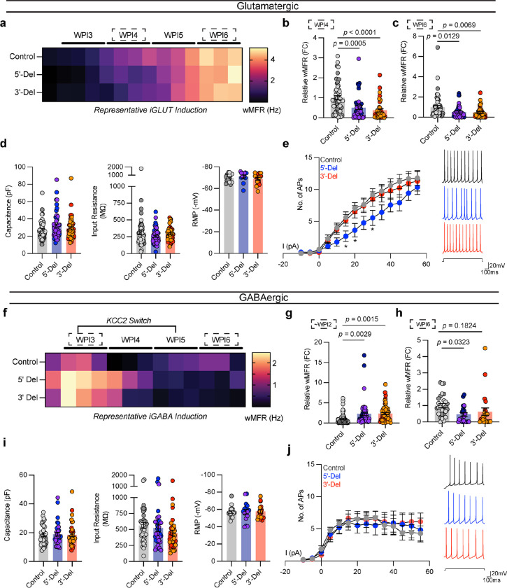
  • Spontaneous activity diverged by cell type: iGLUT neurons from both 5′-Del and 3′-Del showed >40% reduced weighted mean firing rate at WPI4 and WPI6, whereas immature iGABA neurons showed ~2-fold increased activity at ~WPI2 and mature iGABA neurons showed decreased activity at WPI6.
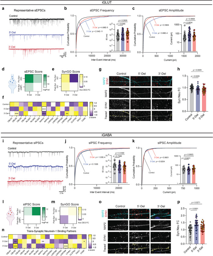
  • Synaptic transmission was decreased in iGLUT neurons (increased sEPSC inter-event intervals for 5′-Del and 3′-Del) but enhanced in iGABA neurons (decreased sIPSC inter-event intervals in 3′-Del), with corresponding bidirectional changes in synapse number.
  • Isogenic shRNA knockdown of wildtype NRXN1α recapitulated 5′-Del-like phenotypes, and knockdown of the mutant 20/24 splice junction reversed 3′-Del-like phenotypes; β-estradiol increased NRXN1α expression (p = 0.0297) and improved 5′-Del iGLUT activity/sEPSC phenotypes, while an ASO reduced mutant isoforms by ~55% (p < 0.001).

Methods

  • Human induced pluripotent stem cells from patient-specific NRXN1+/− 5′-Del, 3′-Del, and healthy control donors were lineage-converted into iGLUT and iGABA neurons, and dorsal/ventral forebrain organoids were generated for scRNA-seq.
  • Functional assays included multi-electrode array recordings, whole-cell patch-clamp electrophysiology for spontaneous and miniature synaptic currents, immunostaining for synaptic puncta, bulk RNA-seq, LeafCutter differential splicing analysis, and single-cell RNA-seq.
  • Causal isogenic manipulations used shRNAs against wildtype exon 9 or the mutant exon 20/24 junction; therapeutic proof-of-principle experiments used β-estradiol and an antisense oligonucleotide targeting the mutant splice junction.

Limitations

  • The paper states that the iGLUT and iGABA populations studied are immature and comprise diverse neuronal subtypes, so discrete effects on subtypes such as SST versus PV GABA neurons remain unresolved.
  • The paper states that future studies in more physiologically relevant, circuit-like models may uncover non-autonomous and activity-dependent phenotypes, and that biochemical/proteomic interactions of NRXN1 remain to be probed, for example with proximity-labelling methods such as BioID.
  • The paper states that it did not resolve whether mutant GOF isoforms shift alternative-splicing stoichiometry against wildtype isoforms or alter trans-synaptic protein–protein interactions; it also notes that the therapeutic findings are proof-of-principle in vitro results requiring in vivo and clinical validation.

Model-written summary. Check it against the paper before relying on it.

Integrity signals

Registry

Crossref · 10.1038/s41586-025-08864-9

Cites

80 of 80 references verified0 citation errors · 0 not checkable

Statistics

Nothing to recompute.

Figures

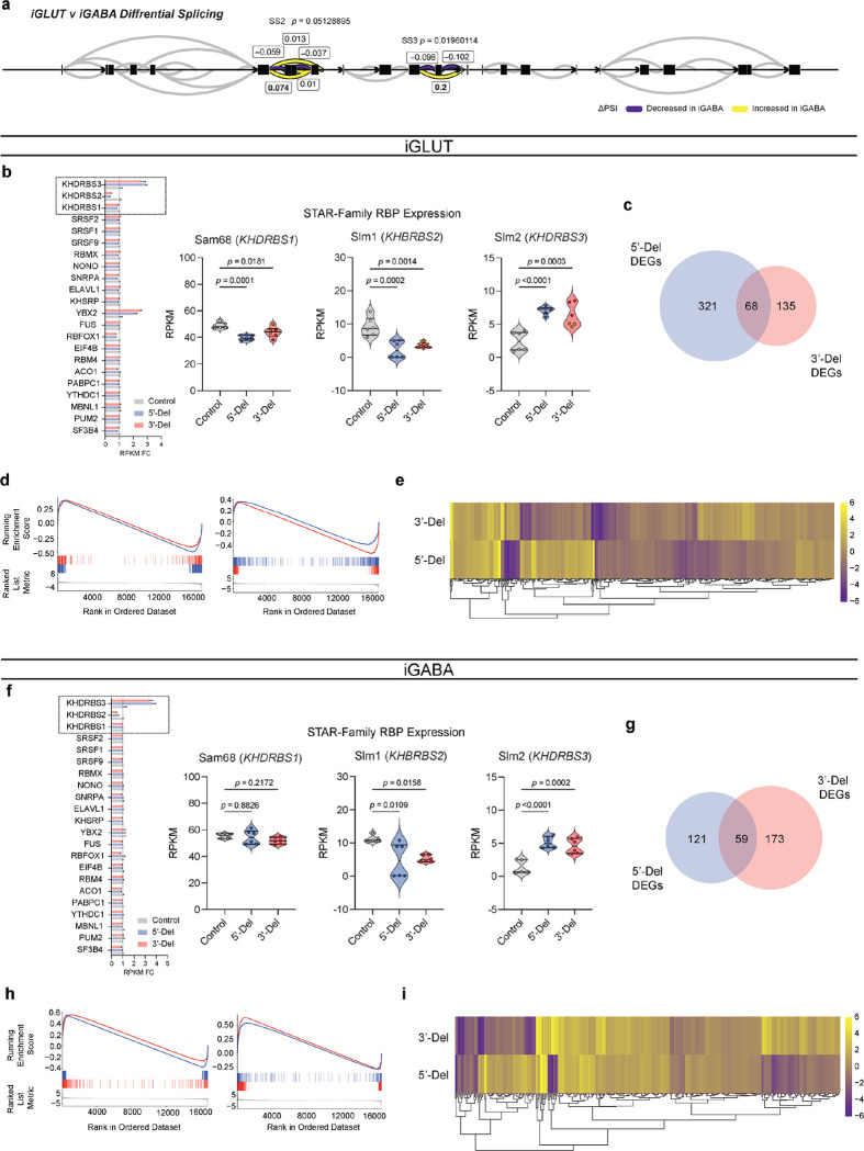
Extended Data Figure 1: (a) Splicegraph displaying significant gene wide splicing clusters at NRXN1 SS3 (p = 0.0196), compared via Dirichlet-multinomial generalized linear model with Bonferroni corrections. (b, f) Gene expression fold-change of select NRXN1 predicted RNA-binding proteins (RBP) across patients and control iGLUT and iGABA neurons, with statistical comparisons for STAR-Family RBPs. (c, g) Overlap of DEGs and (d,h) gene set enrichment analysis (GSEA) between genotypes. (e,i) Distinct gene expression patterns by hierarchal clustering of all patient specific DEGs. Sample information correspond to Fig. 1
Extended Data Figure 1: (a) Splicegraph displaying significant gene wide splicing clusters at NRXN1 SS3 (p = 0.0196), compared via Dirichlet-multinomial generalized linear model with Bonferroni corrections. (b, f) Gene expression fold-change of select NRXN1 predicted RNA-binding proteins (RBP) across patients and control iGLUT and iGABA neurons, with statistical comparisons for STAR-Family RBPs. (c, g) Overlap of DEGs and (d,h) gene set enrichment analysis (GSEA) between genotypes. (e,i) Distinct gene expression patterns by hierarchal clustering of all patient specific DEGs. Sample information correspond to Fig. 1CC BY-NC-ND · PubMed Central
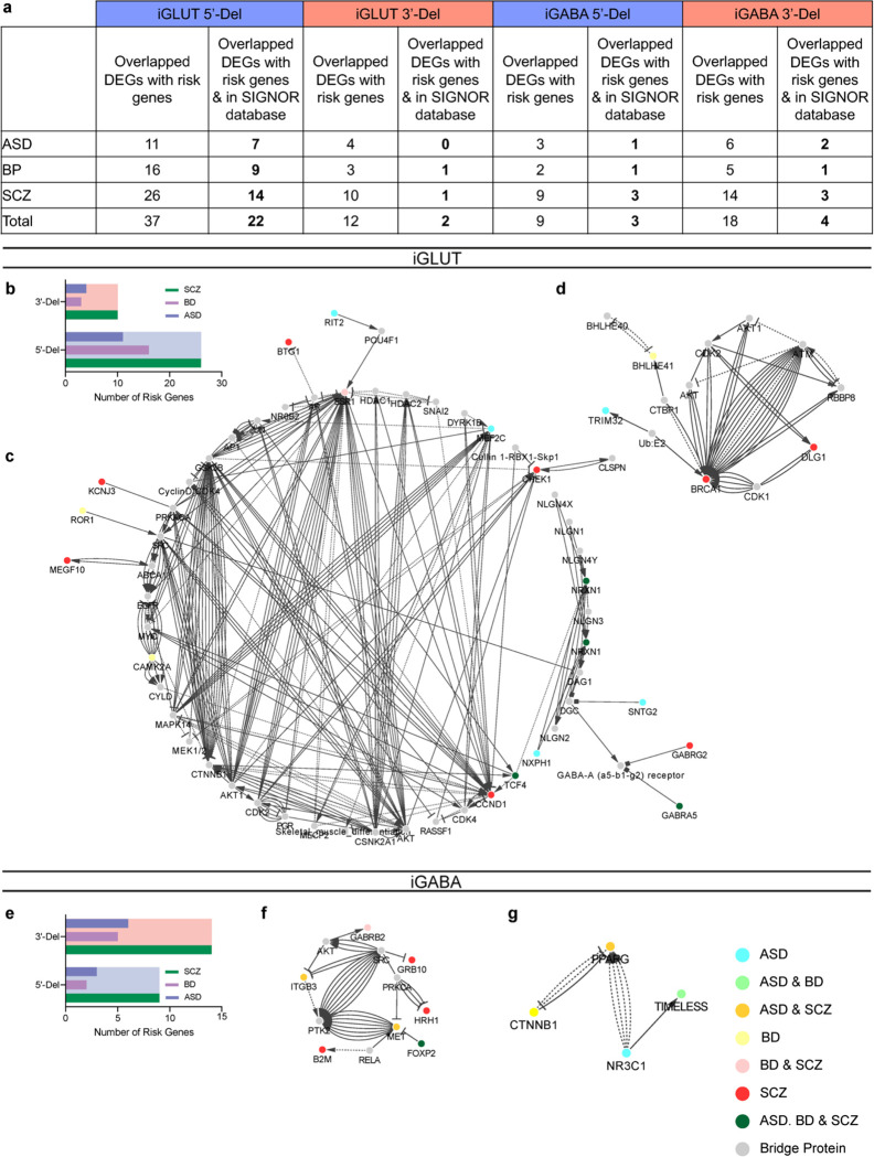
Extended Data Figure 2: Extended transcriptomics analysis on disease risk associated genes. (a) Summary table of overlapping DEGs with risk enrichments across publicly curated datasets for autism (ASD), bipolar disorder (BD) and schizophrenia. (b, e) Enrichment of genes across neuropsychiatric disorders for iGLUT and iGABA neurons. (c) Interaction maps of risk genes for 5’-Del iGLUT, (d) 3’-Del iGLUT, (f) 5’-Del iGABA and (g) 5’-iGABA. Sample information correspond to Fig. 1
Extended Data Figure 2: Extended transcriptomics analysis on disease risk associated genes. (a) Summary table of overlapping DEGs with risk enrichments across publicly curated datasets for autism (ASD), bipolar disorder (BD) and schizophrenia. (b, e) Enrichment of genes across neuropsychiatric disorders for iGLUT and iGABA neurons. (c) Interaction maps of risk genes for 5’-Del iGLUT, (d) 3’-Del iGLUT, (f) 5’-Del iGABA and (g) 5’-iGABA. Sample information correspond to Fig. 1CC BY-NC-ND · PubMed Central
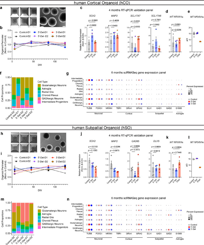
Extended Data Figure 3: Extended data on human organoid generation and characterization. (a,h) Representative images of hiPSC aggregation and immature spheroids post dislodging. (b,i) Normalized organoid perimeters over time (compared to averaged control), hCO (n = 6 donors | 2 batches | 72–161 organoids) and hSO (n = 6 donors | 2 batches | 46–134 organoids). (c, j) RT-qPCR results from 4-month organoids of genes for pluripotency, neuronal, and cell-type specific markers. (d-e, k-l) RT-qPCR results of NRXN1 WT and MT expression hCO (n = 6 donors | 1 representative batch | 12 samples) and hSO (n = 6 donors | 1 representative batch | 12 samples). Statistical tests used were 1-way ANOVAs with Dunnett’s test. (f, m) relative proportions of cell clusters across individual donors, (hCO = 47,460 cells) and (hSO = 35,563 cells). (g, n) Comprehensive gene expression panel across sub-clusters of hCO and hSO samples across neuronal, cortical, subpallial and astroglia markers. Data corresponds to Fig 2.
Extended Data Figure 3: Extended data on human organoid generation and characterization. (a,h) Representative images of hiPSC aggregation and immature spheroids post dislodging. (b,i) Normalized organoid perimeters over time (compared to averaged control), hCO (n = 6 donors | 2 batches | 72–161 organoids) and hSO (n = 6 donors | 2 batches | 46–134 organoids). (c, j) RT-qPCR results from 4-month organoids of genes for pluripotency, neuronal, and cell-type specific markers. (d-e, k-l) RT-qPCR results of NRXN1 WT and MT expression hCO (n = 6 donors | 1 representative batch | 12 samples) and hSO (n = 6 donors | 1 representative batch | 12 samples). Statistical tests used were 1-way ANOVAs with Dunnett’s test. (f, m) relative proportions of cell clusters across individual donors, (hCO = 47,460 cells) and (hSO = 35,563 cells). (g, n) Comprehensive gene expression panel across sub-clusters of hCO and hSO samples across neuronal, cortical, subpallial and astroglia markers. Data corresponds to Fig 2.CC BY-NC-ND · PubMed Central
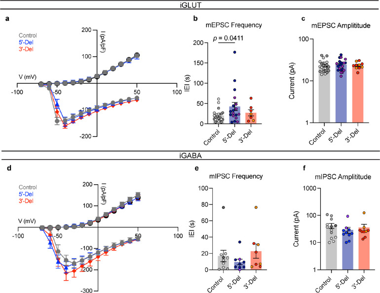
Extended Data Figure 4: Extended data on electrophysiological properties of 5’-Del and 3’-Del neurons. (a) Voltage-gated potassium and channel kinetics across genotypes for iGLUT neurons (n = 6 donors | 2 inductions | 45 neurons). (b) Comparative mEPSC kinetics of IEI and (c) amplitude size from iGLUT neurons (n= 6 donors | 4 inductions | 47 neurons), compared via a 1-way ANOVA with Dunnett’s test. (d) Voltage-gated potassium and channel kinetics across genotypes for iGABA neurons (n = 6 donors | 2 inductions | 34 neurons). (e) Comparative mIPSC kinetics of (a) IEI and (f) amplitude size from iGABA neurons (n= 6 donors | 3 inductions | 27 neurons), compared via a 1-way ANOVA with Dunnett’s test. See Supplementary Table 5 for all summary statistics.
Extended Data Figure 4: Extended data on electrophysiological properties of 5’-Del and 3’-Del neurons. (a) Voltage-gated potassium and channel kinetics across genotypes for iGLUT neurons (n = 6 donors | 2 inductions | 45 neurons). (b) Comparative mEPSC kinetics of IEI and (c) amplitude size from iGLUT neurons (n= 6 donors | 4 inductions | 47 neurons), compared via a 1-way ANOVA with Dunnett’s test. (d) Voltage-gated potassium and channel kinetics across genotypes for iGABA neurons (n = 6 donors | 2 inductions | 34 neurons). (e) Comparative mIPSC kinetics of (a) IEI and (f) amplitude size from iGABA neurons (n= 6 donors | 3 inductions | 27 neurons), compared via a 1-way ANOVA with Dunnett’s test. See Supplementary Table 5 for all summary statistics.CC BY-NC-ND · PubMed Central
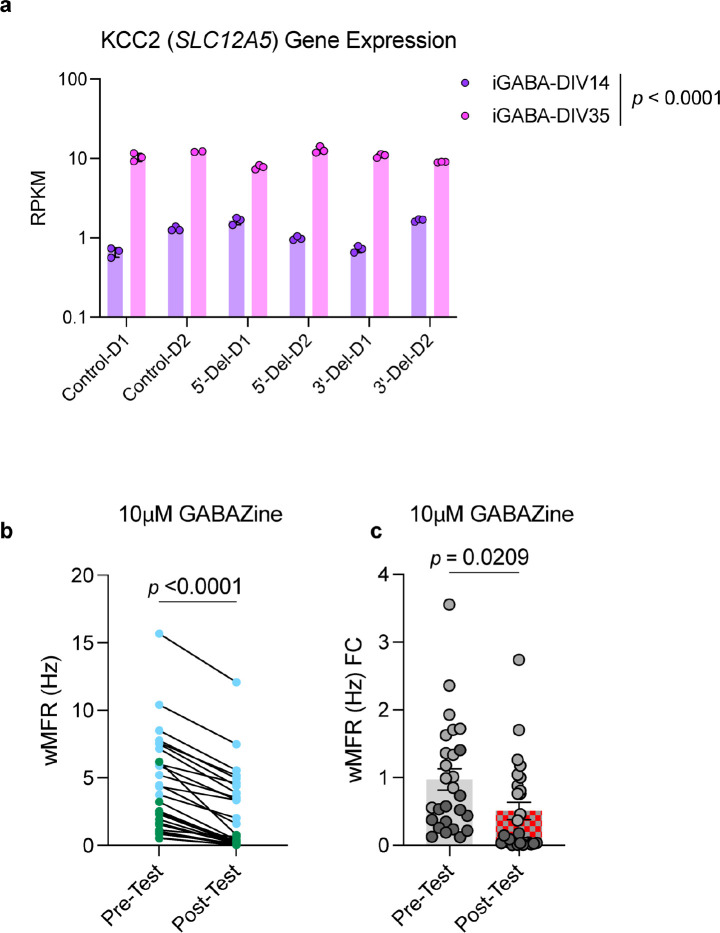
Extended Data Figure 5: Extended KCC2 related data from immature GABA neurons. (a) Transcriptomic comparison of SLC12A5 expression across DIV14 and DIV35 RNASeq timepoints. (b,c) MEA tests from pre- and post-treatment of 10uM GABAzine. (n = 2 donors | 1 representative induction | 28 MEA wells) Statistical tests are paired student’s t-test for time-linked comparison and unpaired student’s t-test for pre/post activity foldchange.
Extended Data Figure 5: Extended KCC2 related data from immature GABA neurons. (a) Transcriptomic comparison of SLC12A5 expression across DIV14 and DIV35 RNASeq timepoints. (b,c) MEA tests from pre- and post-treatment of 10uM GABAzine. (n = 2 donors | 1 representative induction | 28 MEA wells) Statistical tests are paired student’s t-test for time-linked comparison and unpaired student’s t-test for pre/post activity foldchange.CC BY-NC-ND · PubMed Central
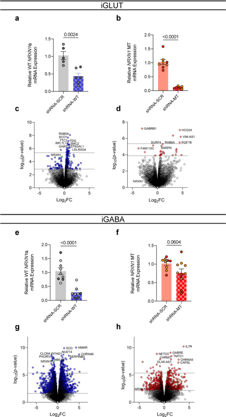
Extended Data Figure 6: shRNA knockdown validation. (a) Extent of shRNA knockdown on WT and (b) MT NRXN1 expression in iGLUT neurons (n = 2 donors | 1–3 inductions). (c) Extent of shRNA knockdown on WT and (d) MT NRXN1 expression in iGABA neurons, (n = 2–3 donors | 1–3 inductions) Statistical tests used were Student’s t-test.
Extended Data Figure 6: shRNA knockdown validation. (a) Extent of shRNA knockdown on WT and (b) MT NRXN1 expression in iGLUT neurons (n = 2 donors | 1–3 inductions). (c) Extent of shRNA knockdown on WT and (d) MT NRXN1 expression in iGABA neurons, (n = 2–3 donors | 1–3 inductions) Statistical tests used were Student’s t-test.CC BY-NC-ND · PubMed Central
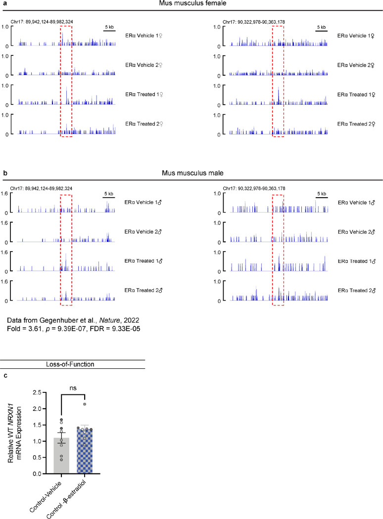
Extended Data Figure 7: ChIP-seq enrichment of ER1 binding at NRXN1 loci in rodent brain. (a) Female and (b) male mus musculus ChIP tracts of NRXN1 locus, with red dashed areas highlighting binding enrichment across vehicle and estradiol treated mice. (c) Effect of beta-estradiol on control donors (n = 16/4 | Representative).
Extended Data Figure 7: ChIP-seq enrichment of ER1 binding at NRXN1 loci in rodent brain. (a) Female and (b) male mus musculus ChIP tracts of NRXN1 locus, with red dashed areas highlighting binding enrichment across vehicle and estradiol treated mice. (c) Effect of beta-estradiol on control donors (n = 16/4 | Representative).CC BY-NC-ND · PubMed Central
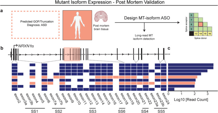
Extended Data Figure 8: In-vivo validation of MT isoform expression from an unrelated autism NRXN1+/− patient. (a) Schematic of novel NRXN1 autism patient, and GOF therapeutic targeting pipeline, with (b) schematic of the NRXN1α isoform structures, with each row representing a unique NRXN1α isoform and each column representing a NRXN1 exon. The colored isoforms (navy, wildtype; peach, patient-specific) are spliced into the transcript while the blank exons are spliced out. (c) The abundance of each NRXN1α isoform by sample.
Extended Data Figure 8: In-vivo validation of MT isoform expression from an unrelated autism NRXN1+/− patient. (a) Schematic of novel NRXN1 autism patient, and GOF therapeutic targeting pipeline, with (b) schematic of the NRXN1α isoform structures, with each row representing a unique NRXN1α isoform and each column representing a NRXN1 exon. The colored isoforms (navy, wildtype; peach, patient-specific) are spliced into the transcript while the blank exons are spliced out. (c) The abundance of each NRXN1α isoform by sample.CC BY-NC-ND · PubMed Central
Figure 1. Aberrant NRXN1 splicing across human iPSC derived glutamatergic (iGLUT) and GABAergic (iGABA) neurons. (a) Brief description of clinical information of all hiPSC lines used in this study, and (b) schematic of NRXN1 gene structure as a splicegraph, denoting splice sites (SS1–6), with red and blue shades corresponding on 3’-Del and 5’-Del genotypes, respectively. Arrows indicate relative internal promoter positions. (c, h) Induction timeline and factors to generate iGLUT and iGABA neurons, with immunostaining validation of neuronal identity (MAP2), glutamate identity (vGLUT1), and GABA identity (vGAT). (d, i). Gene expression panel confirming abundance of neuronal markers and neurotransmitter identity in iGLUT (n: Control = 6/2; 5’-Del = 6/2; 3’-Del = 6/2 | 1 batch) and in iGABA neurons (n: Control = 5/2; 5’-Del = 6/2; 3’-Del = 6/2 | 1 batch). (e, j) Mapped percent of alpha NRXN1 exon reads in iGLUT, compared via a 1-way ANOVA, with Dunnett’s test (F2, 15 = 25.70; 5’-Del p = 0.0031, 3’-Del p = 0.0086), and iGABA neurons (F2, 14 = 92.92; 5’-Del p < 0.001, 3’-Del p = 0.0004), with beta NRXN1 exon reads, calculated by subtracting alpha-specific reads against total reads. (f, k) Splicegraphs displaying significant gene wide splicing clusters, compared via Dirichlet-multinomial generalized linear model with Bonferroni corrections, for 5’-Del iGLUT (SS4 and SS5 cluster p = 0.006) and iGABA neurons (β→α cluster p = 0.0146), and for (g, l) 3’-Del iGLUT (SS4 and SS5 cluster p = 1.09E-19) and iGABA neurons (SS4 and SS5 cluster p = 6.39E-23). n reported as samples/donors | independent batches.
Figure 1. Aberrant NRXN1 splicing across human iPSC derived glutamatergic (iGLUT) and GABAergic (iGABA) neurons. (a) Brief description of clinical information of all hiPSC lines used in this study, and (b) schematic of NRXN1 gene structure as a splicegraph, denoting splice sites (SS1–6), with red and blue shades corresponding on 3’-Del and 5’-Del genotypes, respectively. Arrows indicate relative internal promoter positions. (c, h) Induction timeline and factors to generate iGLUT and iGABA neurons, with immunostaining validation of neuronal identity (MAP2), glutamate identity (vGLUT1), and GABA identity (vGAT). (d, i). Gene expression panel confirming abundance of neuronal markers and neurotransmitter identity in iGLUT (n: Control = 6/2; 5’-Del = 6/2; 3’-Del = 6/2 | 1 batch) and in iGABA neurons (n: Control = 5/2; 5’-Del = 6/2; 3’-Del = 6/2 | 1 batch). (e, j) Mapped percent of alpha NRXN1 exon reads in iGLUT, compared via a 1-way ANOVA, with Dunnett’s test (F2, 15 = 25.70; 5’-Del p = 0.0031, 3’-Del p = 0.0086), and iGABA neurons (F2, 14 = 92.92; 5’-Del p < 0.001, 3’-Del p = 0.0004), with beta NRXN1 exon reads, calculated by subtracting alpha-specific reads against total reads. (f, k) Splicegraphs displaying significant gene wide splicing clusters, compared via Dirichlet-multinomial generalized linear model with Bonferroni corrections, for 5’-Del iGLUT (SS4 and SS5 cluster p = 0.006) and iGABA neurons (β→α cluster p = 0.0146), and for (g, l) 3’-Del iGLUT (SS4 and SS5 cluster p = 1.09E-19) and iGABA neurons (SS4 and SS5 cluster p = 6.39E-23). n reported as samples/donors | independent batches.CC BY-NC-ND · PubMed Central
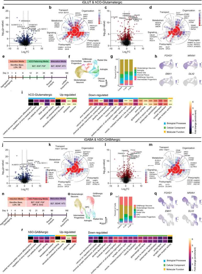
Figure 2: Transcriptomic impact of NRXN1+/− in induced (iGLUT/iGABA) and organoid-derived hCO-glutamatergic and hSO-GABAergic neurons. (a, c) Volcano plots of differential gene expression (DE) analysis across both genotypes in iGLUT and (j, l) iGABA neurons. Vertical dashed lines represent DE genes ±1.5 Log2FC. Horizontal dashed lines represent FDR = 0.1 cutoff (lower) and Bonferroni corrected cutoff (upper). (b, d) Sunburst plots of all FDR corrected DEGs with SynGO annotated synapse function for iGLUT and (k, m) iGABA neurons. (e, n) Timeline of neural organogenesis for hCO and hSOs (f, o), UMAPs of hCO and hSO organoid samples sequenced at 6 months, annotated by cell clusters, and (g, p) relative proportions of cell clusters across genotypes, (hCO = 47,460 cells) and (hSO = 35,563 cells). (h, q) validation of regionalization across forebrain (FOXG1), dorsal (EMX1) and ventral (DLX2) regions, with NRXN1 expression across all cells. (i, r) Gene ontological analysis results using DEGs from scRNASeq. *P < 0.05, **P < 0.01, ***P <0.001, Wilcoxon’s rank sum test, FDR = 0.05. Data represented as mean ± sem. n reported as samples/donors | independent batches.
Figure 2: Transcriptomic impact of NRXN1+/− in induced (iGLUT/iGABA) and organoid-derived hCO-glutamatergic and hSO-GABAergic neurons. (a, c) Volcano plots of differential gene expression (DE) analysis across both genotypes in iGLUT and (j, l) iGABA neurons. Vertical dashed lines represent DE genes ±1.5 Log2FC. Horizontal dashed lines represent FDR = 0.1 cutoff (lower) and Bonferroni corrected cutoff (upper). (b, d) Sunburst plots of all FDR corrected DEGs with SynGO annotated synapse function for iGLUT and (k, m) iGABA neurons. (e, n) Timeline of neural organogenesis for hCO and hSOs (f, o), UMAPs of hCO and hSO organoid samples sequenced at 6 months, annotated by cell clusters, and (g, p) relative proportions of cell clusters across genotypes, (hCO = 47,460 cells) and (hSO = 35,563 cells). (h, q) validation of regionalization across forebrain (FOXG1), dorsal (EMX1) and ventral (DLX2) regions, with NRXN1 expression across all cells. (i, r) Gene ontological analysis results using DEGs from scRNASeq. *P < 0.05, **P < 0.01, ***P <0.001, Wilcoxon’s rank sum test, FDR = 0.05. Data represented as mean ± sem. n reported as samples/donors | independent batches.CC BY-NC-ND · PubMed Central
Figure 3. Spontaneous, passive, and excitable properties are minimally changed from NRXN1+/− induced neurons. (a, f) Timelapse of multi-electrode array recordings every 2–3 days apart starting near ~DIV12 for a single representative induction. Tiles represent averaged wMFR values across genotypes during a single recording session. (b) MEA quantification of iGLUT neuronal activity, compared via a 1-way ANOVA, with Dunnett’s test (n: Control = 52/2; 5’-Del = 42/2; 3’-Del = 38/2 | 3 batches) at WPI4 (F2, 129 = 12.77; 5’-Del p = 0.0005, 3’-Del p < 0.0001), and (c) WPI6 (F2, 129 = 5.737; 5’-Del p = 0.0129, 3’-Del p = 0.0069). (d) intrinsic properties of iGLUT neurons (n: Control = 51/2; 5’-Del = 51/2; 3’-Del = 48/2 | 8 batches): compared via 1-way ANOVA, with Dunnett’s test, Capacitance (F2, 147 = 1.505; 5’-Del p = 0.1565, 3’-Del p = 0.7633), Input resistance (F2, 147 = 1.311; 5’-Del p = 0.1949, 3’-Del p = 0.7906), and RMP (n: Control = 16/2; 5’-Del = 14/2; 3’-Del = 16/2 | 2 batches) compared via 1-way ANOVA, with Dunnett’s test (F2, 43 = 0.8234; 5’-Del p = 0.6378, 3’-Del p = 0.8468). (e) Input-output curves of excitable properties (n: Control = 16/2; 5’-Del = 14/2; 3’-Del = 16/2 | 2 batches), with representative traces (right), compared via Step × Genotype 2-way ANOVA; Dunnett’s Test (F26, 559 = 1.690, 5’-Del p < 0.01 at Step 6, 7 and 9, 3’-Del p = n.s. on all steps). (g) MEA quantification of iGABA neuronal activity, compared via a 1-way ANOVA, with Dunnett’s test (n: Control = 71/2; 5’-Del = 48/2; 3’-Del = 55/2 | 5 batches) at WPI2 (F2, 171 = 7.805; 5’-Del p = 0.0029, 3’-Del p = 0.0015), and (h) WPI5 (n: Control = 27/4; 5’-Del = 23/2; 3’-Del = 23/2 | 2 batches) compared via 1-way ANOVA, with Dunnett’s test (F2, 70 = 3.158; 5’-Del p = 0.0323, 3’-Del p = 0.1824). (i) intrinsic properties of iGABA neurons (n: Control = 39/2; 5’-Del = 34/2; 3’-Del = 37/2 | 6 batches): compared via 1-way ANOVA, with Dunnett’s test, Capacitance (F2, 107 = 0.04256; 5’-Del p = 0.9425, 3’-Del p = 0.9941), Input resistance (n: Control = 39/2; 5’-Del = 34/2; 3’-Del = 36/2 | 6 batches, F2, 106 = 1.451; 5’-Del p = 0.8162, 3’-Del p = 0.1703) and RMP (n: Control = 18/2; 5’-Del = 16/2; 3’-Del = 16/2 | 2 batches) compared via 1-way ANOVA, with Dunnett’s test (F2, 47 = 0.02842; 5’-Del p = 0.9893, 3’-Del p = 0.9601). (j) Input-output curves of excitable properties (n: Control = 16/2; 5’-Del = 14/2; 3’-Del = 16/2 | 2 batches), with representative traces (right), compared via Step × Genotype 2-way ANOVA; Dunnett’s Test (F26, 572 = 0.5137, 5’-Del and 3’-Del p = n.s. on all steps). Data represented as mean ± sem. n reported as samples/donors | independent batches.
Figure 3. Spontaneous, passive, and excitable properties are minimally changed from NRXN1+/− induced neurons. (a, f) Timelapse of multi-electrode array recordings every 2–3 days apart starting near ~DIV12 for a single representative induction. Tiles represent averaged wMFR values across genotypes during a single recording session. (b) MEA quantification of iGLUT neuronal activity, compared via a 1-way ANOVA, with Dunnett’s test (n: Control = 52/2; 5’-Del = 42/2; 3’-Del = 38/2 | 3 batches) at WPI4 (F2, 129 = 12.77; 5’-Del p = 0.0005, 3’-Del p < 0.0001), and (c) WPI6 (F2, 129 = 5.737; 5’-Del p = 0.0129, 3’-Del p = 0.0069). (d) intrinsic properties of iGLUT neurons (n: Control = 51/2; 5’-Del = 51/2; 3’-Del = 48/2 | 8 batches): compared via 1-way ANOVA, with Dunnett’s test, Capacitance (F2, 147 = 1.505; 5’-Del p = 0.1565, 3’-Del p = 0.7633), Input resistance (F2, 147 = 1.311; 5’-Del p = 0.1949, 3’-Del p = 0.7906), and RMP (n: Control = 16/2; 5’-Del = 14/2; 3’-Del = 16/2 | 2 batches) compared via 1-way ANOVA, with Dunnett’s test (F2, 43 = 0.8234; 5’-Del p = 0.6378, 3’-Del p = 0.8468). (e) Input-output curves of excitable properties (n: Control = 16/2; 5’-Del = 14/2; 3’-Del = 16/2 | 2 batches), with representative traces (right), compared via Step × Genotype 2-way ANOVA; Dunnett’s Test (F26, 559 = 1.690, 5’-Del p < 0.01 at Step 6, 7 and 9, 3’-Del p = n.s. on all steps). (g) MEA quantification of iGABA neuronal activity, compared via a 1-way ANOVA, with Dunnett’s test (n: Control = 71/2; 5’-Del = 48/2; 3’-Del = 55/2 | 5 batches) at WPI2 (F2, 171 = 7.805; 5’-Del p = 0.0029, 3’-Del p = 0.0015), and (h) WPI5 (n: Control = 27/4; 5’-Del = 23/2; 3’-Del = 23/2 | 2 batches) compared via 1-way ANOVA, with Dunnett’s test (F2, 70 = 3.158; 5’-Del p = 0.0323, 3’-Del p = 0.1824). (i) intrinsic properties of iGABA neurons (n: Control = 39/2; 5’-Del = 34/2; 3’-Del = 37/2 | 6 batches): compared via 1-way ANOVA, with Dunnett’s test, Capacitance (F2, 107 = 0.04256; 5’-Del p = 0.9425, 3’-Del p = 0.9941), Input resistance (n: Control = 39/2; 5’-Del = 34/2; 3’-Del = 36/2 | 6 batches, F2, 106 = 1.451; 5’-Del p = 0.8162, 3’-Del p = 0.1703) and RMP (n: Control = 18/2; 5’-Del = 16/2; 3’-Del = 16/2 | 2 batches) compared via 1-way ANOVA, with Dunnett’s test (F2, 47 = 0.02842; 5’-Del p = 0.9893, 3’-Del p = 0.9601). (j) Input-output curves of excitable properties (n: Control = 16/2; 5’-Del = 14/2; 3’-Del = 16/2 | 2 batches), with representative traces (right), compared via Step × Genotype 2-way ANOVA; Dunnett’s Test (F26, 572 = 0.5137, 5’-Del and 3’-Del p = n.s. on all steps). Data represented as mean ± sem. n reported as samples/donors | independent batches.CC BY-NC-ND · PubMed Central
Figure 4: Divergent impact on neurotransmission from NRXN1+/− induced neurons. (a) Representative traces of iGLUT sEPSCs. (b) Cumulative probabilities and log-scaled cell averages of inter-event-internals (IEIs) across genotypes (n: Control = 39/4; 5’-Del = 33/3; 3’-Del = 29/2 | 6), compared by Levene’s Test with Bonferroni correction for averaged distributions (5’-Del F = 46.635, df = 1, p = 2.54E-11; 3’-Del F = 17.928, df = 1, p = 4.88E-5), and 1-way ANOVA, with Dunnett’s test for inset (F2, 98 = 3.117; 5’-Del p = 0.0285, 3’-Del p = 0.2929). (c) Cumulative probabilities and log-scaled cell averages of amplitude size across genotypes, compared by Levene’s Test with Bonferroni correction for averaged distributions (5’-Del F = 14.15, df = 1, p = 3.88E-4; 3’-Del F = 1.0851, df = 1, p = 0.5964), and 1-way ANOVA, with Dunnett’s test for inset (F2, 98 = 2.839; 5’-Del p = 0.0634, 3’-Del p = 0.9994). (d) Transformed p-values of Levene’s Test and (e) SynGO gene-set averaged log2FC values across pre- or post-synaptic genes. (f) Gene expression panel (z-scores), of canonical NRXN1 binding partners in iGLUT neurons, boxes indicate reaching genome wide significance. (g) Representative images of iGLUT synaptic puncta traced (SYN1) onto dendrites (MAP2), and (h) normalized fold change of SYN1 puncta to length of MAP2 ratio (Syn:Neu), (n: Control = 40/2; 5’-Del: 39/2; 3’-Del: 40/2), compared via 1-way ANOVA, with Dunnett’s test (F2, 116 = 7.538, 5’-Del p = 0.0205, 3’-Del p = 0.0005). (i) Representative traces of iGABA sIPSCs. (j) Cumulative probabilities and log-scaled cell averages of inter-event-internals (IEIs) across genotypes (n: Control = 26/2; 5’-Del = 25/3; 3’-Del = 22/2 | 4), compared by Levene’s Test with Bonferroni correction for averaged distributions (5’-Del F = 3.4002, df = 1, p = 0.1306; 3’-Del F = 16.501, df = 1, p = 1.00E-04), and 1-way ANOVA, with Dunnett’s test for inset (F2, 70 = 0.8296; 5’-Del p = 0.7986, 3’-Del p = 0.339). (k) Cumulative probabilities and log-scaled cell averages of amplitude size across genotypes, compared by Levene’s Test for averaged distributions (5’-Del F = 0.1, df = 1, p = 0.9204; 3’-Del F = 0.0006, df = 1, p = 0.9801), and 1-way ANOVA, with Dunnett’s test for inset (F2, 70 = 0.08143; 5’-Del p = 0.9431, 3’-Del p = 0.9904). (l) Transformed p-values of Levene’s Test and (m) SynGO gene-set averaged log2FC values across pre- or post-synaptic genes. (n) Gene expression panel (z-scores), of canonical NRXN1 binding partners in iGABA neurons, boxes indicate reaching genome wide significance. (o) Representative images of iGABA synaptic puncta traced (SYN1) onto dendrites (MAP2), and (p) normalized fold change of SYN1 puncta to length of MAP2 ratio (Syn:Neu), (n: Control = 33/2; 5’-Del: 36/2; 3’-Del: 40/2), compared via 1-way ANOVA, with Dunnett’s test (F2, 101 = 12.59, 5’-Del p < 0.0001, 3’-Del p = 0.0271). *p < 0.05, **p < 0.01, ***p <0.001, Wilcoxon’s rank sum test, FDR = 0.05. Data represented as mean ± sem. n reported as samples/donors | independent batches.
Figure 4: Divergent impact on neurotransmission from NRXN1+/− induced neurons. (a) Representative traces of iGLUT sEPSCs. (b) Cumulative probabilities and log-scaled cell averages of inter-event-internals (IEIs) across genotypes (n: Control = 39/4; 5’-Del = 33/3; 3’-Del = 29/2 | 6), compared by Levene’s Test with Bonferroni correction for averaged distributions (5’-Del F = 46.635, df = 1, p = 2.54E-11; 3’-Del F = 17.928, df = 1, p = 4.88E-5), and 1-way ANOVA, with Dunnett’s test for inset (F2, 98 = 3.117; 5’-Del p = 0.0285, 3’-Del p = 0.2929). (c) Cumulative probabilities and log-scaled cell averages of amplitude size across genotypes, compared by Levene’s Test with Bonferroni correction for averaged distributions (5’-Del F = 14.15, df = 1, p = 3.88E-4; 3’-Del F = 1.0851, df = 1, p = 0.5964), and 1-way ANOVA, with Dunnett’s test for inset (F2, 98 = 2.839; 5’-Del p = 0.0634, 3’-Del p = 0.9994). (d) Transformed p-values of Levene’s Test and (e) SynGO gene-set averaged log2FC values across pre- or post-synaptic genes. (f) Gene expression panel (z-scores), of canonical NRXN1 binding partners in iGLUT neurons, boxes indicate reaching genome wide significance. (g) Representative images of iGLUT synaptic puncta traced (SYN1) onto dendrites (MAP2), and (h) normalized fold change of SYN1 puncta to length of MAP2 ratio (Syn:Neu), (n: Control = 40/2; 5’-Del: 39/2; 3’-Del: 40/2), compared via 1-way ANOVA, with Dunnett’s test (F2, 116 = 7.538, 5’-Del p = 0.0205, 3’-Del p = 0.0005). (i) Representative traces of iGABA sIPSCs. (j) Cumulative probabilities and log-scaled cell averages of inter-event-internals (IEIs) across genotypes (n: Control = 26/2; 5’-Del = 25/3; 3’-Del = 22/2 | 4), compared by Levene’s Test with Bonferroni correction for averaged distributions (5’-Del F = 3.4002, df = 1, p = 0.1306; 3’-Del F = 16.501, df = 1, p = 1.00E-04), and 1-way ANOVA, with Dunnett’s test for inset (F2, 70 = 0.8296; 5’-Del p = 0.7986, 3’-Del p = 0.339). (k) Cumulative probabilities and log-scaled cell averages of amplitude size across genotypes, compared by Levene’s Test for averaged distributions (5’-Del F = 0.1, df = 1, p = 0.9204; 3’-Del F = 0.0006, df = 1, p = 0.9801), and 1-way ANOVA, with Dunnett’s test for inset (F2, 70 = 0.08143; 5’-Del p = 0.9431, 3’-Del p = 0.9904). (l) Transformed p-values of Levene’s Test and (m) SynGO gene-set averaged log2FC values across pre- or post-synaptic genes. (n) Gene expression panel (z-scores), of canonical NRXN1 binding partners in iGABA neurons, boxes indicate reaching genome wide significance. (o) Representative images of iGABA synaptic puncta traced (SYN1) onto dendrites (MAP2), and (p) normalized fold change of SYN1 puncta to length of MAP2 ratio (Syn:Neu), (n: Control = 33/2; 5’-Del: 36/2; 3’-Del: 40/2), compared via 1-way ANOVA, with Dunnett’s test (F2, 101 = 12.59, 5’-Del p < 0.0001, 3’-Del p = 0.0271). *p < 0.05, **p < 0.01, ***p <0.001, Wilcoxon’s rank sum test, FDR = 0.05. Data represented as mean ± sem. n reported as samples/donors | independent batches.CC BY-NC-ND · PubMed Central
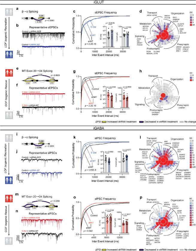
Figure 5: Isogenic recapitulation and rescue of neurotransmission phenotypes. (a) Differential splicing of β→α cluster in shWT, compared via Dirichlet-multinomial generalized linear model (p = 0.0083) in iGLUT neurons. (b) Representative traces of iGLUT WT knockdown effects, with (c) cumulative probabilities of sEPSC IEI distributions, with insets of cell-averaged IEI and amplitude measures (n: shNT = 12/1; shWT = 7/1 | 2 batches). Curves were compared via a Levene’s test (F = 70.78, df =1, p < 2.2E-16), and insets were compared via a Student’s t-test (IEI t=1.166, df=19, p = 0.2579; AMP t=0.5661, df=19, p = 0.578). (d) SynGO (biological process) sunburst plots showing enrichment of DEGs associated with synaptic function for iGLUT WT-KD compared to a brain expressed background via Fisher’s exact test (1.274161-fold, p = 0.1049). (e) Splicing of MT exon 20→24 cluster in shMT, compared via Dirichlet-multinomial generalized linear model (p = 0.0623). (f) Representative traces of iGLUT MT knockdown effects, with (g) cumulative probabilities of sEPSC IEI distributions, with insets of cell-averaged IEI and amplitude measures (n:shNT = 14/1; shMT = 18/1 | 2 batches). Curves were compared via a Levene’s test (F = 230.6; df = 1, p < 2.2E-16), and insets were compared via a Student’s t-test (IEI t=2.471, df=30, p = 0.0194; AMP t=1.812, df=30, p = 0.08). (h) SynGO (biological process) sunburst plots showing enrichment of DEGs associated with synaptic function for iGLUT MT-KD compared to a brain expressed background via Fisher’s exact test (1.214533-fold, p = 0.4541). (i) Differential splicing of β→α cluster in shWT, compared via Dirichlet-multinomial generalized linear model (p = 0.0203) in iGABA neurons. (j) Representative traces of iGABA WT knockdown effects, with (k) cumulative probabilities of sIPSC IEI distributions, with insets of cell-averaged, IEI and amplitude measures (shNT = 16/3; shWT = 19/3 | 2 batches). Curves were compared via a Levene’s test (F = 30.879, df =1, p < 3.66E-08), and insets were compared via a Student’s t-test (IEI t=2.768, df=33, p = 0.0092; AMP t=0.7856, df=33, p = 0.4377). (l) SynGO (biological process) sunburst plots for iGABA WT-KD compared to a brain expressed background via Fisher’s exact test (1.234861-fold, p = 1.307E-6). (m) Splicing of MT exon 20→24 cluster in shMT, compared via Dirichlet-multinomial generalized linear model (p = 0.0258) in iGABA neurons. (n) Representative traces of iGABA MT knockdown effects, with (o) cumulative probabilities of sIPSC IEI distributions, with insets of cell-averaged, IEI and amplitude measures (shNT = 16/1; shMT = 19/1 | 2 batches). Curves were compared via a Levene’s test (F = 4.1324; df = 1, p < 0.04226), and insets were compared via a Student’s t-test (IEI t=1.305, df=25, p = 0.2038; AMP t=0.9292, df=25, p = 0.3617). (p) SynGO (biological process) sunburst plots for iGABA WT-KD compared to a brain expressed background via Fisher’s exact test (1.347225-fold, p = 2.694E-4). Data represented as mean ± sem. n reported as samples/donors | independent batches.
Figure 5: Isogenic recapitulation and rescue of neurotransmission phenotypes. (a) Differential splicing of β→α cluster in shWT, compared via Dirichlet-multinomial generalized linear model (p = 0.0083) in iGLUT neurons. (b) Representative traces of iGLUT WT knockdown effects, with (c) cumulative probabilities of sEPSC IEI distributions, with insets of cell-averaged IEI and amplitude measures (n: shNT = 12/1; shWT = 7/1 | 2 batches). Curves were compared via a Levene’s test (F = 70.78, df =1, p < 2.2E-16), and insets were compared via a Student’s t-test (IEI t=1.166, df=19, p = 0.2579; AMP t=0.5661, df=19, p = 0.578). (d) SynGO (biological process) sunburst plots showing enrichment of DEGs associated with synaptic function for iGLUT WT-KD compared to a brain expressed background via Fisher’s exact test (1.274161-fold, p = 0.1049). (e) Splicing of MT exon 20→24 cluster in shMT, compared via Dirichlet-multinomial generalized linear model (p = 0.0623). (f) Representative traces of iGLUT MT knockdown effects, with (g) cumulative probabilities of sEPSC IEI distributions, with insets of cell-averaged IEI and amplitude measures (n:shNT = 14/1; shMT = 18/1 | 2 batches). Curves were compared via a Levene’s test (F = 230.6; df = 1, p < 2.2E-16), and insets were compared via a Student’s t-test (IEI t=2.471, df=30, p = 0.0194; AMP t=1.812, df=30, p = 0.08). (h) SynGO (biological process) sunburst plots showing enrichment of DEGs associated with synaptic function for iGLUT MT-KD compared to a brain expressed background via Fisher’s exact test (1.214533-fold, p = 0.4541). (i) Differential splicing of β→α cluster in shWT, compared via Dirichlet-multinomial generalized linear model (p = 0.0203) in iGABA neurons. (j) Representative traces of iGABA WT knockdown effects, with (k) cumulative probabilities of sIPSC IEI distributions, with insets of cell-averaged, IEI and amplitude measures (shNT = 16/3; shWT = 19/3 | 2 batches). Curves were compared via a Levene’s test (F = 30.879, df =1, p < 3.66E-08), and insets were compared via a Student’s t-test (IEI t=2.768, df=33, p = 0.0092; AMP t=0.7856, df=33, p = 0.4377). (l) SynGO (biological process) sunburst plots for iGABA WT-KD compared to a brain expressed background via Fisher’s exact test (1.234861-fold, p = 1.307E-6). (m) Splicing of MT exon 20→24 cluster in shMT, compared via Dirichlet-multinomial generalized linear model (p = 0.0258) in iGABA neurons. (n) Representative traces of iGABA MT knockdown effects, with (o) cumulative probabilities of sIPSC IEI distributions, with insets of cell-averaged, IEI and amplitude measures (shNT = 16/1; shMT = 19/1 | 2 batches). Curves were compared via a Levene’s test (F = 4.1324; df = 1, p < 0.04226), and insets were compared via a Student’s t-test (IEI t=1.305, df=25, p = 0.2038; AMP t=0.9292, df=25, p = 0.3617). (p) SynGO (biological process) sunburst plots for iGABA WT-KD compared to a brain expressed background via Fisher’s exact test (1.347225-fold, p = 2.694E-4). Data represented as mean ± sem. n reported as samples/donors | independent batches.CC BY-NC-ND · PubMed Central
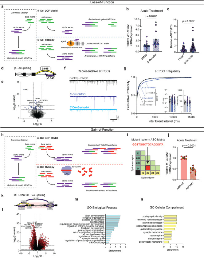
Figure 6: Precise therapeutic targeting of stratified GOF- and LOF-NRXN1+/− in iGLUT neurons. (a) Model and proposed mechanism of rescue for LOF patients ameliorating loss of wildtype isoforms. (b) RT-qPCR of WT NRXN1 gene expression in 5-Del patients, post-acute treatment (3–5 days) (n: DMSO = 19/2; β-estradiol = 18/2 | 3 batches) compared via Student’s t-test t=2.293, df=35, p = 0.028. (c) Quantification of iGLUT neuronal activity at WPI3 across vehicle and treatment (n: DMSO = 72/3; β-estradiol = 74/3 | 3 batches) compared via Student’s t-test (t=2.804, df=144, p = 0.0057). (d) Splicing of β→α cluster and (e) volcano plot of DEGs in β-estradiol treated 5’-Del iGLUT neurons, compared to vehicle treated 5’-Del iGLUT neurons. (f) Representative patch-clamp traces and (h) cumulative probabilities of sEPSC IEI distributions, with insets of cell-averaged, IEI and amplitude measures (n: Control + DMSO = 18/1; 5’-Del + DMSO = 33/2 5’-Del + β-estradiol = 35/2). Curves were compared via Levene’s test with Bonferroni corrections (Control + DMSO v. 5’-Del + DMSO F = 13.151; df = 1, p = 8.763E-4; 5’Del + DMSO v 5’-Del + β-estradiol F = 14.359; df = 1, p = 4.62E-04; Control + DMSO v 5’-Del + β-estradiol, F = 0.0141; df = 1, p = 0.955). Insets were compared via Student’s t-test (IEI t=1.161, df=66, p = 0.2499). (h) Model and proposed mechanism of rescue for GOF patients expressing mutant isoforms. (i) Schematic of a NRXN1 ASO matrix, with the selective sequence targeting 20/24 splice junction. (j) RT-qPCR of MT NRXN1 gene expression in 3’-Del patients’ post-acute treatment for 72hrs (ASO-NT = 8/2; ASO-MT = 10/2 | batches), compared via Student’s t-test t=12.91, df=16 p < 0.0001. (k) Splicing of MT Exon 20→24 cluster and (l) volcano plot of DEGs in ASO-MT treated 3’-Del iGLUT neurons, compared to ASO-NT treated 3’-Del iGLUT neurons.(m) Biological process and (n) cellular compartment GO terms demonstrating enrichment (−log10[adj p value]) of selected synapse related pathways. Data represented as mean ± sem. n reported as samples/donors | independent batches.
Figure 6: Precise therapeutic targeting of stratified GOF- and LOF-NRXN1+/− in iGLUT neurons. (a) Model and proposed mechanism of rescue for LOF patients ameliorating loss of wildtype isoforms. (b) RT-qPCR of WT NRXN1 gene expression in 5-Del patients, post-acute treatment (3–5 days) (n: DMSO = 19/2; β-estradiol = 18/2 | 3 batches) compared via Student’s t-test t=2.293, df=35, p = 0.028. (c) Quantification of iGLUT neuronal activity at WPI3 across vehicle and treatment (n: DMSO = 72/3; β-estradiol = 74/3 | 3 batches) compared via Student’s t-test (t=2.804, df=144, p = 0.0057). (d) Splicing of β→α cluster and (e) volcano plot of DEGs in β-estradiol treated 5’-Del iGLUT neurons, compared to vehicle treated 5’-Del iGLUT neurons. (f) Representative patch-clamp traces and (h) cumulative probabilities of sEPSC IEI distributions, with insets of cell-averaged, IEI and amplitude measures (n: Control + DMSO = 18/1; 5’-Del + DMSO = 33/2 5’-Del + β-estradiol = 35/2). Curves were compared via Levene’s test with Bonferroni corrections (Control + DMSO v. 5’-Del + DMSO F = 13.151; df = 1, p = 8.763E-4; 5’Del + DMSO v 5’-Del + β-estradiol F = 14.359; df = 1, p = 4.62E-04; Control + DMSO v 5’-Del + β-estradiol, F = 0.0141; df = 1, p = 0.955). Insets were compared via Student’s t-test (IEI t=1.161, df=66, p = 0.2499). (h) Model and proposed mechanism of rescue for GOF patients expressing mutant isoforms. (i) Schematic of a NRXN1 ASO matrix, with the selective sequence targeting 20/24 splice junction. (j) RT-qPCR of MT NRXN1 gene expression in 3’-Del patients’ post-acute treatment for 72hrs (ASO-NT = 8/2; ASO-MT = 10/2 | batches), compared via Student’s t-test t=12.91, df=16 p < 0.0001. (k) Splicing of MT Exon 20→24 cluster and (l) volcano plot of DEGs in ASO-MT treated 3’-Del iGLUT neurons, compared to ASO-NT treated 3’-Del iGLUT neurons.(m) Biological process and (n) cellular compartment GO terms demonstrating enrichment (−log10[adj p value]) of selected synapse related pathways. Data represented as mean ± sem. n reported as samples/donors | independent batches.CC BY-NC-ND · PubMed Central

Verification

Claims the paper makes

0 Valid · 1 Not valid · 38 Not checked · 100 Own results and interpretations

52 more claims

Create a free account to see all 139 claims and to verify the 46 references still open.

See all claims — free account
  • It may state two separate claims as one. 56
  • The quote only partly states this claim. 1
  • Probably not a substantial claim. 42

Create a free account to see these claims.

Self-check

Percentages, sums and reported differences re-computed from the paper’s own numbers.

Each contradiction between two passages listed here was confirmed by an independent check.

1 of 1

Citations

Cites

Show all 80 references

Cited by

See all 30

Metadata

Authors
Fernando, M. B., Fan, Y., Zhang, Y., Tokolyi, A., Murphy, A. N., Kammourh, S., Deans, P. J. M., Ghorbani, S., Onatzevitch, R., Pero, A., Padilla, C., Williams, S. E., Flaherty, E. K., Prytkova, I. A., Cao, L., Knowles, D. A., Fang, G., Slesinger, P. A., & Brennand, K. J.
Affiliations
  • Yale University (US)
  • Icahn School of Medicine at Mount Sinai (US)
  • New York Genome Center (US)
  • Haukeland University Hospital (NO)
Corresponding author
  • Fang, Gang
  • Slesinger, Paul A.
  • Brennand, Kristen J.
Venue
Nature
Published
2025-04-09
Volume
642
Issue
8068
Pages
710-720
ISSN
  • 0028-0836 (ISSN-L)
  • 1476-4687
OpenAlex
W4409283565
Cited by
30
References
80
Citations by year
2026: 16 · 2025: 11
FWCI
10.54
Type
journal article
Language
en
Open access
Locations
  • Nature (publishedVersion)
  • bioRxiv (Cold Spring Harbor Laboratory) (submittedVersion, cc-by-nc-nd)
  • PubMed (publishedVersion)
  • PubMed Central (submittedVersion, cc-by-nc-nd)
Funders
  • National Science Foundation (2146398)
  • Icahn School of Medicine at Mount Sinai
  • Johns Hopkins University
  • National Institute on Alcohol Abuse and Alcoholism (F31AA027949)
  • National Institute of Mental Health (R01MH121074, R01MH123155, RM1MH132648, R01MH125579)
Topics
  • Life Sciences › Biochemistry, Genetics and Molecular Biology › Genetics › Genetics and Neurodevelopmental Disorders
  • Life Sciences › Neuroscience › Cellular and Molecular Neuroscience › Neuroscience and Neuropharmacology Research
  • Life Sciences › Biochemistry, Genetics and Molecular Biology › Genetics › Genomic variations and chromosomal abnormalities
Keywords
NRXN1, copy number variation, alternative splicing, neuropsychiatric disorders, loss-of-function mutations, gain-of-function mutations, human induced pluripotent stem cells, glutamatergic neurons, GABAergic neurons, synaptic activity, cell type-specific effects, precision medicine
MeSH terms
Loss of Function Mutation, Animals, Calcium-Binding Proteins, Female, Heterozygote, Humans, Male, Phenotype, Synapses, Cell Adhesion Molecules, Neuronal, Gene Deletion, Sequence Deletion, Alternative Splicing, Neural Cell Adhesion Molecules, Mice, DNA Copy Number Variations, Induced Pluripotent Stem Cells, GABAergic Neurons

About these checks

Registry, retraction, Cites and statistics results are computed, not judged, with one exception: a printed reference that no index lists is read once by a model before it counts as a citation error. Claims are extracted by a model and kept only when their quote appears verbatim in the text; each cited source is then checked, with a model’s help, for whether it says what the claim relies on. Those verdicts can be wrong — read the claim’s quote and the cited source before relying on one.

A language model drew the claims from the paper on 2026-11-13, under version 3 of the claim definition (model deepseek-v4.1-flash, prompt extract-v3).

A second model labelled each claim on 2026-11-13 (model jev-1.13-20260917, definition revision gold-guideline-v0.2).

Statistics and arithmetic are recomputed, not judged.

Confidence bands are shown only for fields calibrated on our annotated test set. None is calibrated yet, so no band is shown.

What counts as a claim ›How we measure confidence ›How we check this ›

Are you an author, or do you see a problem with this page? Request a review

How to cite

Cite CoChat for the checks on this page, and the paper for its own findings.

This page
CoChat. (2026). Citation check: Phenotypic complexities of rare heterozygous neurexin-1 deletions. https://app.cochat.ai/papers/10.1038/s41586-025-08864-9
The paper
Fernando, M. B., Fan, Y., Zhang, Y., Tokolyi, A., Murphy, A. N., Kammourh, S., Deans, P. J. M., Ghorbani, S., Onatzevitch, R., Pero, A., Padilla, C., Williams, S. E., Flaherty, E. K., Prytkova, I. A., Cao, L., Knowles, D. A., Fang, G., Slesinger, P. A., & Brennand, K. J. (2025). Phenotypic complexities of rare heterozygous neurexin-1 deletions. Nature. https://doi.org/10.1038/s41586-025-08864-9

Data sources

Bibliographic data: Semantic Scholar, OpenAlex, Crossref.

Contains information from the Semantic Scholar Academic Graph Datasets, made available under the ODC Attribution License (ODC-BY 1.0).

Semantic Scholar’s data is described in: Kinney, R., et al. (2023). The Semantic Scholar Open Data Platform. arXiv:2301.10140.