

The DOI matches its Crossref record; 87 of the 88 references it cites were found in a registry or an index.
Cerebrospinal fluid proteomics define the natural history of autosomal dominant Alzheimer’s disease
Open accessAbstract
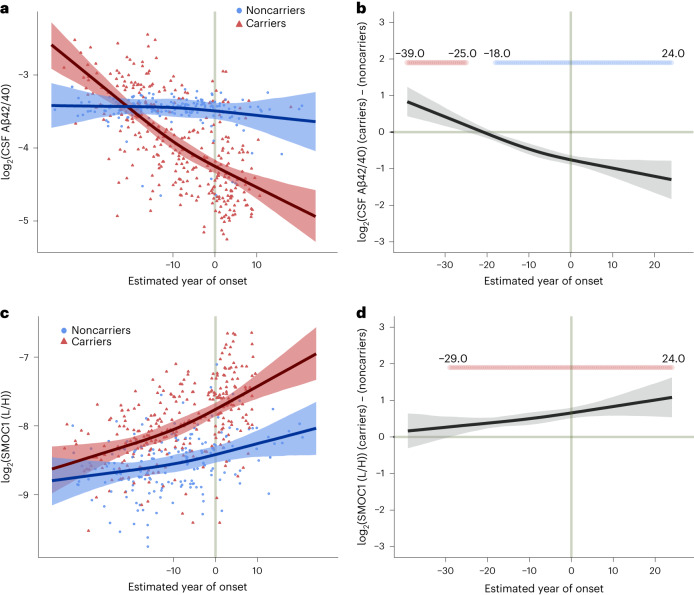
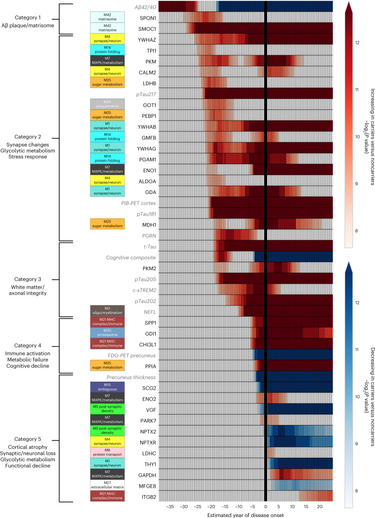
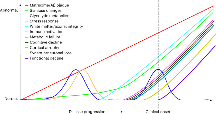
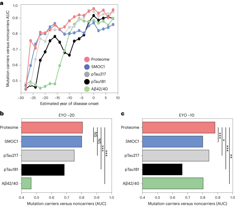
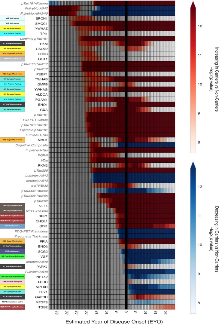
Alzheimer’s disease (AD) pathology develops many years before the onset of cognitive symptoms. Two pathological processes—aggregation of the amyloid-β (Aβ) peptide into plaques and the microtubule protein tau into neurofibrillary tangles (NFTs)—are hallmarks of the disease. However, other pathological brain processes are thought to be key disease mediators of Aβ plaque and NFT pathology. How these additional pathologies evolve over the course of the disease is currently unknown. Here we show that proteomic measurements in autosomal dominant AD cerebrospinal fluid (CSF) linked to brain protein coexpression can be used to characterize the evolution of AD pathology over a timescale spanning six decades. SMOC1 and SPON1 proteins associated with Aβ plaques were elevated in AD CSF nearly 30 years before the onset of symptoms, followed by changes in synaptic proteins, metabolic proteins, axonal proteins, inflammatory proteins and finally decreases in neurosecretory proteins. The proteome discriminated mutation carriers from noncarriers before symptom onset as well or better than Aβ and tau measures. Our results highlight the multifaceted landscape of AD pathophysiology and its temporal evolution. Such knowledge will be critical for developing precision therapeutic interventions and biomarkers for AD beyond those associated with Aβ and tau.
Overview
The claims and statistics of this paper haven’t been checked yet.
Run the check — free, no account needed. It reads the open-access text and checks the sources it cites.
Integrity signals
Registry
Cites
Statistics
Figures
Verification
Claims the paper makes
The claims and statistics of this paper haven’t been checked yet.
Run the check — free, no account needed. It reads the open-access text and checks the sources it cites.
Claims other papers make about this one
Each is checked against this paper’s own text: does it say what the citing paper says it does?
The source backs part of the claim, not all of it — or backs it for a different population, condition or comparison.
In the citing paper
“Synapse degeneration is an insidious process, occurring before neuron loss and long before disease diagnosis 4,17 .”
From NAD+ hydrolase Sarm1 is a key driver of synapse degeneration and memory loss in Alzheimer’s disease (2025) · cites this paper as [17]
Experimental — accuracy not yet measuredThe source backs part of the claim, not all of it — or backs it for a different population, condition or comparison.
In the citing paper
“While Aβ disrupts synaptic transmission 32,34,35 and induces synapse loss 17”
From NAD+ hydrolase Sarm1 is a key driver of synapse degeneration and memory loss in Alzheimer’s disease (2025) · cites this paper as [17]
Experimental — accuracy not yet measured
Self-check
Percentages, sums and reported differences re-computed from the paper’s own numbers.
The self-check needs the paper’s full text.
Citations
Cites
Folstein, M. F., Folstein, S. E., & McHugh, P. R. (1975) · Journal of Psychiatric Research · doi:10.1016/0022-3956(75)90026-6 · Cited by 88,282
Hirono, N., Mori, E., Ikejiri, Y., Imamura, T., Shimomura, T., Hashimoto, M., Yamashita, H., & Ikeda, M. (1997) · PubMed · Cited by 21,012
Kok, J. S., Berg, I. J., & Scherder, E. J. A. (2013) · Dementia and Geriatric Cognitive Disorders Extra · doi:10.1159/000353441 · Cited by 20,589
Older Adults' Reasons for Using Technology while Aging in Place
Peek, S. T. M., Luijkx, K. G., Rijnaard, M. D., Nieboer, M. E., van der Voort, C. S., Aarts, S., van Hoof, J., Vrijhoef, H. J., & Wouters, E. (2015) · Gerontology · doi:10.1159/000430949 · Cited by 19,852
Sensitivity and False Alarm Rate of a Fall Sensor in Long-Term Fall Detection in the Elderly
Kangas, M., Korpelainen, R., Vikman, I., Nyberg, L., & Jämsä, T. J. (2014) · Gerontology · doi:10.1159/000362720 · Cited by 19,007
NIA‐AA Research Framework: Toward a biological definition of Alzheimer's disease
Jack, C. R., Bennett, D. A., Blennow, K., Carrillo, M. C., Dunn, B., Haeberlein, S. B., Holtzman, D. M., Jagust, W., Jessen, F., Karlawish, J., et al. (2018) · Alzheimer's & Dementia · doi:10.1016/j.jalz.2018.02.018 · Cited by 10,537
The Clinical Dementia Rating (CDR): Current version and scoring rules
Morris, J. C. (1993) · Neurology · doi:10.1212/wnl.43.11.2412-a · Cited by 9,882
Self-citationbrms : An R Package for Bayesian Multilevel Models Using Stan
Bürkner, P.-C. (2017) · Journal of Statistical Software · doi:10.18637/jss.v080.i01 · Cited by 9,476 · cited 1×
“Therefore, we used Bayesian mixed-effect ordinal regression models with a cumulative link function to model the two categorical outcomes (Formula 3) 74,75 .”
MethodNichols, E., Steinmetz, J. D., Vollset, S. E., Fukutaki, K., Chalek, J., Abd-Allah, F., Abdoli, A., Abualhasan, A., Abu-Gharbieh, E., Akram, T. T., et al. (2022) · The Lancet Public Health · doi:10.1016/s2468-2667(21)00249-8 · Cited by 5,575
Clinical and biomarker changes in dominantly inherited Alzheimer's disease
Bateman, R. J., Xiong, C., Benzinger, T. L. S., Fagan, A. M., Goate, A., Fox, N. C., Marcus, D. S., Cairns, N. J., Xie, X., Blazey, T. M., et al. (2012) · New England Journal of Medicine · doi:10.1056/nejmoa1202753 · Cited by 3,814
Self-citation
Cited by
Sorted by citations
Items 101–101 of 101
NAD+ hydrolase Sarm1 is a key driver of synapse degeneration and memory loss in Alzheimer’s disease
Fan, F., Joshi, P., Liu, X., Kotturu, S. K., Abiola, M., Liu, Q., & Lou, X. (2025) · doi:10.64898/2025.12.21.695860
Metadata
- Authors
- Johnson, E. C. B., Bian, S., Haque, R. U., Carter, E. K., Watson, C. M., Gordon, B. A., Ping, L., Duong, D. M., Epstein, M. P., McDade, E., Barthélemy, N. R., Karch, C. M., Xiong, C., Cruchaga, C., Perrin, R. J., Wingo, A. P., Wingo, T. S., Chhatwal, J. P., Day, G. S., Noble, J. M., Berman, S. B., Martins, R., Graff-Radford, N. R., Schofield, P. R., Ikeuchi, T., Mori, H., Levin, J., Farlow, M., Lah, J. J., Haass, C., Jucker, M., Morris, J. C., Benzinger, T. L. S., Roberts, B. R., Bateman, R. J., Fagan, A. M., Seyfried, N. T., Levey, A. I., the Dominantly Inherited Alzheimer Network, Vöglein, J., Allegri, R., Mendez, P. C., Surace, E., Berman, S. B., Ikonomovic, S., Nadkarni, N., Lopera, F., Ramirez, L., Aguillon, D., Leon, Y., Ramos, C., Alzate, D., Baena, A., Londono, N., Moreno, S., Laske, C., Kuder-Buletta, E., Graber-Sultan, S., Preische, O., Hofmann, A., Kasuga, K., Niimi, Y., Ishii, K., Senda, M., Sanchez-Valle, R., Rosa-Neto, P., Fox, N., Cash, D., Lee, J.-H., Roh, J. H., Riddle, M., Menard, W., Bodge, C., Surti, M., Takada, L. T., Sanchez-Gonzalez, V. J., Orozco-Barajas, M., Goate, A., Renton, A., Esposito, B., Marsh, J., Cruchaga, C., Fernandez, V., Jerome, G., Herries, E., Llibre-Guerra, J., Brooks, W., Bechara, J., Hassenstab, J., Franklin, E., Chen, A., Chen, C., Flores, S., Friedrichsen, N., Hantler, N., Hornbeck, R., Jarman, S., Keefe, S., Koudelis, D., Massoumzadeh, P., McCullough, A., McKay, N., Nicklaus, J., Pulizos, C., Wang, Q., Mishall, S., Sabaredzovic, E., Deng, E., Candela, M., Smith, H., Hobbs, D., Scott, J., Wang, P., Xu, X., Li, Y., Gremminger, E., Ma, Y., Bui, R., Lu, R., Ortiz, A. L. S., Daniels, A., Courtney, L., Supnet-Bell, C., Xu, J., & Ringman, J.
- Affiliations
- Emory University (US)
- Washington University in St. Louis (US)
- Brigham and Women's Hospital (US)
- Mayo Clinic in Florida (US)
- Columbia University Irving Medical Center (US)
- Edith Cowan University (AU)
- UNSW Sydney (AU)
- Brain Research Institute, Niigata University (JP)
- Osaka Metropolitan University (JP)
- Ludwig-Maximilians-Universität München (DE)
- Indiana University Bloomington (US)
- German Center for Neurodegenerative Diseases (DE)
- Fundación para la Lucha contra las Enfermedades Neurológicas de la Infancia (AR)
- University of Pittsburgh (US)
- Universidad de Antioquia (CO)
- University of Tokyo Hospital (JP)
- Tokyo Metropolitan Institute of Gerontology (JP)
- Hospital Clínic de Barcelona (ES)
- UK Dementia Research Institute (GB)
- Korea University (KR)
- Brown University (US)
- Universidade de São Paulo (BR)
- Icahn School of Medicine at Mount Sinai (US)
- University of Southern California (US)
- Corresponding author
- Johnson, Erik C. B.
- ORCID
- Johnson, Erik C. B.
- Bian, Shijia
- Haque, Rafi U.
- Carter, E. Kathleen
- Watson, Caroline M.
- Gordon, Brian A.
- Ping, Lingyan
- Duong, Duc M.
- Epstein, Michael P.
- McDade, Eric
- Barthélemy, Nicolas R.
- Karch, Celeste M.
- Xiong, Chengjie
- Perrin, Richard J.
- Wingo, Aliza P.
- Wingo, Thomas S.
- Chhatwal, Jasmeer P.
- Day, Gregory S.
- Noble, James M.
- Martins, Ralph
- Schofield, Peter R.
- Ikeuchi, Takeshi
- Mori, Hiroshi
- Levin, Johannes
- Farlow, Martin
- Lah, James J.
- Haass, Christian
- Jucker, Mathias
- Morris, John C.
- Benzinger, Tammie L. S.
- Roberts, Blaine R.
- Bateman, Randall J.
- Fagan, Anne M.
- Seyfried, Nicholas T.
- Levey, Allan I.
- Vöglein, Jonathan
- Allegri, Ricardo
- Surace, Ezequiel
- Nadkarni, Neelesh
- Lopera, Francisco
- Aguillon, David
- Baena, Ana
- Laske, Christoph
- Kasuga, Kensaku
- Niimi, Yoshiki
- Ishii, Kenji
- Senda, Michio
- Fox, Nick
- Lee, Jae-Hong
- Roh, Jee Hoon
- Riddle, Meghan
- Takada, Leonel Tadao
- Goate, Alison
- Renton, Alan
- Marsh, Jacob
- Brooks, William
- Bechara, Jacob
- Hassenstab, Jason
- Franklin, Erin
- Chen, Allison
- Chen, Charles
- Flores, Shaney
- Hornbeck, Russ
- Keefe, Sarah
- Massoumzadeh, Parinaz
- McCullough, Austin
- McKay, Nicole
- Nicklaus, Joyce
- Wang, Qing
- Deng, Emily
- Smith, Hunter
- Hobbs, Diana
- Scott, Jalen
- Wang, Peter
- Li, Yan
- Ma, Yinjiao
- Bui, Ryan
- Lu, Ruijin
- Daniels, Alisha
- Xu, Jinbin
- Ringman, John
- Venue
- Nature Medicine
- Published
- 2023-08
- Volume
- 29
- Issue
- 8
- Pages
- 1979-1988
- ISSN
- 1078-8956 (ISSN-L)
- 1546-170X
- PMC
- PMC10427428
- OpenAlex
- W4385639069
- Cited by
- 170
- References
- 88
- Citations by year
- 2026: 44 · 2025: 64 · 2024: 55 · 2023: 6
- FWCI
- 30.19
- Type
- journal article
- Language
- en
- Open access
- hybrid
- doi.org
- Locations
- Nature Medicine (publishedVersion, cc-by)
- PubMed Central (publishedVersion, cc-by)
- PubMed (publishedVersion)
- UCL Discovery (University College London) (submittedVersion, cc-by)
- Open access LMU (Ludwid Maxmilian's Universitat Munchen) (submittedVersion, cc-by)
- eScholarship (California Digital Library) (submittedVersion, cc-by)
- DZNE Pub (submittedVersion, cc-by)
- Digital Commons@Becker (Washington University School of Medicine) (submittedVersion, cc-by)
- Australasian Journal of Paramedicine (submittedVersion, cc-by)
- Greater South Information System
- Funders
- Alzheimer's Association (SG-20-690363-DIAN)
- Emory University (U01AG061357)
- Washington University in St. Louis
- Fondation Brain Canada
- Japan Agency for Medical Research and Development (17929884, 22dk0207049h0003)
- Deutsche Forschungsgemeinschaft (HA1737/16‐1, EXC 2145, Koselleck Project HA1737/16-1, EXC 2145SyNergy, 390857198)
- Korea Health Industry Development Institute
- Deutsches Zentrum für Neurodegenerative Erkrankungen
- National Institutes of Health (1s10od025214-01, 1rf1ag071170-01)
- Canadian Institutes of Health Research
- Goizueta Business School, Emory University
- Instituto de Salud Carlos III
- National Institute on Aging (R01AG056533, RF1 AG071170, AG 024904, AG066444, UF1AG032438, U01AG061357, P01AG003991, U19AG032438, K08AG068604, P30AG066444, U19AG024904, R01AG072120, R01AG075827, P01AG026276, P30AG066511)
- National Institute of Neurological Disorders and Stroke (R01NS092865)
- Topics
- Life Sciences › Neuroscience › Cellular and Molecular Neuroscience › Alzheimer's disease research and treatments
- Health Sciences › Medicine › Psychiatry and Mental health › Dementia and Cognitive Impairment Research
- Health Sciences › Medicine › Clinical Neurology › Cerebrospinal fluid and hydrocephalus
- Keywords
- autosomal dominant Alzheimer's disease, cerebrospinal fluid proteomics, natural history of disease, amyloid plaques, neurofibrillary tangles, SMOC1, SPON1, presymptomatic biomarkers, mutation carriers, synaptic proteins, neuroinflammation, disease progression
- MeSH terms
- Adult, Alzheimer Disease, Female, Humans, Male, Middle Aged, Mutation, Biomarkers, Osteonectin, Age of Onset, Proteomics
About these checks
Registry, retraction, Cites and statistics results are computed, not judged, with one exception: a printed reference that no index lists is read once by a model before it counts as a citation error. Claims are extracted by a model and kept only when their quote appears verbatim in the text; each cited source is then checked, with a model’s help, for whether it says what the claim relies on. Those verdicts can be wrong — read the claim’s quote and the cited source before relying on one.
Statistics and arithmetic are recomputed, not judged.
Confidence bands are shown only for fields calibrated on our annotated test set. None is calibrated yet, so no band is shown.
What counts as a claim ›How we measure confidence ›How we check this ›
Are you an author, or do you see a problem with this page? Request a review
