

The DOI matches its Crossref record; 103 of the 103 references it cites were found in a registry or an index.
Location Sensitive Deep Convolutional Neural Networks for Segmentation of White Matter Hyperintensities
Open accessAbstract
The anatomical location of imaging features is of crucial importance for accurate diagnosis in many medical tasks. Convolutional neural networks (CNN) have had huge successes in computer vision, but they lack the natural ability to incorporate the anatomical location in their decision making process, hindering success in some medical image analysis tasks. In this paper, to integrate the anatomical location information into the network, we propose several deep CNN architectures that consider multi-scale patches or take explicit location features while training. We apply and compare the proposed architectures for segmentation of white matter hyperintensities in brain MR images on a large dataset. As a result, we observe that the CNNs that incorporate location information substantially outperform a conventional segmentation method with handcrafted features as well as CNNs that do not integrate location information. On a test set of 50 scans, the best configuration of our networks obtained a Dice score of 0.792, compared to 0.805 for an independent human observer. Performance levels of the machine and the independent human observer were not statistically significantly different (p-value = 0.06).
Overview
The claims and statistics of this paper haven’t been checked yet.
Run the check — free, no account needed. It reads the open-access text and checks the sources it cites.
Integrity signals
Registry
Cites
Statistics
Figures
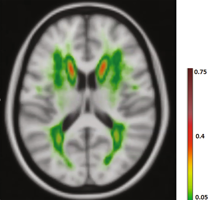
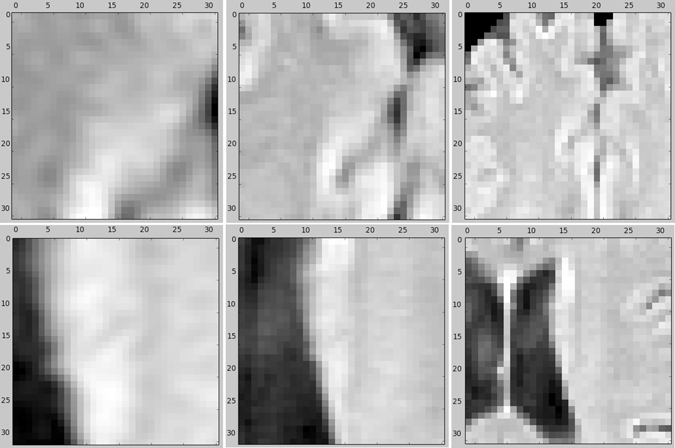
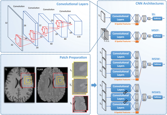
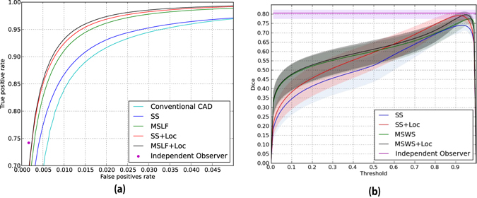
Verification
Claims the paper makes
The claims and statistics of this paper haven’t been checked yet.
Run the check — free, no account needed. It reads the open-access text and checks the sources it cites.
Claims other papers make about this one
Each is checked against this paper’s own text: does it say what the citing paper says it does?
In the citing paper
“Noteworthy, the improved segmentation performance is mainly due to the contribution of handcrafted features rather than the multi-scale paradigm.”
From Embracing imperfect datasets: A review of deep learning solutions for medical image segmentation (2020) · cites this paper as Ghafoorian et al. (2017)
In the citing paper
“On a test set of 46 cases, their multi-scale architecture equipped with the hand-crafted features achieves 6% increase in Dice over their single scale baseline that does not incorporate the handcrafted features.”
From Embracing imperfect datasets: A review of deep learning solutions for medical image segmentation (2020) · cites this paper as Ghafoorian et al. (2017)
Self-check
Percentages, sums and reported differences re-computed from the paper’s own numbers.
The self-check needs the paper’s full text.
Citations
Cites
ImageNet classification with deep convolutional neural networks
Krizhevsky, A., Sutskever, I., & Hinton, G. E. (2017) · Communications of the ACM · doi:10.1145/3065386 · Cited by 112,155
U-Net: Convolutional Networks for Biomedical Image Segmentation
Ronneberger, O., Fischer, P., & Brox, T. (2015) · Lecture notes in computer science · doi:10.1007/978-3-319-24574-4_28 · Cited by 99,404
LeCun, Y., Bengio, Y., & Hinton, G. (2015) · Nature · doi:10.1038/nature14539 · Cited by 84,126
ImageNet: A large-scale hierarchical image database
Deng, J., Dong, W., Socher, R., Li, L.-J., Li, K., & Fei-Fei, L. (2009) · 2009 IEEE Conference on Computer Vision and Pattern Recognition · doi:10.1109/cvpr.2009.5206848 · Cited by 65,333
Gradient-based learning applied to document recognition
Lecun, Y., Bottou, L., Bengio, Y., & Haffner, P. (1998) · Proceedings of the IEEE · doi:10.1109/5.726791 · Cited by 58,805
Fully convolutional networks for semantic segmentation
Long, J., Shelhamer, E., & Darrell, T. (2015) · 2015 IEEE Conference on Computer Vision and Pattern Recognition (CVPR) · doi:10.1109/cvpr.2015.7298965 · Cited by 38,834
(2014) · Cited by 33,419
Advances in neural information processing systems 7
Cios, K. J., & Shields, M. E. (1997) · Neurocomputing · doi:10.1016/s0925-2312(97)90018-7 · Cited by 22,093
Delving Deep into Rectifiers: Surpassing Human-Level Performance on ImageNet Classification
He, K., Zhang, X., Ren, S., & Sun, J. (2015) · 2015 IEEE International Conference on Computer Vision (ICCV) · doi:10.1109/iccv.2015.123 · Cited by 19,741
Deep learning in neural networks: an overview
Schmidhuber, J. (2015) · Neural Networks · doi:10.1016/j.neunet.2014.09.003 · Cited by 18,570
Research works printed but not linked: 3
- Ghafoorian, M. et al. Transfer learning for domain adaptation in mri: Application in brain lesion segmentation. arXiv preprint arXiv:1702.07841 (2017). · arXiv
- Chen, H., Dou, Q., Yu, L. & Heng, P.-A. Voxresnet: Deep voxelwise residual networks for volumetric brain segmentation. arXiv preprint arXiv:1608.05895 (2016). · arXiv
- Maas, A. L., Hannun, A. Y. & Ng, A. Y. Rectifier nonlinearities improve neural network acoustic models. In Proc. ICML, vol. 30 (2013).
Cited by
Sorted by citations
A survey on deep learning in medical image analysis
Litjens, G., Kooi, T., Bejnordi, B. E., Setio, A. A. A., Ciompi, F., Ghafoorian, M., van der Laak, J. A. W. M., van Ginneken, B., & Sánchez, C. I. (2017) · Medical Image Analysis · doi:10.1016/j.media.2017.07.005 · Cited by 15,332
Self-citationArtificial intelligence in radiology
Hosny, A., Parmar, C., Quackenbush, J., Schwartz, L. H., & Aerts, H. J. W. L. (2018) · Nature Reviews Cancer · doi:10.1038/s41568-018-0016-5 · Cited by 3,803
Embracing imperfect datasets: A review of deep learning solutions for medical image segmentation
Tajbakhsh, N., Jeyaseelan, L., Li, Q., Chiang, J. N., Wu, Z., & Ding, X. (2020) · Medical Image Analysis · doi:10.1016/j.media.2020.101693 · Cited by 1,095
“3D model with a 3D representation augmented with handcrafted features Ghafoorian et al. (2017). We explain these methods in more detail as follows.”
MethodInfluentialDeep learning in medical imaging and radiation therapy
Sahiner, B., Pezeshk, A. X., Hadjiiski, L. M., Wang, X., Drukker, K., Cha, K. H., Summers, R. M., & Giger, M. L. (2018) · Medical Physics · doi:10.1002/mp.13264 · Cited by 763
Deep learning for lung cancer prognostication: A retrospective multi-cohort radiomics study
Hosny, A., Parmar, C., Coroller, T. P., Grossmann, P., Zeleznik, R., Kumar, A., Bussink, J., Gillies, R. J., Mak, R. H., & Aerts, H. J. W. L. (2018) · PLOS Medicine · doi:10.1371/journal.pmed.1002711 · Cited by 579
Holzinger, A., Carrington, A., & Müller, H. (2020) · KI - Künstliche Intelligenz · doi:10.1007/s13218-020-00636-z · Cited by 406
Transfer Learning for Domain Adaptation in MRI: Application in Brain Lesion Segmentation
Ghafoorian, M., Mehrtash, A., Kapur, T., Karssemeijer, N., Marchiori, E., Pesteie, M., Guttmann, C. R. G., de Leeuw, F., Tempany, C. M., van Ginneken, B., et al. (2017) · Lecture notes in computer science · doi:10.1007/978-3-319-66179-7_59 · Cited by 337
Self-citationKuijf, H. J., Casamitjana, A., Collins, D. L., Dadar, M., Georgiou, A., Ghafoorian, M., Jin, D., Khademi, A., Knight, J., Li, H., et al. (2019) · IEEE Transactions on Medical Imaging · doi:10.1109/tmi.2019.2905770 · Cited by 322
Self-citationCuadrado‐Godia, E., Dwivedi, P., Sharma, S., Ois, Á., Roquer, J., Balcells, M., Laird, J. R., Turk, M., Suri, H. S., Nicolaides, A. N., et al. (2018) · Journal of Stroke · doi:10.5853/jos.2017.02922 · Cited by 297
Deep learning based diagnosis of Parkinson’s disease using convolutional neural network
Sivaranjini, S., & Sujatha, C. M. (2019) · Multimedia Tools and Applications · doi:10.1007/s11042-019-7469-8 · Cited by 282
QuickNAT: A fully convolutional network for quick and accurate segmentation of neuroanatomy
Roy, A. G., Conjeti, S., Navab, N., & Wachinger, C. (2018) · NeuroImage · doi:10.1016/j.neuroimage.2018.11.042 · Cited by 280
A brief review on multi-task learning
Thung, K., & Chong-yaw Wee, A. (2018) · Multimedia Tools and Applications · doi:10.1007/s11042-018-6463-x · Cited by 266
Artificial intelligence in medical imaging of the liver
Zhou, L., Wang, J., Yu, S.-Y., Wu, G.-G., Wei, Q., Deng, Y., Wu, X., Cui, X., & Dietrich, C. F. (2019) · World Journal of Gastroenterology · doi:10.3748/wjg.v25.i6.672 · Cited by 282
Variability and reproducibility in deep learning for medical image segmentation
Renard, F., Guedria, S., Palma, N. D., & Vuillerme, N. (2020) · Scientific Reports · doi:10.1038/s41598-020-69920-0 · Cited by 255
Interactive machine learning: experimental evidence for the human in the algorithmic loop
Holzinger, A., Plass, M., Kickmeier-Rust, M., Holzinger, K., Crişan, G. C., Pintea, C.-M., & Palade, V. (2018) · Applied Intelligence · doi:10.1007/s10489-018-1361-5 · Cited by 235
Guerrero, R., Qin, C., Oktay, O., Bowles, C. T., Chen, L., Joules, R., Wolz, R., Valdés-Hernández, M. C., Dickie, D. A., Wardlaw, J. M., et al. (2017) · NeuroImage Clinical · doi:10.1016/j.nicl.2017.12.022 · Cited by 222
Hirani, R., Noruzi, K. A., Khuram, H., Hussaini, A. S., Aifuwa, E., Ely, K., Lewis, J. M., Gabr, A. E., Smiley, A., Tiwari, R. K., et al. (2024) · Life · doi:10.3390/life14050557 · Cited by 280
Fully convolutional network ensembles for white matter hyperintensities segmentation in MR images
Li, H., Jiang, G., Zhang, J., Wang, R., Wang, Z., Zheng, W., & Menze, B. H. (2018) · NeuroImage · doi:10.1016/j.neuroimage.2018.07.005 · Cited by 209
Transfer Learning for Domain Adaptation in MRI: Application in Brain Lesion Segmentation
Ghafoorian, M., Mehrtash, A., Kapur, T., Karssemeijer, N., Marchiori, E., Pesteie, M., Guttmann, C. R. G., Leeuw, F.-E. D., Tempany, C. M., van Ginneken, B., et al. (2017) · Radboud Repository (Radboud University) · Cited by 192
Self-citationAdvancing Patient Care: How Artificial Intelligence Is Transforming Healthcare
Poalelungi, D. G., Mușat, C. L., Fulga, A., Neagu, M., Neagu, A., Piraianu, A. I., & Fulga, I. (2023) · Journal of Personalized Medicine · doi:10.3390/jpm13081214 · Cited by 191
Addressing class imbalance in deep learning for small lesion detection on medical images
Bria, A., Marrocco, C., & Tortorella, F. (2020) · Computers in Biology and Medicine · doi:10.1016/j.compbiomed.2020.103735 · Cited by 158
The roles of supervised machine learning in systems neuroscience
Glaser, J. I., Benjamin, A. S., Farhoodi, R., & Kording, K. P. (2019) · Progress in Neurobiology · doi:10.1016/j.pneurobio.2019.01.008 · Cited by 163
Machine learning in GI endoscopy: practical guidance in how to interpret a novel field
van der Sommen, F., de Groof, J., Struyvenberg, M. R., van der Putten, J. A., Boers, T. G. W., Fockens, K. N., Schoon, E. J., Curvers, W. L., de With, P. H. N., Mori, Y., et al. (2020) · Gut · doi:10.1136/gutjnl-2019-320466 · Cited by 139
Kushibar, K., Valverde, S., González-Villà, S., Bernal, J., Cabezas, M., Oliver, A., & Lladó, X. (2018) · Medical Image Analysis · doi:10.1016/j.media.2018.06.006 · Cited by 121
Artificial intelligence in breast ultrasound
Wu, G.-G., Zhou, L., Xu, J., Wang, J., Wei, Q., Deng, Y., Cui, X., & Dietrich, C. F. (2019) · World Journal of Radiology · doi:10.4329/wjr.v11.i2.19 · Cited by 121
Moeskops, P., de Bresser, J., Kuijf, H. J., Mendrik, A. M., Biessels, G. J., Pluim, J. P. W., & Išgum, I. (2017) · NeuroImage Clinical · doi:10.1016/j.nicl.2017.10.007 · Cited by 111
Surianarayanan, C., Lawrence, J. J., Chelliah, P. R., Prakash, E., & Hewage, C. T. E. R. (2023) · Sensors · doi:10.3390/s23063062 · Cited by 127
Diagnosis of Parkinson’s disease using deep CNN with transfer learning and data augmentation
Kaur, S., Aggarwal, H., & Rani, R. (2020) · Multimedia Tools and Applications · doi:10.1007/s11042-020-10114-1 · Cited by 110
Dadar, M., Pascoal, T. A., Manitsirikul, S., Misquitta, K., Fonov, V., Tartaglia, M. C., Breitner, J. C. S., Rosa‐Neto, P., Carmichael, O. T., DeCarli, C. S., et al. (2017) · IEEE Transactions on Medical Imaging · doi:10.1109/tmi.2017.2693978 · Cited by 107
Polyp-Net: A Multimodel Fusion Network for Polyp Segmentation
Banik, D., Roy, K., Bhattacharjee, D., Nasipuri, M., & Krejcar, O. (2020) · IEEE Transactions on Instrumentation and Measurement · doi:10.1109/tim.2020.3015607 · Cited by 109
Qin, W., Wu, J., Han, F., Yuan, Y., Zhao, W., Ibragimov, B., Gu, J., & Xing, L. (2018) · Physics in Medicine and Biology · doi:10.1088/1361-6560/aabd19 · Cited by 106
Multi-Receptive-Field CNN for Semantic Segmentation of Medical Images
Liu, L., Wu, F., Wang, Y., & Wang, J. (2020) · IEEE Journal of Biomedical and Health Informatics · doi:10.1109/jbhi.2020.3016306 · Cited by 106
Vania, M., Mureja, D., & Lee, D. (2019) · Journal of Computational Design and Engineering · doi:10.1016/j.jcde.2018.05.002 · Cited by 105
Dadar, M., Maranzano, J., Misquitta, K., Anor, C. J., Fonov, V., Tartaglia, M. C., Carmichael, O. T., DeCarli, C. S., & Collins, D. L. (2017) · NeuroImage · doi:10.1016/j.neuroimage.2017.06.009 · Cited by 100
Automatic Choroid Layer Segmentation from Optical Coherence Tomography Images Using Deep Learning
Masood, S., Fang, R., Li, P., Li, H., Sheng, B., Mathavan, A., Wang, X., Yang, P., Wu, Q. E., Qin, J., et al. (2019) · Scientific Reports · doi:10.1038/s41598-019-39795-x · Cited by 96
Characterization of White Matter Hyperintensities in Large-Scale MRI-Studies
Frey, B. M., Petersen, M., Mayer, C., Schulz, M., Cheng, B., & Thomalla, G. (2019) · Frontiers in Neurology · doi:10.3389/fneur.2019.00238 · Cited by 91
Gabr, R. E., Coronado, I., Robinson, M., Sujit, S. J., Datta, S., Sun, X., Allen, W. J., Lublin, F. D., Wolinsky, J. S., & Narayana, P. A. (2020) · Multiple Sclerosis Journal · doi:10.1177/1352458519856843 · Cited by 92
Piraianu, A. I., Fulga, A., Mușat, C. L., Ciobotaru, O. R., Poalelungi, D. G., Stamate, E., Ciobotaru, O. C., & Fulga, I. (2023) · Diagnostics · doi:10.3390/diagnostics13182992 · Cited by 99
Parkinson’s Disease Detection Using Hybrid LSTM-GRU Deep Learning Model
Rehman, A. U., Saba, T., Mujahid, M., Alamri, F. S., & ElHakim, N. (2023) · Electronics · doi:10.3390/electronics12132856 · Cited by 89
Tissue classification and segmentation of pressure injuries using convolutional neural networks
Zahia, S., Sierra-Sosa, D., García-Zapirain, B., & Elmaghraby, A. S. (2018) · Computer Methods and Programs in Biomedicine · doi:10.1016/j.cmpb.2018.02.018 · Cited by 87
Mehrtash, A., Sedghi, A., Ghafoorian, M., Taghipour, M., Tempany, C. M., Wells, W. M., Kapur, T., Mousavi, P., Abolmaesumi, P., & Fedorov, A. (2017) · Proceedings of SPIE, the International Society for Optical Engineering/Proceedings of SPIE · doi:10.1117/12.2277123 · Cited by 85
Self-citationFully automated detection of breast cancer in screening MRI using convolutional neural networks
Dalmış, M. U., Vreemann, S., Kooi, T., Mann, R. M., Karssemeijer, N., & Gubern‐Mérida, A. (2018) · Journal of medical imaging · doi:10.1117/1.jmi.5.1.014502 · Cited by 81
Self-citationRoy, K. C., Hasan, S., Culotta, A., & Eluru, N. (2021) · Transportation Research Part C Emerging Technologies · doi:10.1016/j.trc.2021.103339 · Cited by 85
TW3-Based Fully Automated Bone Age Assessment System Using Deep Neural Networks
Son, S. J., Song, Y.-M., Kim, N., Do, Y., Kwak, N., Lee, M. S., & Lee, B.-D. (2019) · IEEE Access · doi:10.1109/access.2019.2903131 · Cited by 80
Triplanar ensemble U-Net model for white matter hyperintensities segmentation on MR images
Sundaresan, V., Zamboni, G., Rothwell, P. M., Jenkinson, M., & Griffanti, L. (2021) · Medical Image Analysis · doi:10.1016/j.media.2021.102184 · Cited by 76
Application of Convolutional Neural Network in Segmenting Brain Regions from MRI Data
Ali, H. M., Kaiser, M. S., & Mahmud, M. (2019) · Lecture notes in computer science · doi:10.1007/978-3-030-37078-7_14 · Cited by 76
Automated detection of white matter hyperintensities of all sizes in cerebral small vessel disease
Ghafoorian, M., Karssemeijer, N., van Uden, I. W. M., de Leeuw, F., Heskes, T., Marchiori, E., & Platel, B. (2016) · Medical Physics · doi:10.1118/1.4966029 · Cited by 75
Self-citationDeep Learning in Nuclear Medicine and Molecular Imaging: Current Perspectives and Future Directions
Choi, H. (2017) · Nuclear Medicine and Molecular Imaging · doi:10.1007/s13139-017-0504-7 · Cited by 75
Segmenting brain tumors from FLAIR MRI using fully convolutional neural networks
Lorenzo, P. R., Nalepa, J., Bobek‐Billewicz, B., Wawrzyniak, P., Mrukwa, G., Kawulok, M., Ulrych, P., & Hayball, M. P. (2019) · Computer Methods and Programs in Biomedicine · doi:10.1016/j.cmpb.2019.05.006 · Cited by 71
Qiu, B., Guo, J., Kraeima, J., Glas, H. H., Borra, R., Witjes, M. J. H., & van Ooijen, P. M. A. (2019) · Physics in Medicine and Biology · doi:10.1088/1361-6560/ab2c95 · Cited by 72
Toward human intervention-free clinical diagnosis of intracranial aneurysm via deep neural network
Bo, Z.-H., Qiao, H., Tian, C., Guo, Y., Li, W., Liang, T., Li, D., Liao, D., Zeng, X., Mei, L., et al. (2021) · Patterns · doi:10.1016/j.patter.2020.100197 · Cited by 71
Mehrtash, A., Ghafoorian, M., Pernelle, G., Ziaei, A., Heslinga, F. G., Tuncali, K., Fedorov, A., Kikinis, R., Tempany, C. M., Wells, W. M., et al. (2018) · IEEE Transactions on Medical Imaging · doi:10.1109/tmi.2018.2876796 · Cited by 69
Self-citationKim, K.-W., Kim, S., Lee, S. H., Lee, S. H., Lee, H. S., & Kim, S. J. (2018) · Scientific Reports · doi:10.1038/s41598-018-31486-3 · Cited by 68
Mak, R. H., Endres, M. G., Paik, J. H., Sergeev, R. A., Aerts, H. J. W. L., Williams, C. L., Lakhani, K. R., & Guinan, E. C. (2019) · JAMA Oncology · doi:10.1001/jamaoncol.2019.0159 · Cited by 65
Zaffino, P., Pernelle, G., Mastmeyer, A., Mehrtash, A., Zhang, H., Kikinis, R., Kapur, T., & Spadea, M. F. (2019) · Physics in Medicine and Biology · doi:10.1088/1361-6560/ab2f47 · Cited by 66
Balakrishnan, R., Hernández, M. V., & Farrall, A. (2021) · Computerized Medical Imaging and Graphics · doi:10.1016/j.compmedimag.2021.101867 · Cited by 63
Baghdadi, A., Aldhaam, N. A., Elsayed, A. S., Hussein, A. A., Cavuoto, L. A., KAUFFMAN, E. C., & Guru, K. A. (2020) · British Journal of Urology · doi:10.1111/bju.14985 · Cited by 63
Zhu, W., Huang, H., Zhou, Y., Shi, F., Shen, H., Chen, R., Hua, R., Wang, W., Xu, S., & Luo, X. (2022) · Frontiers in Aging Neuroscience · doi:10.3389/fnagi.2022.915009 · Cited by 62
MIMoSA: An Automated Method for Intermodal Segmentation Analysis of Multiple Sclerosis Brain Lesions
Valcarcel, A. M., Linn, K. A., Vandekar, S., Satterthwaite, T. D., Muschelli, J., Calabresi, P. A., Pham, D. L., Martin, M. L., & Shinohara, R. T. (2018) · Journal of Neuroimaging · doi:10.1111/jon.12506 · Cited by 60
Multiple Sclerosis Lesion Segmentation from Brain MRI via Fully Convolutional Neural NetworksPreprint
Roy, S., Butman, J. A., Reich, D. S., Calabresi, P. A., & Pham, D. L. (2018) · arXiv (Cornell University) · doi:10.48550/arxiv.1803.09172 · Cited by 81
Skip Connection U-Net for White Matter Hyperintensities Segmentation From MRI
Wu, J., Zhang, Y., Wang, K., & Tang, X. (2019) · IEEE Access · doi:10.1109/access.2019.2948476 · Cited by 58
Deep convolutional neural network in medical image processing
Mohapatra, S., Swarnkar, T., & Das, J. (2020) · Elsevier eBooks · doi:10.1016/b978-0-12-823014-5.00006-5 · Cited by 54
Deep learning models for ischemic stroke lesion segmentation in medical images: A survey
Luo, J., Dai, P., He, Z., Huang, Z., Liao, S., & Liu, K. (2024) · Computers in Biology and Medicine · doi:10.1016/j.compbiomed.2024.108509 · Cited by 55
Synthetic Image Rendering Solves Annotation Problem in Deep Learning Nanoparticle Segmentation
Mill, L., Wolff, D., Gerrits, N., Philipp, P., Kling, L., Vollnhals, F., Ignatenko, A., Jaremenko, C., Huang, Y., Castro, O. D., et al. (2021) · Small Methods · doi:10.1002/smtd.202100223 · Cited by 49
Shephard, A., Bashir, R. M. S., Mahmood, H., Jahanifar, M., Minhas, F., Raza, S. E. A., McCombe, K. D., Craig, S. G., James, J. A., Brooks, J. M., et al. (2024) · npj Precision Oncology · doi:10.1038/s41698-024-00624-8 · Cited by 56
Bernal, J., Kushibar, K., Cabezas, M., Valverde, S., Oliver, A., & Lladó, X. (2019) · IEEE Access · doi:10.1109/access.2019.2926697 · Cited by 46
Deep Learning: A Breakthrough in Medical Imaging
Ahmad, H. M., Khan, M. J., Yousaf, A., Ghuffar, S., & Khurshid, K. (2019) · Current Medical Imaging Formerly Current Medical Imaging Reviews · doi:10.2174/1573405615666191219100824 · Cited by 46
Clever Hans effect found in a widely used brain tumour MRI dataset
Wallis, D., & Buvat, I. (2022) · Medical Image Analysis · doi:10.1016/j.media.2022.102368 · Cited by 48
Fletcher, E. M., DeCarli, C. S., Fan, A. P., & Knaack, A. (2021) · Frontiers in Neuroscience · doi:10.3389/fnins.2021.683426 · Cited by 50
A propagation-DNN: Deep combination learning of multi-level features for MR prostate segmentation
Yan, K., Wang, X., Kim, J., Khadra, M. H., FULHAM, M. J., & Feng, D. D. (2018) · Computer Methods and Programs in Biomedicine · doi:10.1016/j.cmpb.2018.12.031 · Cited by 42
Wallis, D., Soussan, M., Lacroix, M., Akl, P., Duboucher, C., & Buvat, I. (2021) · European Journal of Nuclear Medicine and Molecular Imaging · doi:10.1007/s00259-021-05513-x · Cited by 44
Laso, P., Cerri, S., Sorby‐Adams, A., Guo, J. D., Mateen, F. J., Goebl, P., Wu, J., Liu, P., Li, H., Young, S. I., et al. (2024) · IEEE International Symposium on Biomedical Imaging (ISBI) · doi:10.1109/isbi56570.2024.10635502 · Cited by 39
Deep Learning for Differentiating Benign From Malignant Parotid Lesions on MR Images
Xia, X., Feng, B., Wang, J., Hua, Q., Yang, Y., Liang, S., Mou, Y., & Hu, W. (2021) · Frontiers in Oncology · doi:10.3389/fonc.2021.632104 · Cited by 39
Wavelet-enhanced convolutional neural network: a new idea in a deep learning paradigm
Savareh, B. A., Emami, H., Hajiabadi, M., Azimi, S. M., & Ghafoori, M. (2018) · Biomedizinische Technik/Biomedical Engineering · doi:10.1515/bmt-2017-0178 · Cited by 37
Liu, L., Chen, S., Zhu, X., Zhao, X., Wu, F., & Wang, J. (2019) · Neurocomputing · doi:10.1016/j.neucom.2019.12.050 · Cited by 36
Recurrent Convolutional Neural Networks for 3D Mandible Segmentation in Computed Tomography
Qiu, B., Guo, J., Kraeima, J., Glas, H. H., Zhang, W., Borra, R., Witjes, M. J. H., & van Ooijen, P. M. A. (2021) · Journal of Personalized Medicine · doi:10.3390/jpm11060492 · Cited by 37
Sundaresan, V., Zamboni, G., Dinsdale, N. K., Rothwell, P. M., Griffanti, L., & Jenkinson, M. (2021) · Medical Image Analysis · doi:10.1016/j.media.2021.102215 · Cited by 36
Detection of white matter lesion regions in MRI using SLIC0 and convolutional neural network
Diniz, P. H. B., Valente, T. L. A., Diniz, J. O. B., Silva, A. C., Gattass, M., Ventura, N., Muniz, B. C., & Gasparetto, E. L. (2018) · Computer Methods and Programs in Biomedicine · doi:10.1016/j.cmpb.2018.04.011 · Cited by 34
Novosad, P., Fonov, V., Collins, D. L., & Initiative, A. D. N. (2019) · Human Brain Mapping · doi:10.1002/hbm.24803 · Cited by 32
Amer, M., & Maul, T. (2020) · Neural Processing Letters · doi:10.1007/s11063-020-10273-0 · Cited by 32
Improving Lesion Segmentation for Diabetic Retinopathy Using Adversarial Learning
Xiao, Q., Zou, J., Yang, M., Gaudio, A., Kitani, K., Smailagic, A., Costa, P., & Xu, M. (2019) · Lecture notes in computer science · doi:10.1007/978-3-030-27272-2_29 · Cited by 32
Hotz, I., Deschwanden, P. F., Liem, F., Mérillat, S., Malagurski, B., Kollias, S., & Jäncke, L. (2021) · Human Brain Mapping · doi:10.1002/hbm.25739 · Cited by 33
Automatic and Robust Segmentation of Multiple Sclerosis Lesions with Convolutional Neural Networks
Afzal, H. M. R., Luo, S., Ramadan, S. A., Lechner‐Scott, J., Amin, M. R., Li, J., & Afzal, M. K. (2020) · Computers, materials & continua/Computers, materials & continua (Print) · doi:10.32604/cmc.2020.012448 · Cited by 31
Zero-Shot Sim-to-Real Transfer of Tactile Control Policies for Aggressive Swing-Up Manipulation
Bi, T., Sferrazza, C., & Raffaello D’andrea, B. (2021) · IEEE Robotics and Automation Letters · doi:10.1109/lra.2021.3084880 · Cited by 31
Recent Advances in Deep Learning Techniques and Its Applications: An Overview
Hazra, A., Choudhary, P., & Singh, M. S. (2020) · Lecture notes in bioengineering · doi:10.1007/978-981-15-6329-4_10 · Cited by 33
Fetal Cortical Plate Segmentation Using Fully Convolutional Networks With Multiple Plane Aggregation
Hong, J., Yun, H. J., Park, G., Kim, S., Laurentys, C. T., Siqueira, L. C., Tarui, T., Rollins, C. K., Ortinau, C. M., Grant, P. E., et al. (2020) · Frontiers in Neuroscience · doi:10.3389/fnins.2020.591683 · Cited by 29
Qiu, B., van der Wel, H., Kraeima, J., Glas, H. H., Guo, J., Borra, R., Witjes, M. J. H., & van Ooijen, P. M. A. (2021) · Journal of Personalized Medicine · doi:10.3390/jpm11050364 · Cited by 30
Modeling and diagnosis Parkinson disease by using hand drawing: deep learning model
Aldhyani, T. H. H., Al-Nefaie, A. H. A., & Koundal, D. (2024) · AIMS Mathematics · doi:10.3934/math.2024334 · Cited by 29
Qiu, B., van der Wel, H., Kraeima, J., Glas, H. H., Guo, J., Borra, R., Witjes, M. J. H., & van Ooijen, P. M. A. (2021) · Journal of Personalized Medicine · doi:10.3390/jpm11060560 · Cited by 30
Orenuga, S., Jordache, P., Mirzai, D. A., Monteros, T., Gonzalez, E., Madkoor, A., Hirani, R., Tiwari, R. K., & Etienne, M. (2025) · Life · doi:10.3390/life15030424 · Cited by 31
Deep-learning-based automatic facial bone segmentation using a two-dimensional U-Net
Morita, D., Soufi, M., Tsujiko, S., Otake, Y., Sato, Y., & Numajiri, T. (2022) · International Journal of Oral and Maxillofacial Surgery · doi:10.1016/j.ijom.2022.10.015 · Cited by 27
Vu, M. T., & Jardani, A. (2022) · Journal of Hydrology · doi:10.1016/j.jhydrol.2022.127752 · Cited by 25
Jung, K.-H., Park, H., & Hwang, W. (2017) · Hanyang Medical Reviews · doi:10.7599/hmr.2017.37.2.61 · Cited by 26
Park, B., Lee, M. J., Lee, S.-H., Cha, J., Chung, C., Kim, S. T., & Park, H. (2018) · NeuroImage Clinical · doi:10.1016/j.nicl.2018.02.033 · Cited by 25
Fatima, G., Ashiquzzaman, A., Kim, S. S., Kim, Y. R., Kwon, H.-S., & Chung, E. (2025) · Neurobiology of Disease · doi:10.1016/j.nbd.2025.106877 · Cited by 26
Primary Categorizing and Masking Cerebral Small Vessel Disease Based on “Deep Learning System”
Duan, Y., Shan, W., Liu, L., Wang, Q., Wu, Z., Liu, P., Ji, J., Liu, Y., He, K., & Wang, Y. (2020) · Frontiers in Neuroinformatics · doi:10.3389/fninf.2020.00017 · Cited by 24
Automatic brain lesion segmentation on standard magnetic resonance images: a scoping review
Gryska, E. A., Schneiderman, J. F., Björkman‐Burtscher, I. M., & Heckemann, R. A. (2021) · BMJ Open · doi:10.1136/bmjopen-2020-042660 · Cited by 24
Adamson, P. M., Bhattbhatt, V., Principi, S., Beriwal, S., Strain, L. S., Offe, M., Wang, A., Vo, N., Schmidt, T. G., & Jordan, P. (2022) · Medical Physics · doi:10.1002/mp.15521 · Cited by 24
Deep learning for neural decoding in motor cortex
Liu, F., Meamardoost, S., Gunawan, R., Komiyama, T., Mewes, C. K. A., Zhang, Y., Hwang, E. J., & Wang, L. (2022) · Journal of Neural Engineering · doi:10.1088/1741-2552/ac8fb5 · Cited by 27
Multiple Sclerosis Lesion Segmentation - A Survey of Supervised CNN-Based Methods
Zhang, H., & Oğuz, İ. (2021) · Lecture notes in computer science · doi:10.1007/978-3-030-72084-1_2 · Cited by 24
Metadata
- Authors
- Ghafoorian, M., Karssemeijer, N., Heskes, T., van Uden, I. W. M., Sanchez, C. I., Litjens, G., de Leeuw, F.-E., van Ginneken, B., Marchiori, E., & Platel, B.
- Affiliations
- Radboud University Nijmegen (NL)
- Radboud University Medical Center (NL)
- Corresponding author
- Ghafoorian, Mohsen
- Venue
- Scientific Reports
- Published
- 2017-07-11
- Volume
- 7
- Issue
- 1
- Pages
- 5110-5110
- ISSN
- 2045-2322 (ISSN-L)
- PMC
- PMC5505987
- OpenAlex
- W2532750509
- Cited by
- 236
- References
- 98
- Citations by year
- 2026: 7 · 2025: 18 · 2024: 18 · 2023: 18 · 2022: 30 · 2021: 41 · 2020: 25 · 2019: 32 · 2018: 33 · 2017: 12
- FWCI
- 11.58
- Type
- journal article
- Language
- en
- Open access
- gold
- doi.org
- Locations
- Scientific Reports (publishedVersion, cc-by)
- PubMed Central (publishedVersion, cc-by)
- PubMed (publishedVersion)
- DOAJ (DOAJ: Directory of Open Access Journals) (submittedVersion, cc-by-sa)
- Radboud Repository (Radboud University) (submittedVersion)
- Radboud Repository (Radboud University) (submittedVersion, other-oa)
- Funders
- Nederlandse Organisatie voor Wetenschappelijk Onderzoek (016‐126‐351)
- Topics
- Physical Sciences › Computer Science › Computer Vision and Pattern Recognition › Medical Image Segmentation Techniques
- Physical Sciences › Computer Science › Computer Vision and Pattern Recognition › Advanced Neural Network Applications
- Physical Sciences › Computer Science › Computer Vision and Pattern Recognition › Brain Tumor Detection and Classification
- Keywords
- white matter hyperintensities, segmentation, brain MRI, deep convolutional neural networks, medical image analysis
- MeSH terms
- Aged, Aged, 80 and over, Decision Making, Female, Humans, Image Processing, Computer-Assisted, Magnetic Resonance Imaging, Male, Middle Aged, Neural Networks, Computer, Neuroimaging, White Matter
About these checks
Registry, retraction, Cites and statistics results are computed, not judged, with one exception: a printed reference that no index lists is read once by a model before it counts as a citation error. Claims are extracted by a model and kept only when their quote appears verbatim in the text; each cited source is then checked, with a model’s help, for whether it says what the claim relies on. Those verdicts can be wrong — read the claim’s quote and the cited source before relying on one.
Statistics and arithmetic are recomputed, not judged.
Confidence bands are shown only for fields calibrated on our annotated test set. None is calibrated yet, so no band is shown.
What counts as a claim ›How we measure confidence ›How we check this ›
Are you an author, or do you see a problem with this page? Request a review
