

The DOI matches its Crossref record; 103 of the 103 references it cites were found in a registry or an index.
Location Sensitive Deep Convolutional Neural Networks for Segmentation of White Matter Hyperintensities
Open accessAbstract
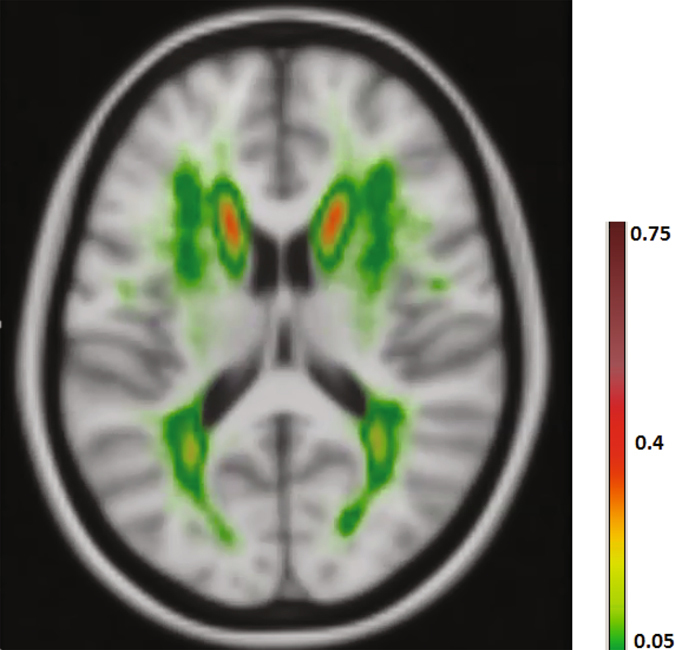
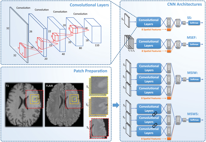
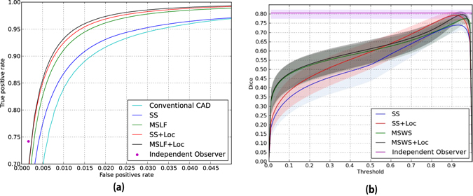
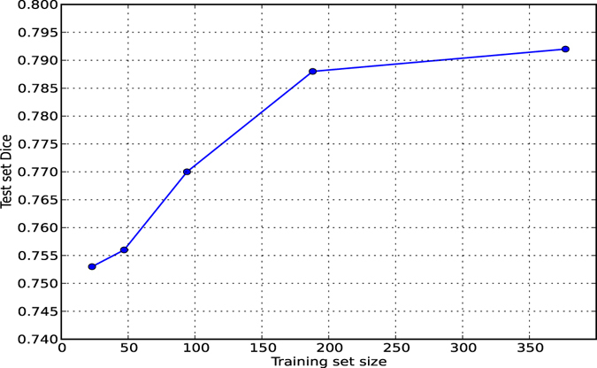
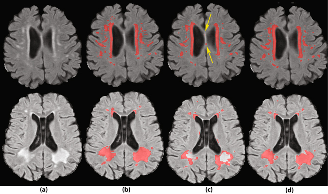
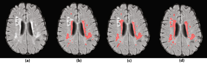
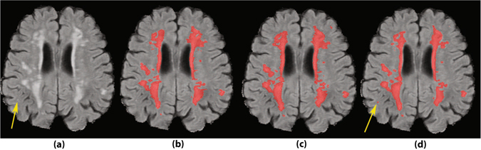
The anatomical location of imaging features is of crucial importance for accurate diagnosis in many medical tasks. Convolutional neural networks (CNN) have had huge successes in computer vision, but they lack the natural ability to incorporate the anatomical location in their decision making process, hindering success in some medical image analysis tasks. In this paper, to integrate the anatomical location information into the network, we propose several deep CNN architectures that consider multi-scale patches or take explicit location features while training. We apply and compare the proposed architectures for segmentation of white matter hyperintensities in brain MR images on a large dataset. As a result, we observe that the CNNs that incorporate location information substantially outperform a conventional segmentation method with handcrafted features as well as CNNs that do not integrate location information. On a test set of 50 scans, the best configuration of our networks obtained a Dice score of 0.792, compared to 0.805 for an independent human observer. Performance levels of the machine and the independent human observer were not statistically significantly different (p-value = 0.06).
Overview
The claims and statistics of this paper haven’t been checked yet.
Run the check — free, no account needed. It reads the open-access text and checks the sources it cites.
Integrity signals
Registry
Cites
Statistics
Figures
Verification
Claims the paper makes
The claims and statistics of this paper haven’t been checked yet.
Run the check — free, no account needed. It reads the open-access text and checks the sources it cites.
Claims other papers make about this one
Each is checked against this paper’s own text: does it say what the citing paper says it does?
In the citing paper
“Noteworthy, the improved segmentation performance is mainly due to the contribution of handcrafted features rather than the multi-scale paradigm.”
From Embracing imperfect datasets: A review of deep learning solutions for medical image segmentation (2020) · cites this paper as Ghafoorian et al. (2017)
In the citing paper
“On a test set of 46 cases, their multi-scale architecture equipped with the hand-crafted features achieves 6% increase in Dice over their single scale baseline that does not incorporate the handcrafted features.”
From Embracing imperfect datasets: A review of deep learning solutions for medical image segmentation (2020) · cites this paper as Ghafoorian et al. (2017)
Self-check
Percentages, sums and reported differences re-computed from the paper’s own numbers.
The self-check needs the paper’s full text.
Citations
Cites
ImageNet classification with deep convolutional neural networks
Krizhevsky, A., Sutskever, I., & Hinton, G. E. (2017) · Communications of the ACM · doi:10.1145/3065386 · Cited by 112,155
U-Net: Convolutional Networks for Biomedical Image Segmentation
Ronneberger, O., Fischer, P., & Brox, T. (2015) · Lecture notes in computer science · doi:10.1007/978-3-319-24574-4_28 · Cited by 99,404
LeCun, Y., Bengio, Y., & Hinton, G. (2015) · Nature · doi:10.1038/nature14539 · Cited by 84,126
ImageNet: A large-scale hierarchical image database
Deng, J., Dong, W., Socher, R., Li, L.-J., Li, K., & Fei-Fei, L. (2009) · 2009 IEEE Conference on Computer Vision and Pattern Recognition · doi:10.1109/cvpr.2009.5206848 · Cited by 65,333
Gradient-based learning applied to document recognition
Lecun, Y., Bottou, L., Bengio, Y., & Haffner, P. (1998) · Proceedings of the IEEE · doi:10.1109/5.726791 · Cited by 58,805
Fully convolutional networks for semantic segmentation
Long, J., Shelhamer, E., & Darrell, T. (2015) · 2015 IEEE Conference on Computer Vision and Pattern Recognition (CVPR) · doi:10.1109/cvpr.2015.7298965 · Cited by 38,834
(2014) · Cited by 33,419
Advances in neural information processing systems 7
Cios, K. J., & Shields, M. E. (1997) · Neurocomputing · doi:10.1016/s0925-2312(97)90018-7 · Cited by 22,093
Delving Deep into Rectifiers: Surpassing Human-Level Performance on ImageNet Classification
He, K., Zhang, X., Ren, S., & Sun, J. (2015) · 2015 IEEE International Conference on Computer Vision (ICCV) · doi:10.1109/iccv.2015.123 · Cited by 19,741
Deep learning in neural networks: an overview
Schmidhuber, J. (2015) · Neural Networks · doi:10.1016/j.neunet.2014.09.003 · Cited by 18,570
A survey on deep learning in medical image analysis
Litjens, G., Kooi, T., Bejnordi, B. E., Setio, A. A. A., Ciompi, F., Ghafoorian, M., van der Laak, J. A. W. M., van Ginneken, B., & Sánchez, C. I. (2017) · Medical Image Analysis · doi:10.1016/j.media.2017.07.005 · Cited by 15,332
Self-citation(2010) · Cited by 14,134
Receptive fields, binocular interaction and functional architecture in the cat’s visual cortex
Hubel, D. H., & Wiesel, T. N. (1962) · The Journal of Physiology · doi:10.1113/jphysiol.1962.sp006837 · Cited by 13,680
Fully convolutional networks for semantic segmentation
Shelhamer, E., Long, J., & Darrell, T. (2017) · IEEE Transactions on Pattern Analysis and Machine Intelligence · doi:10.1109/tpami.2016.2572683 · Cited by 11,898
Jenkinson, M., Beckmann, C. F., Behrens, T. E. J., Woolrich, M. W., & Smith, S. M. (2012) · NeuroImage · doi:10.1016/j.neuroimage.2011.09.015 · Cited by 11,314
Fast robust automated brain extraction
Smith, S. M. (2002) · Human Brain Mapping · doi:10.1002/hbm.10062 · Cited by 10,496
Diagnostic criteria for multiple sclerosis: 2010 revisions to the McDonald criteria
Polman, C. H., Reingold, S. C., Banwell, B., Clanet, M., Cohen, J. A., Filippi, M., Fujihara, K., Havrdova, E., Hutchinson, M., Kappos, L., et al. (2011) · Annals of Neurology · doi:10.1002/ana.22366 · Cited by 9,802
Zhang, Y., Brady, M., & Smith, S. M. (2001) · IEEE Transactions on Medical Imaging · doi:10.1109/42.906424 · Cited by 7,628
A global optimisation method for robust affine registration of brain images
Jenkinson, M., & Smith, S. M. (2001) · Medical Image Analysis · doi:10.1016/s1361-8415(01)00036-6 · Cited by 7,018
DeepFace: Closing the Gap to Human-Level Performance in Face Verification
Taigman, Y., Yang, M., Ranzato, M., & Wolf, L. (2014) · 2014 IEEE Conference on Computer Vision and Pattern Recognition · doi:10.1109/cvpr.2014.220 · Cited by 6,542
Large-Scale Machine Learning with Stochastic Gradient Descent
Bottou, L. (2010) · Proceedings of COMPSTAT'2010 · doi:10.1007/978-3-7908-2604-3_16 · Cited by 5,795
Fukushima, K. (1980) · Biological Cybernetics · doi:10.1007/bf00344251 · Cited by 5,759
Wardlaw, J. M., Smith, E. E., Biessels, G. J., Cordonnier, C., Fazekas, F., Frayne, R., Lindley, R. I., O'Brien, J. T., Barkhof, F., Benavente, O. R., et al. (2013) · The Lancet Neurology · doi:10.1016/s1474-4422(13)70124-8 · Cited by 5,544
Diagnostic criteria for multiple sclerosis: 2005 revisions to the “McDonald Criteria”
Polman, C. H., Reingold, S. C., Edan, G., Filippi, M., Hartung, H., Kappos, L., Lublin, F. D., Metz, L. M., McFarland, H. F., O'Connor, P. W., et al. (2005) · Annals of Neurology · doi:10.1002/ana.20703 · Cited by 5,017
Efficient multi-scale 3D CNN with fully connected CRF for accurate brain lesion segmentation
Kamnitsas, K., Ledig, C., Newcombe, V. F. J., Simpson, J. P., Kane, A. D., Menon, D. K., Rueckert, D., & Glocker, B. (2017) · Medical Image Analysis · doi:10.1016/j.media.2016.10.004 · Cited by 3,498
Brain tumor segmentation with Deep Neural Networks
Havaei, M., Davy, A., Warde-Farley, D., Biard, A., Courville, A., Bengio, Y., Pal, C., Jodoin, P.-M., & Larochelle, H. (2017) · Medical Image Analysis · doi:10.1016/j.media.2016.05.004 · Cited by 3,278
Brain Tumor Segmentation Using Convolutional Neural Networks in MRI Images
Pereira, S., Pinto, A., Alves, V., & Silva, C. A. (2016) · IEEE Transactions on Medical Imaging · doi:10.1109/tmi.2016.2538465 · Cited by 2,735
Learning Hierarchical Features for Scene Labeling
Farabet, C., Couprie, C., Najman, L., & LeCun, Y. (2013) · IEEE Transactions on Pattern Analysis and Machine Intelligence · doi:10.1109/tpami.2012.231 · Cited by 2,620
Stochastic Gradient Descent Tricks
Bottou, L. (2012) · Lecture notes in computer science · doi:10.1007/978-3-642-35289-8_25 · Cited by 2,168
Greenspan, H. K., van Ginneken, B., & Summers, R. M. (2016) · IEEE Transactions on Medical Imaging · doi:10.1109/tmi.2016.2553401 · Cited by 1,725
Evaluation of Pooling Operations in Convolutional Architectures for Object Recognition
Scherer, D., Müller, A., & Behnke, S. (2010) · Lecture notes in computer science · doi:10.1007/978-3-642-15825-4_10 · Cited by 1,603
Learning Rich Features from RGB-D Images for Object Detection and Segmentation
Gupta, S., Girshick, R., Arbeláez, P., & Malik, J. (2014) · Lecture Notes in Computer Science · doi:10.1007/978-3-319-10584-0_23 · Cited by 1,528
de Leeuw, F.-E. (2001) · Journal of Neurology, Neurosurgery & Psychiatry · doi:10.1136/jnnp.70.1.9 · Cited by 1,291
(2012) · neural information processing systems · Cited by 1,291
An automated tool for detection of FLAIR-hyperintense white-matter lesions in Multiple Sclerosis
Schmidt, P. J., Christian Gaser, B., Arsić, M., Buck, D., Förschler, A., Berthele, A., Hoshi, M., Ilg, R., Schmid, V. J., Zimmer, C., et al. (2011) · NeuroImage · doi:10.1016/j.neuroimage.2011.11.032 · Cited by 1,219
Simultaneous Detection and Segmentation
Hariharan, B., Arbeláez, P., Girshick, R., & Malik, J. (2014) · Lecture notes in computer science · doi:10.1007/978-3-319-10584-0_20 · Cited by 1,189
U-Net: Convolutional Networks for Biomedical Image SegmentationPreprint
Ronneberger, O., Fischer, P., & Brox, T. (2015) · arXiv (Cornell University) · doi:10.48550/arxiv.1505.04597 · Cited by 1,079
Multi-column deep neural network for traffic sign classification
Cireşan, D., Meier, U., Masci, J., & Schmidhuber, J. (2012) · Neural Networks · doi:10.1016/j.neunet.2012.02.023 · Cited by 1,057
Delving Deep into Rectifiers: Surpassing Human-Level Performance on ImageNet ClassificationPreprint
He, K., Zhang, X., Ren, S., et al. (2015) · arXiv (Cornell University) · doi:10.48550/arxiv.1502.01852 · Cited by 1,000
Large scale deep learning for computer aided detection of mammographic lesions
Kooi, T., Litjens, G., van Ginneken, B., Gubern-Mérida, A., Sánchez, C. I., Mann, R., den Heeten, A., & Karssemeijer, N. (2017) · Medical Image Analysis · doi:10.1016/j.media.2016.07.007 · Cited by 965
Self-citationAutomatic Segmentation of MR Brain Images With a Convolutional Neural Network
Moeskops, P., Viergever, M. A., Mendrik, A. M., de Vries, L. S., Benders, M. J. N. L., & Isgum, I. (2016) · IEEE Transactions on Medical Imaging · doi:10.1109/tmi.2016.2548501 · Cited by 861
Deep convolutional neural networks for multi-modality isointense infant brain image segmentation
Zhang, W., Li, R., Deng, H., Wang, L., Lin, W., Ji, S., & Shen, D. (2015) · NeuroImage · doi:10.1016/j.neuroimage.2014.12.061 · Cited by 817
VoxResNet: Deep voxelwise residual networks for brain segmentation from 3D MR images
Chen, H., Dou, Q., Yu, L., Qin, J., & Heng, P. (2017) · NeuroImage · doi:10.1016/j.neuroimage.2017.04.041 · Cited by 781
Automatic "pipeline" analysis of 3-D MRI data for clinical trials: application to multiple sclerosis
Zijdenbos, A. P., Forghani, R., & Evans, A. C. (2002) · IEEE Transactions on Medical Imaging · doi:10.1109/tmi.2002.806283 · Cited by 755
Automatic Detection of Cerebral Microbleeds From MR Images via 3D Convolutional Neural Networks
Dou, Q., Chen, H., Yu, L., Zhao, L., Qin, J., Wang, D., Mok, V. C., Shi, L., & Heng, P.-A. (2016) · IEEE Transactions on Medical Imaging · doi:10.1109/tmi.2016.2528129 · Cited by 679
Deep MRI brain extraction: A 3D convolutional neural network for skull stripping
Kleesiek, J., Urban, G., Hubert, A., Schwarz, D., Maier-Hein, K., Bendszus, M., & Biller, A. (2016) · NeuroImage · doi:10.1016/j.neuroimage.2016.01.024 · Cited by 505
Automated segmentation of multiple sclerosis lesions by model outlier detection
Leemput, K. V., Maes, F., Vandermeulen, D., Colchester, A. C. F., & Suetens, P. (2001) · IEEE Transactions on Medical Imaging · doi:10.1109/42.938237 · Cited by 487
Brosch, T., Tang, L. Y. W., Yoo, Y., Li, D. K. B., Traboulsee, A., & Tam, R. (2016) · IEEE Transactions on Medical Imaging · doi:10.1109/tmi.2016.2528821 · Cited by 460
White matter hyperintensities in late life depression: a systematic review
Herrmann, L. L., Le Masurier, M., & Ebmeier, K. P. (2007) · Journal of Neurology, Neurosurgery & Psychiatry · doi:10.1136/jnnp.2007.124651 · Cited by 456
Hough-CNN: Deep learning for segmentation of deep brain regions in MRI and ultrasound
Milletarì, F., Ahmadi, S., Kroll, C., Plate, A., Rozanski, V. E., Maiostre, J., Levin, J., Dietrich, O., Ertl-Wagner, B. B., Bötzel, K., et al. (2017) · Computer Vision and Image Understanding · doi:10.1016/j.cviu.2017.04.002 · Cited by 406
A Four-Dimensional Probabilistic Atlas of the Human Brain
Mazziotta, J. C., Toga, A. W., Evans, A. C., Fox, P. T., Lancaster, J. L., Zilles, K., Woods, R. P., Paus, T., Simpson, G., Pike, G. B., et al. (2001) · Journal of the American Medical Informatics Association · doi:10.1136/jamia.2001.0080401 · Cited by 384
Au, R., Massaro, J. M., Wolf, P. A., Young, M. E., Beiser, A., Seshadri, S., D’Agostino, R. B., & DeCarli, C. (2006) · Archives of Neurology · doi:10.1001/archneur.63.2.246 · Cited by 381
García-Lorenzo, D., Francis, S., Narayanan, S., Arnold, D. L., & Collins, D. L. (2012) · Medical Image Analysis · doi:10.1016/j.media.2012.09.004 · Cited by 364
A topology-preserving approach to the segmentation of brain images with multiple sclerosis lesions
Shiee, N., Bazin, P., Ozturk, A., Reich, D., Calabresi, P., & Pham, D. (2009) · NeuroImage · doi:10.1016/j.neuroimage.2009.09.005 · Cited by 355
HeMIS: Hetero-Modal Image Segmentation
Havaei, M., Guizard, N., Chapados, N., & Bengio, Y. (2016) · Lecture Notes in Computer Science · doi:10.1007/978-3-319-46723-8_54 · Cited by 332
White matter lesion extension to automatic brain tissue segmentation on MRI
de Boer, R., Vrooman, H. A., van der Lijn, F., Vernooij, M. W., Ikram, M. A., van der Lugt, A., Breteler, M. M. B., & Niessen, W. J. (2009) · NeuroImage · doi:10.1016/j.neuroimage.2009.01.011 · Cited by 329
Probabilistic segmentation of white matter lesions in MR imaging
Anbeek, P., Vincken, K. L., van Osch, M. J. P., Bisschops, R. H. C., & van der Grond, J. (2004) · NeuroImage · doi:10.1016/j.neuroimage.2003.10.012 · Cited by 323
van Straaten, E. C. W., Fazekas, F., Rostrup, E., Scheltens, P., Schmidt, R., Pantoni, L., Inzitari, D., Waldemar, G., Erkinjuntti, T., Mäntylä, R., et al. (2006) · Stroke · doi:10.1161/01.str.0000202585.26325.74 · Cited by 314
Fully convolutional networks for multi-modality isointense infant brain image segmentation
Nie, D., Wang, L., Gao, Y., & Shen, D. (2016) · 2016 IEEE 13th International Symposium on Biomedical Imaging (ISBI) · doi:10.1109/isbi.2016.7493515 · Cited by 304
A prospective study of cerebral white matter abnormalities in older people with gait dysfunction
Whitman, G. T., Tang, T., Lin, A., & Baloh, R. W. (2001) · Neurology · doi:10.1212/wnl.57.6.990 · Cited by 298
Computer-Assisted Segmentation of White Matter Lesions in 3D MR Images Using Support Vector Machine
Lao, Z., Shen, D. G., Liu, D., Jawad, A. F., Melhem, E. R., Launer, L. J., Bryan, R. N., & Davatzikos, C. A. (2008) · Academic Radiology · doi:10.1016/j.acra.2007.10.012 · Cited by 242
Spatial decision forests for MS lesion segmentation in multi-channel magnetic resonance images
Geremia, E., Olivier Clatz, A., Menze, B. H., Ender Konukoglu, A., Criminisi, A., & Ayache, N. (2011) · NeuroImage · doi:10.1016/j.neuroimage.2011.03.080 · Cited by 232
Fully automatic segmentation of white matter hyperintensities in MR images of the elderly
Behloul, F., van den Heuvel, D. M. J., Olofsen, H., van Osch, M. J. P., van der Grond, J., van Buchem, M. A., & Reiber, J. H. C. (2005) · NeuroImage · doi:10.1016/j.neuroimage.2005.06.061 · Cited by 231
Automatic segmentation and volumetry of multiple sclerosis brain lesions from MR images
Jain, S., Sima, D. M., Ribbens, A., Cambron, M., Maertens, A., Hecke, W. V., Mey, J. D., Barkhof, F., Steenwijk, M. D., Daams, M., et al. (2015) · NeuroImage Clinical · doi:10.1016/j.nicl.2015.05.003 · Cited by 220
Multi-Column Deep Neural Networks for offline handwritten Chinese character classification
Ciresan, D. C., & Meier, U. (2015) · International Joint Conference on Neural Networks (IJCNN) · doi:10.1109/ijcnn.2015.7280516 · Cited by 214
Model-based 3-D segmentation of multiple sclerosis lesions in magnetic resonance brain images
Kamber, M., Shinghal, R., Collins, D. L., Francis, G., & Evans, A. C. (1995) · IEEE Transactions on Medical Imaging · doi:10.1109/42.414608 · Cited by 211
Grimaud, J., Lai, M., Thorpe, J. W., Adeleine, P., Wang, L., Barker, G. J., Plummer, D. L., Tofts, P. S., McDonald, W. I., & Miller, D. (1996) · Magnetic Resonance Imaging · doi:10.1016/0730-725x(96)00018-5 · Cited by 204
van Norden, A. G., de Laat, K. F., Gons, R. A. R., van Uden, I. W., van Dijk, E. J., van Oudheusden, L. J. B., Esselink, R. A. J., Bloem, B. R., van Engelen, B. G. M., Zwarts, M. J., et al. (2011) · BMC Neurology · doi:10.1186/1471-2377-11-29 · Cited by 193
Firbank, M., Wiseman, R., Burton, E. J., Saxby, B. K., O'Brien, J. T., & Ford, G. A. (2007) · Journal of Neurology · doi:10.1007/s00415-006-0238-4 · Cited by 189
Impact of White Matter Changes on Clinical Manifestation of Alzheimer’s Disease
Hirono, N., Kitagaki, H., Kazui, H., Hashimoto, M., & Mori, E. (2000) · Stroke · doi:10.1161/01.str.31.9.2182 · Cited by 186
(2015) · arXiv (Cornell University) · Cited by 182
Equilibrated adaptive learning rates for non-convex optimizationPreprint
Dauphin, Y., de Vries, H., & Bengio, Y. (2015) · arXiv (Cornell University) · doi:10.48550/arxiv.1502.04390 · Cited by 172
Caligiuri, M. E., Perrotta, P., Augimeri, A., Rocca, F., Quattrone, A., & Cherubini, A. (2015) · Neuroinformatics · doi:10.1007/s12021-015-9260-y · Cited by 151
Multiscale CNNs for Brain Tumor Segmentation and Diagnosis
Zhao, L., & Jia, K. (2016) · Computational and Mathematical Methods in Medicine · doi:10.1155/2016/8356294 · Cited by 151
Ghafoorian, M., Karssemeijer, N., Heskes, T., Bergkamp, M. I., Wissink, J., Obels, J., Keizer, K., de Leeuw, F., van Ginneken, B., Marchiori, E., et al. (2017) · NeuroImage Clinical · doi:10.1016/j.nicl.2017.01.033 · Cited by 151
Self-citationWhite matter volumes and periventricular white matter hyperintensities in aging and dementia
Smith, C. D., Snowdon, D. A., Wang, H., & Markesbery, W. R. (2000) · Neurology · doi:10.1212/wnl.54.4.838 · Cited by 137
Deep Convolutional Encoder Networks for Multiple Sclerosis Lesion Segmentation
Brosch, T., Yoo, Y., Tang, L. Y. W., Li, D. K. B., Traboulsee, A. L., & Tam, R. C. (2015) · Lecture notes in computer science · doi:10.1007/978-3-319-24574-4_1 · Cited by 127
Sub-cortical brain structure segmentation using F-CNN'S
Shakeri, M., Tsogkas, S., Ferrante, E., Lippé, S., Kadoury, S., Nikos Paragios, A., & Kokkinos, I. (2016) · IEEE International Symposium on Biomedical Imaging (ISBI) · doi:10.1109/isbi.2016.7493261 · Cited by 108
Schoonheim, M. M., Vigeveno, R. M., Lopes, F. C. R., Pouwels, P. J. W., Polman, C. H., Barkhof, F., & Geurts, J. J. G. (2013) · Human Brain Mapping · doi:10.1002/hbm.22332 · Cited by 92
Ithapu, V. K., Singh, V., Lindner, C., Austin, B. P., Chris Hinrichs, A., Carlsson, C. M., Bendlin, B. B., & Johnson, S. C. (2014) · Human Brain Mapping · doi:10.1002/hbm.22472 · Cited by 83
Ghafoorian, M., Karssemeijer, N., Heskes, T., van Uder, I. W. M., de Leeuw, F., Marchiori, E., van Ginneken, B., & Platel, B. (2016) · IEEE International Symposium on Biomedical Imaging (ISBI) · doi:10.1109/isbi.2016.7493532 · Cited by 75
Self-citationAutomated detection of white matter hyperintensities of all sizes in cerebral small vessel disease
Ghafoorian, M., Karssemeijer, N., van Uden, I. W. M., de Leeuw, F., Heskes, T., Marchiori, E., & Platel, B. (2016) · Medical Physics · doi:10.1118/1.4966029 · Cited by 75
Self-citationShape and Volume of Lacunar Infarcts
Hervé, D., Mangin, J., Molko, N., Bousser, M., & Chabriat, H. (2005) · Stroke · doi:10.1161/01.str.0000185678.26296.38 · Cited by 68
Robust White Matter Lesion Segmentation in FLAIR MRI
Khademi, A., Venetsanopoulos, A., & Moody, A. R. (2011) · IEEE Transactions on Biomedical Engineering · doi:10.1109/tbme.2011.2181167 · Cited by 57
A comparison of different automated methods for the detection of white matter lesions in MRI data
Klöppel, S., Abdulkadir, A., Hadjidemetriou, S., Issleib, S., Frings, L., Nguyen, T. T., Mader, I., Teipel, S. J., Hüll, M., & Ronneberger, O. (2011) · NeuroImage · doi:10.1016/j.neuroimage.2011.04.053 · Cited by 56
Brain Imaging and Cognitive Predictors of Stroke and Alzheimer Disease in the Framingham Heart Study
Weinstein, G., Beiser, A., DeCarli, C. S., Au, R., Wolf, P. A., & Seshadri, S. S. (2013) · Stroke · doi:10.1161/strokeaha.113.000947 · Cited by 56
Segmentation of age-related white matter changes in a clinical multi-center study
Dyrby, T. B., Rostrup, E., Baaré, W. F. C., van Straaten, E. C. W., Barkhof, F., Vrenken, H., Ropele, S., Schmidt, R., Erkinjuntti, T., Wahlund, L., et al. (2008) · NeuroImage · doi:10.1016/j.neuroimage.2008.02.024 · Cited by 53
White Matter Integrity and Depressive Symptoms in Cerebral Small Vessel Disease: The RUN DMC Study
van Uden, I. W. M., Tuladhar, A. M., de Laat, K. F., van Norden, A. G. W., Norris, D. G., van Dijk, E. J., Tendolkar, I., & de Leeuw, F. (2014) · American Journal of Geriatric Psychiatry · doi:10.1016/j.jagp.2014.07.002 · Cited by 53
Advances in the Medical Sciences
(1939) · Science · doi:10.1126/science.90.2329.8-t · Cited by 53
F-Measure as the Error Function to Train Neural Networks
Pastor-Pellicer, J., Zamora-Martínez, F., España-Boquera, S., & Castro-Bleda, M. J. (2013) · Lecture notes in computer science · doi:10.1007/978-3-642-38679-4_37 · Cited by 50
Automatic segmentation of cerebral white matter hyperintensities using only 3D FLAIR images
Simões, R., Mönninghoff, C., Jokisch, M., Weimar, C., Wanke, I., van Walsum, A. V. C., & Slump, C. H. (2013) · Magnetic Resonance Imaging · doi:10.1016/j.mri.2012.12.004 · Cited by 50
Multi-Column Deep Neural Networks for Offline Handwritten Chinese Character ClassificationPreprint
Cireşan, D., & Schmidhuber, J. (2013) · arXiv (Cornell University) · doi:10.48550/arxiv.1309.0261 · Cited by 46
Shi, L., Wang, D., Liu, S., Pu, Y., Wang, Y., Chu, C. W., Ahuja, A. T., & Wang, Y. (2012) · Journal of Neuroscience Methods · doi:10.1016/j.jneumeth.2012.12.014 · Cited by 44
White matter hyperintensities and cortical acetylcholinesterase activity in parkinsonian dementia
Marshall, G. A., Shchelchkov, E., Kaufer, D., Ivanco, L. S., & Bohnen, N. I. (2005) · Acta Neurologica Scandinavica · doi:10.1111/j.1600-0404.2005.00553.x · Cited by 37
Dou, Q., Chen, H., Yu, L., Shi, L., Wang, D., Mok, V., & Heng, P. (2015) · Annual International Conference of the IEEE Engineering in Medicine and Biology Society (EMBC) · doi:10.1109/embc.2015.7320232 · Cited by 32
Genetic Data Sharing and Privacy
The TRACK TBI Investigators, Sorani, M. D., Yue, J. K., Sharma, S., Manley, G. T., Ferguson, A. R., Cooper, S. R., Dams-O’Connor, K., Gordon, W. A., Lingsma, H. F., et al. (2014) · Neuroinformatics · doi:10.1007/s12021-014-9248-z · Cited by 31
Hough-CNN: Deep Learning for Segmentation of Deep Brain Regions in MRI and UltrasoundPreprint
Milletarì, F., Ahmadi, S., Kroll, C., et al. (2016) · arXiv (Cornell University) · doi:10.48550/arxiv.1601.07014 · Cited by 31
Automated Bayesian Segmentation of Microvascular White-Matter Lesions in the ACCORD-MIND Study
Herskovits, E., Bryan, R. N., & Yang, F. (2008) · Advances in Medical Sciences · doi:10.2478/v10039-008-0039-3 · Cited by 22
Small white matter lesion detection in cerebral small vessel disease
Ghafoorian, M., Karssemeijer, N., van Uden, I., de Leeuw, F., Heskes, T., Marchiori, E., & Platel, B. (2015) · Proceedings of SPIE, the International Society for Optical Engineering/Proceedings of SPIE · doi:10.1117/12.2081597 · Cited by 10
Self-citationSub-cortical brain structure segmentation using F-CNN's
Shakeri, M., Tsogkas, S., Ferrante, E., et al. (2016) · HAL (Le Centre pour la Communication Scientifique Directe) · doi:10.48550/arxiv.1602.02130 · Cited by 4
Solano (2000) · doi:10.1002/1531-8249(200002)47:2 · Cited by 3
HeMIS: Hetero-Modal Image SegmentationPreprint
Havaei, M., Guizard, N., Chapados, N., et al. (2016) · arXiv (Cornell University) · doi:10.48550/arxiv.1607.05194 · Cited by 2
Vijverberg, K., Ghafoorian, M., van Uden, I. W. M., de Leeuw, F., Platel, B., & Heskes, T. (2016) · Proceedings of SPIE, the International Society for Optical Engineering/Proceedings of SPIE · doi:10.1117/12.2216409 · Cited by 1
Self-citation
Research works printed but not linked: 3
- Ghafoorian, M. et al. Transfer learning for domain adaptation in mri: Application in brain lesion segmentation. arXiv preprint arXiv:1702.07841 (2017). · arXiv
- Chen, H., Dou, Q., Yu, L. & Heng, P.-A. Voxresnet: Deep voxelwise residual networks for volumetric brain segmentation. arXiv preprint arXiv:1608.05895 (2016). · arXiv
- Maas, A. L., Hannun, A. Y. & Ng, A. Y. Rectifier nonlinearities improve neural network acoustic models. In Proc. ICML, vol. 30 (2013).
Cited by
Sorted by citations
A survey on deep learning in medical image analysis
Litjens, G., Kooi, T., Bejnordi, B. E., Setio, A. A. A., Ciompi, F., Ghafoorian, M., van der Laak, J. A. W. M., van Ginneken, B., & Sánchez, C. I. (2017) · Medical Image Analysis · doi:10.1016/j.media.2017.07.005 · Cited by 15,332
Self-citationArtificial intelligence in radiology
Hosny, A., Parmar, C., Quackenbush, J., Schwartz, L. H., & Aerts, H. J. W. L. (2018) · Nature Reviews Cancer · doi:10.1038/s41568-018-0016-5 · Cited by 3,803
Embracing imperfect datasets: A review of deep learning solutions for medical image segmentation
Tajbakhsh, N., Jeyaseelan, L., Li, Q., Chiang, J. N., Wu, Z., & Ding, X. (2020) · Medical Image Analysis · doi:10.1016/j.media.2020.101693 · Cited by 1,095
“3D model with a 3D representation augmented with handcrafted features Ghafoorian et al. (2017). We explain these methods in more detail as follows.”
MethodInfluentialDeep learning in medical imaging and radiation therapy
Sahiner, B., Pezeshk, A. X., Hadjiiski, L. M., Wang, X., Drukker, K., Cha, K. H., Summers, R. M., & Giger, M. L. (2018) · Medical Physics · doi:10.1002/mp.13264 · Cited by 763
Deep learning for lung cancer prognostication: A retrospective multi-cohort radiomics study
Hosny, A., Parmar, C., Coroller, T. P., Grossmann, P., Zeleznik, R., Kumar, A., Bussink, J., Gillies, R. J., Mak, R. H., & Aerts, H. J. W. L. (2018) · PLOS Medicine · doi:10.1371/journal.pmed.1002711 · Cited by 579
Metadata
- Authors
- Ghafoorian, M., Karssemeijer, N., Heskes, T., van Uden, I. W. M., Sanchez, C. I., Litjens, G., de Leeuw, F.-E., van Ginneken, B., Marchiori, E., & Platel, B.
- Affiliations
- Radboud University Nijmegen (NL)
- Radboud University Medical Center (NL)
- Corresponding author
- Ghafoorian, Mohsen
- Venue
- Scientific Reports
- Published
- 2017-07-11
- Volume
- 7
- Issue
- 1
- Pages
- 5110-5110
- ISSN
- 2045-2322 (ISSN-L)
- PMC
- PMC5505987
- OpenAlex
- W2532750509
- Cited by
- 236
- References
- 98
- Citations by year
- 2026: 7 · 2025: 18 · 2024: 18 · 2023: 18 · 2022: 30 · 2021: 41 · 2020: 25 · 2019: 32 · 2018: 33 · 2017: 12
- FWCI
- 11.58
- Type
- journal article
- Language
- en
- Open access
- gold
- doi.org
- Locations
- Scientific Reports (publishedVersion, cc-by)
- PubMed Central (publishedVersion, cc-by)
- PubMed (publishedVersion)
- DOAJ (DOAJ: Directory of Open Access Journals) (submittedVersion, cc-by-sa)
- Radboud Repository (Radboud University) (submittedVersion)
- Radboud Repository (Radboud University) (submittedVersion, other-oa)
- Funders
- Nederlandse Organisatie voor Wetenschappelijk Onderzoek (016‐126‐351)
- Topics
- Physical Sciences › Computer Science › Computer Vision and Pattern Recognition › Medical Image Segmentation Techniques
- Physical Sciences › Computer Science › Computer Vision and Pattern Recognition › Advanced Neural Network Applications
- Physical Sciences › Computer Science › Computer Vision and Pattern Recognition › Brain Tumor Detection and Classification
- Keywords
- white matter hyperintensities, segmentation, brain MRI, deep convolutional neural networks, medical image analysis
- MeSH terms
- Aged, Aged, 80 and over, Decision Making, Female, Humans, Image Processing, Computer-Assisted, Magnetic Resonance Imaging, Male, Middle Aged, Neural Networks, Computer, Neuroimaging, White Matter
About these checks
Registry, retraction, Cites and statistics results are computed, not judged, with one exception: a printed reference that no index lists is read once by a model before it counts as a citation error. Claims are extracted by a model and kept only when their quote appears verbatim in the text; each cited source is then checked, with a model’s help, for whether it says what the claim relies on. Those verdicts can be wrong — read the claim’s quote and the cited source before relying on one.
Statistics and arithmetic are recomputed, not judged.
Confidence bands are shown only for fields calibrated on our annotated test set. None is calibrated yet, so no band is shown.
What counts as a claim ›How we measure confidence ›How we check this ›
Are you an author, or do you see a problem with this page? Request a review
