

The DOI matches its Crossref record; 37 of the 37 references it cites were found in a registry or an index.
QuPath: Open source software for digital pathology image analysis
Open accessPublished version of a preprint
Abstract
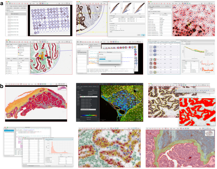
QuPath is new bioimage analysis software designed to meet the growing need for a user-friendly, extensible, open-source solution for digital pathology and whole slide image analysis. In addition to offering a comprehensive panel of tumor identification and high-throughput biomarker evaluation tools, QuPath provides researchers with powerful batch-processing and scripting functionality, and an extensible platform with which to develop and share new algorithms to analyze complex tissue images. Furthermore, QuPath's flexible design makes it suitable for a wide range of additional image analysis applications across biomedical research.
Overview
The claims and statistics of this paper haven’t been checked yet.
Run the check — free, no account needed. It reads the open-access text and checks the sources it cites.
Integrity signals
Registry
Cites
Statistics
Figures
Verification
Claims the paper makes
The claims and statistics of this paper haven’t been checked yet.
Run the check — free, no account needed. It reads the open-access text and checks the sources it cites.
Claims other papers make about this one
Each is checked against this paper’s own text: does it say what the citing paper says it does?
The source backs part of the claim, not all of it — or backs it for a different population, condition or comparison.
In the citing paper
“On the software side, frameworks such as TensorFlow and PyTorch support AI model development, while tools like QuPath enable image annotation and analysis [44].”
From Artificial Intelligence in Dermatopathology: An Update and Review of the Current Literature (2026) · cites this paper as [44]
Experimental — accuracy not yet measuredThe cited source says what the claim attributes to it.
In the citing paper
“Emerging solutions include standardized open-source tools (e.g., QuPath) [44], collaborative multi-institutional annotation efforts, and AI-assisted annotation workflows that reduce manual burden [45].”
From Artificial Intelligence in Dermatopathology: An Update and Review of the Current Literature (2026) · cites this paper as [44]
The cited source says what the claim attributes to it.
In the citing paper
“The microglial ultrastructural analyses was performed using QuPath (v.0.4.4) [71] on images blinded to both animal and group, following an adaptation from our previous work [56, 64].”
From Sex chromosomes and sex hormones differently shape microglial properties during normal physiological conditions in the adult mouse hippocampus (2025) · cites this paper as [71]
Self-check
Percentages, sums and reported differences re-computed from the paper’s own numbers.
The self-check needs the paper’s full text.
Citations
Cites
Breiman, L. (2001) · Machine Learning · doi:10.1023/a:1010933404324 · Cited by 133,538
Fiji: an open-source platform for biological-image analysis
Schindelin, J., Arganda-Carreras, I., Frise, E., Kaynig, V., Longair, M., Pietzsch, T., Preibisch, S., Rueden, C., Saalfeld, S., Schmid, B., et al. (2012) · Nature Methods · doi:10.1038/nmeth.2019 · Cited by 66,890
NIH Image to ImageJ: 25 years of image analysis
Schneider, C. A., Rasband, W. S., & Eliceiri, K. W. (2012) · Nature Methods · doi:10.1038/nmeth.2089 · Cited by 64,638
PD-1 and PD-L1 Immune Checkpoint Blockade to Treat Breast Cancer
Hartkopf, A. D., Taran, F., Wallwiener, M., Walter, C. B., Krämer, B., Grischke, E., & Brucker, S. Y. (2016) · Breast Care · doi:10.1159/000453569 · Cited by 30,989
Textural Features for Image Classification
Haralick, R. M., Shanmugam, K., & Dinstein, I. (1973) · IEEE Transactions on Systems, Man, and Cybernetics · doi:10.1109/tsmc.1973.4309314 · Cited by 22,886
Adverse Renal Effects of Immune Checkpoint Inhibitors: A Narrative Review
Wanchoo, R., Karam, S., Uppal, N. N., Barta, V. S., Deray, G., Devoe, C., Launay‐Vacher, V., Jhaveri, K. D., & on behalf of Cancer and Kidney International Network Workgroup on Immune Checkpoint Inhibitors (2017) · American Journal of Nephrology · doi:10.1159/000455014 · Cited by 15,320
SLIC Superpixels Compared to State-of-the-Art Superpixel Methods
Achanta, R., Shaji, A., Smith, K., Lucchi, A., Fua, P., & Susstrunk, S. E. (2012) · IEEE Transactions on Pattern Analysis and Machine Intelligence · doi:10.1109/tpami.2012.120 · Cited by 9,451
Type, Density, and Location of Immune Cells Within Human Colorectal Tumors Predict Clinical Outcome
Galon, J., Costes, A., Sanchez-Cabo, F., Kirilovsky, A., Mlecnik, B., Lagorce-Pagès, C., Tosolini, M., Camus, M., Berger, A., Wind, P., et al. (2006) · Science · doi:10.1126/science.1129139 · Cited by 6,516
Pembrolizumab for the Treatment of Non–Small-Cell Lung Cancer
Garon, E. B., Rizvi, N. A., Hui, R., Leighl, N., Balmanoukian, A. S., Eder, J. P., Patnaik, A., Aggarwal, C., Gubens, M., Horn, L., et al. (2015) · New England Journal of Medicine · doi:10.1056/nejmoa1501824 · Cited by 6,198
Quantification of histochemical staining by color deconvolution.
Ruifrok, A. C. C., & Johnston, D. A. (2001) · PubMed · Cited by 2,029
Cited by
Sorted by citations
The human tumor microbiome is composed of tumor type–specific intracellular bacteria
Nejman, D., Livyatan, I., Fuks, G., Gavert, N., Zwang, Y., Geller, L. T., Rotter-Maskowitz, A., Weiser, R., Mallel, G., Gigi, E., et al. (2020) · Science · doi:10.1126/science.aay9189 · Cited by 2,464
CellProfiler 4: improvements in speed, utility and usability
Stirling, D. R., Swain-Bowden, M. J., Lucas, A. M., Carpenter, A. E., Cimini, B. A., & Goodman, A. (2021) · BMC Bioinformatics · doi:10.1186/s12859-021-04344-9 · Cited by 2,109
Resolving the fibrotic niche of human liver cirrhosis at single-cell level
Ramachandran, P., Dobie, R., Wilson-Kanamori, J. R., Dora, E. F., Henderson, B. E. P., Luu, N. T., Portman, J. R., Matchett, K. P., Brice, M., Marwick, J. A., et al. (2019) · Nature · doi:10.1038/s41586-019-1631-3 · Cited by 1,784
Hover-Net: Simultaneous segmentation and classification of nuclei in multi-tissue histology images
Graham, S., Vu, Q. D., Raza, S. E. A., Azam, A. S., Tsang, Y. W., Kwak, J. T., & Rajpoot, N. (2019) · Medical Image Analysis · doi:10.1016/j.media.2019.101563 · Cited by 1,516
Kather, J. N., Krisam, J., Charoentong, P., Luedde, T., Herpel, E., Weis, C.-A., Gaiser, T., Marx, A., Valous, N. A., Ferber, D., et al. (2019) · PLOS Medicine · doi:10.1371/journal.pmed.1002730 · Cited by 1,082
Metadata
- Authors
- Bankhead, P., Loughrey, M. B., Fernández, J. A., Dombrowski, Y., McArt, D. G., Dunne, P. D., McQuaid, S., Gray, R. T., Murray, L. J., Coleman, H. G., James, J. A., Salto-Tellez, M., & Hamilton, P. W.
- Affiliations
- Queen's University Belfast (GB)
- Corresponding author
- Hamilton, Peter W.
- ORCID
- Venue
- Scientific Reports
- Published
- 2017-12-04
- Volume
- 7
- Issue
- 1
- Pages
- 16878-16878
- ISSN
- 2045-2322 (ISSN-L)
- PMC
- PMC5715110
- OpenAlex
- W2952481429
- Cited by
- 8,908
- References
- 37
- Citations by year
- 2026: 1616 · 2025: 1896 · 2024: 1681 · 2023: 1359 · 2022: 1023 · 2021: 704 · 2020: 396 · 2019: 171 · 2018: 49 · 2017: 4
- FWCI
- 99.74
- Type
- journal article
- Language
- en
- Open access
- gold
- doi.org
- Locations
- Scientific Reports (publishedVersion, cc-by)
- bioRxiv (Cold Spring Harbor Laboratory) (acceptedVersion, cc-by-nc-nd)
- Europe PMC (PubMed Central) (publishedVersion, cc-by)
- PubMed Central (publishedVersion, cc-by)
- PubMed (publishedVersion)
- DOAJ (DOAJ: Directory of Open Access Journals) (submittedVersion)
- Edinburgh Research Explorer (University of Edinburgh) (submittedVersion, cc-by)
- Funders
- Invest Northern Ireland (RDO0712612)
- Friends of the Cancer Centre
- Cancer Research UK (20256, A15333, C37703/A15333, C11512, A20256, C11512/A20256, C50104/A17592, 15333)
- Public Health Agency (SPI/3315/06, EAT/4905/13)
- Health and Social Care Research and Development Division
- Topics
- Physical Sciences › Computer Science › Artificial Intelligence › AI in cancer detection
- Life Sciences › Biochemistry, Genetics and Molecular Biology › Biophysics › Cell Image Analysis Techniques
- Health Sciences › Medicine › Radiology, Nuclear Medicine and Imaging › Radiomics and Machine Learning in Medical Imaging
- Keywords
- QuPath, digital pathology, whole slide image analysis, open source software, tumor detection, biomarker analysis, batch processing, scripting, tissue image analysis
- MeSH terms
- Algorithms, Colonic Neoplasms, Humans, Image Interpretation, Computer-Assisted, Biomarkers, Tumor, User-Computer Interface, Kaplan-Meier Estimate, Programmed Cell Death 1 Ligand 2 Protein
About these checks
Registry, retraction, Cites and statistics results are computed, not judged, with one exception: a printed reference that no index lists is read once by a model before it counts as a citation error. Claims are extracted by a model and kept only when their quote appears verbatim in the text; each cited source is then checked, with a model’s help, for whether it says what the claim relies on. Those verdicts can be wrong — read the claim’s quote and the cited source before relying on one.
Statistics and arithmetic are recomputed, not judged.
Confidence bands are shown only for fields calibrated on our annotated test set. None is calibrated yet, so no band is shown.
What counts as a claim ›How we measure confidence ›How we check this ›
Are you an author, or do you see a problem with this page? Request a review
