

The DOI matches its Crossref record; 1 of the 1 references it cites were found in a registry or an index.
TiO2 nanoparticles induce omphalocele in chicken embryo by disrupting Wnt signaling pathway
Abstract
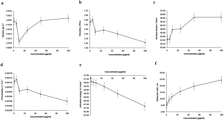
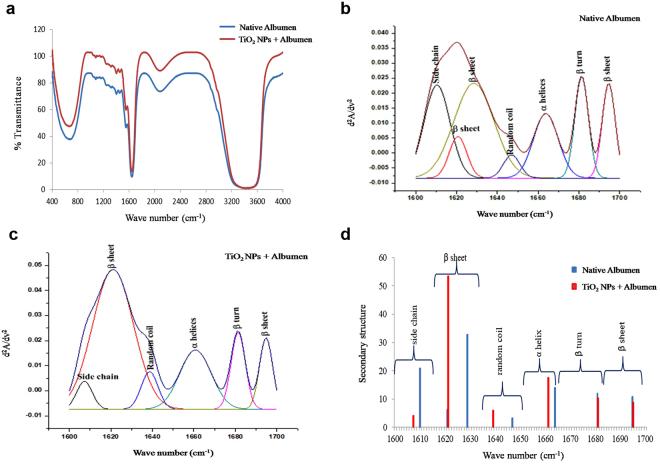
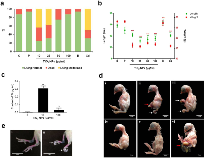
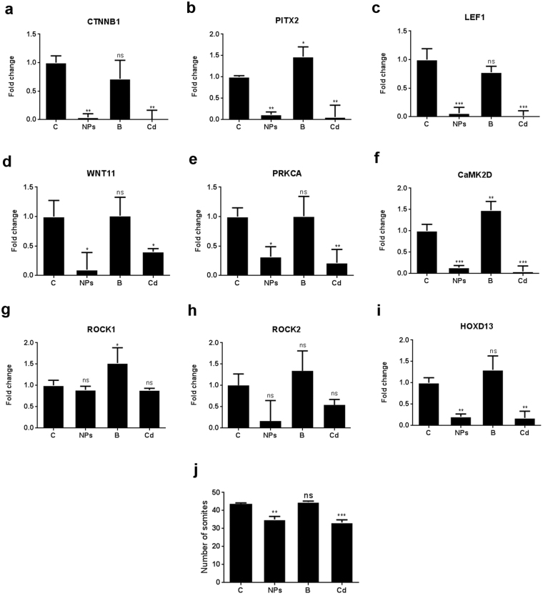
Titanium dioxide nanoparticles (TiO 2 NPs) are among abundantly used metal oxide NPs but their interactions with biomolecules and subsequent embryonic toxicity in higher vertebrates is not extensively reported. Physicochemical interactions of TiO 2 NPs with egg albumen reveals that lower doses of TiO 2 NPs (10 and 25 µg/ml) accounted for higher friccohesity and activation energy but an increment in molecular radii was recorded at higher doses (50 and 100 µg/ml). FTIR analysis revealed conformational changes in secondary structure of egg albumen as a result of electrostratic interactions between egg albumen and TiO 2 NPs. The morphometric data of chicken embryo recorded a reduction at all the doses of TiO 2 NPs, but toxicity and developmental deformity (omphalocele and flexed limbs) were recorded at lower doses only. Inductively coupled plasma optical emission spectrometry (ICP-OES) confirmed presence of Ti in chicken embryos. mRNA levels of genes involved in canonical and non-canonical Wnt signaling were lowered following TiO 2 NPs treatment resulting in free radical mediated disruption of lateral plate mesoderm and somite myogenesis. Conformational changes in egg albumen and subsequent developmental deformity in chicken embryo following TiO 2 NPs treatment warrants detailed studies of NP toxicity at lower doses prior to their biomedical applications.
Overview
The claims and statistics of this paper haven’t been checked yet.
Run the check — free, no account needed. It reads the open-access text and checks the sources it cites.
Integrity signals
Registry
Cites
Statistics
Figures
Verification
Claims the paper makes
The claims and statistics of this paper haven’t been checked yet.
Run the check — free, no account needed. It reads the open-access text and checks the sources it cites.
Claims other papers make about this one
Each is checked against this paper’s own text: does it say what the citing paper says it does?
In the citing paper
“the chicken embryo model has an advantage that the direct effect of NPs on the embryonic development can be evaluated by removing the indirect effect mediated by maternal factors easily, as reported for the results of developmental toxicity of TiO2-NP [185] and carbon NPs [186–188].”
From Particle toxicology and health - where are we? (2019) · cites this paper as [185]
Self-check
Percentages, sums and reported differences re-computed from the paper’s own numbers.
The self-check needs the paper’s full text.
Citations
Cites
Walkey, C. D., & Chan, W. C. W. (2012) · Chem. Soc. Rev. · doi:10.1039/c1cs15233e · Cited by 1,758
Research works printed but not linked: 50
- Oberdörster, G., Oberdörster, E. & Oberdörster, J. Nanotoxicology: an emerging discipline evolving from studies of ultrafine particles. Environmental health perspectives 113, 823 (2005). · DOI · Paper page
- Nel, A., Xia, T., Mädler, L. & Li, N. Toxic potential of materials at the nanolevel. science 311, 622–627 (2006). · DOI · Paper page
- Vance, M. E. et al. Nanotechnology in the real world: Redeveloping the nanomaterial consumer products inventory. Beilstein journal of nanotechnology 6, 1769 (2015). · DOI · Paper page
- Dalai, S., Pakrashi, S., Chandrasekaran, N. & Mukherjee, A. Acute toxicity of TiO2 nanoparticles to Ceriodaphnia dubia under visible light and dark conditions in a freshwater system. PLoS One 8, e62970 (2013). · DOI
- Maurer-Jones, M. A., Gunsolus, I. L., Murphy, C. J. & Haynes, C. L. Toxicity of engineered nanoparticles in the environment. Analytical chemistry 85, 3036–3049 (2013). · DOI
- Shukla, R. K. et al. TiO2 nanoparticles induce oxidative DNA damage and apoptosis in human liver cells. Nanotoxicology 7, 48–60 (2013). · DOI
- Lai, J. C. et al. Exposure to titanium dioxide and other metallic oxide nanoparticles induces cytotoxicity on human neural cells and fibroblasts. International journal of nanomedicine 3, 533 (2008).
- Hussain, S., Hess, K., Gearhart, J., Geiss, K. & Schlager, J. In vitro toxicity of nanoparticles in BRL 3A rat liver cells. Toxicology in vitro 19, 975–983 (2005). · DOI
- Li, Y. et al. Molecular control of TiO2-NPs toxicity formation at predicted environmental relevant concentrations by Mn-SODs proteins. PLoS One 7, e44688 (2012). · DOI
- Hu, C. et al. Toxicological effects of TiO 2 and ZnO nanoparticles in soil on earthworm Eisenia fetida. Soil Biology and Biochemistry 42, 586–591 (2010). · DOI
- Gupta, G. S., Kumar, A., Shanker, R. & Dhawan, A. Assessment of agglomeration, co-sedimentation and trophic transfer of titanium dioxide nanoparticles in a laboratory-scale predator-prey model system. Scientific reports 6, 31422 (2016). · DOI
- Clemente, Z., Castro, V., Moura, M., Jonsson, C. & Fraceto, L. Toxicity assessment of TiO 2 nanoparticles in zebrafish embryos under different exposure conditions. Aquatic toxicology 147, 129–139 (2014). · DOI
- Linhua, H., Zhenyu, W. & Baoshan, X. Effect of sub-acute exposure to TiO2 nanoparticles on oxidative stress and histopathological changes in Juvenile Carp (Cyprinus carpio). Journal of Environmental Sciences 21, 1459–1466 (2009). · DOI
- Zhu, X., Zhou, J. & Cai, Z. TiO2 nanoparticles in the marine environment: Impact on the toxicity of tributyltin to abalone (Haliotis diversicolor supertexta) embryos. Environmental science & technology 45, 3753–3758 (2011). · DOI
- Mohammadipour, A. et al. Maternal exposure to titanium dioxide nanoparticles during pregnancy; impaired memory and decreased hippocampal cell proliferation in rat offspring. Environmental toxicology and pharmacology 37, 617–625 (2014). · DOI
- Wierzbicki, M., Sawosz, E. & Grodzik, M. Caveolin-1 localization in chicken embryo chorioallantoic membrane treated with diamond and graphite nanoparticles. Brzozowski m., głogowski r., grzeszczak-pytlak a. Reproduc-tive efficiency of mink females, selected for weaned litter size after first season of repro-duction 5, 133 (2012).
- Pineda, L., Sawosz, E., Vadalasetty, K. & Chwalibog, A. Effect of copper nanoparticles on metabolic rate and development of chicken embryos. Animal feed science and technology 186, 125–129 (2013). · DOI
- Wierzbicki, M. et al. Carbon nanoparticles downregulate expression of basic fibroblast growth factor in the heart during embryogenesis. International journal of nanomedicine 8, 3427 (2013).
- Prasek, M. et al. Influence of nanoparticles of platinum on chicken embryo development and brain morphology. Nanoscale research letters 8, 251 (2013). · DOI
- Szmidt, M. et al. Toxicity of different forms of graphene in a chicken embryo model. Environmental Science and Pollution Research 23, 19940–19948 (2016). · DOI
- Hotowy, A. et al. Silver nanoparticles administered to chicken affect VEGFA and FGF2 gene expression in breast muscle and heart. Nanoscale research letters 7, 418 (2012). · DOI
- Patel, S. et al. DNA binding and dispersion activities of titanium dioxide nanoparticles with UV/vis spectrophotometry, fluorescence spectroscopy and physicochemical analysis at physiological temperature. Journal of Molecular Liquids 213, 304–311 (2016). · DOI
- Willems, E., Decuypere, E., Buyse, J. & Everaert, N. Importance of albumen during embryonic development in avian species, with emphasis on domestic chicken. World’s poultry science journal 70, 503–518 (2014). · DOI
- Singh, M. Study of apparent molal volume and viscosity of mutual citric acid and disodium hydrogen orthophosphate aqueous systems. Journal of Chemical Sciences 118, 269–274 (2006). · DOI
- Venyaminov, S. Y. & Kalnin, N. Quantitative IR spectrophotometry of peptide compounds in water (H2O) solutions. II. Amide absorption bands of polypeptides and fibrous proteins in α‐, β‐, and random coil conformations. Biopolymers 30, 1259–1271 (1990). · DOI
- Brosnan, J. T. & Brosnan, M. E. Glutamate: a truly functional amino acid. Amino acids 45, 413–418 (2013). · DOI
- Rollerova, E. et al. Titanium dioxide nanoparticles: some aspects of toxicity/focus on the development. Endocrine regulations 49, 97–112 (2015). · DOI
- Doi, T., Puri, P., Bannigan, J. & Thompson, J. Altered PITX2 and LEF1 gene expression in the cadmium-induced omphalocele in the chick model. Pediatric Surgery International 27, 495–499 (2011). · DOI
- Thompson, J. M. & Bannigan, J. G. Omphalocele induction in the chick embryo by administration of cadmium. Journal of pediatric surgery 42, 1703–1709 (2007). · DOI
- Pampfer, S. & Streffer, C. Prenatal death and malformations after irradiation of mouse zygotes with neutrons or X‐rays. Teratology 37, 599–607 (1988). · DOI
- Wangikar, P., Dwivedi, P. & Sinha, N. Effect in rats of simultaneous prenatal exposure to ochratoxin A and aflatoxin B1. I. Maternal toxicity and fetal malformations. Birth defects research Part B: Developmental and reproductive toxicology 71, 343–351 (2004). · DOI
- Doi, T., Puri, P., Bannigan, J. & Thompson, J. Disruption of noncanonical Wnt/CA 2+ pathway in the cadmium-induced omphalocele in the chick model. Journal of pediatric surgery 45, 1645–1649 (2010). · DOI
- Kioussi, C. et al. Identification of a Wnt/Dvl/β-Catenin → Pitx2 pathway mediating cell-type-specific proliferation during development. Cell 111, 673–685 (2002). · DOI
- Vadlamudi, U. et al. PITX2, β-catenin and LEF-1 interact to synergistically regulate the LEF-1 promoter. Journal of cell science 118, 1129–1137 (2005). · DOI
- Burke, A. C. & Nowicki, J. A new view of patterning domains in the vertebrate mesoderm. Developmental cell 4, 159–165 (2003). · DOI
- Shih, H. P., Gross, M. K. & Kioussi, C. Cranial muscle defects of Pitx2 mutants result from specification defects in the first branchial arch. Proceedings of the National Academy of Sciences 104, 5907–5912 (2007). · DOI
- Geetha-Loganathan, P., Nimmagadda, S., Huang, R., Christ, B. & Scaal, M. Regulation of ectodermal Wnt6 expression by the neural tube is transduced by dermomyotomal Wnt11: a mechanism of dermomyotomal lip sustainment. Development 133, 2897–2904 (2006). · DOI
- Ozaki, M., Ogita, H. & Takai, Y. Involvement of integrin‐induced activation of protein kinase C in the formation of adherens junctions. Genes to Cells 12, 651–662 (2007). · DOI
- Shi, J. & Wei, L. Rho kinase in the regulation of cell death and survival. Archivum immunologiae et therapiae experimentalis 55, 61–75 (2007). · DOI · Paper page
- Shimizu, Y. et al. ROCK-I regulates closure of the eyelids and ventral body wall by inducing assembly of actomyosin bundles. J Cell Biol 168, 941–953 (2005). · DOI
- Matsumaru, D. et al. Genetic analysis of Hedgehog signaling in ventral body wall development and the onset of omphalocele formation. PloS one 6, e16260 (2011). · DOI
- Goff, D. J. & Tabin, C. J. Analysis of Hoxd-13 and Hoxd-11 misexpression in chick limb buds reveals that Hox genes affect both bone condensation and growth. Development 124, 627–636 (1997). · DOI
- Wu, S., Weng, Z., Liu, X., Yeung, K. & Chu, P. Functionalized TiO2 based nanomaterials for biomedical applications. Advanced Functional Materials 24, 5464–5481 (2014). · DOI
- Yin, Z. F., Wu, L., Yang, H. G. & Su, Y. H. Recent progress in biomedical applications of titanium dioxide. Physical Chemistry Chemical Physics 15, 4844–4858 (2013). · DOI
- Chandra, A., Patidar, V., Singh, M. & Kale, R. Physicochemical and friccohesity study of glycine, L-alanine and L-phenylalanine with aqueous methyltrioctylammonium and cetylpyridinium chloride from T = (293.15 to 308.15) K. The Journal of Chemical Thermodynamics 65, 18–28 (2013). · DOI
- Hamburger, V. & Hamilton, H. L. A series of normal stages in the development of the chick embryo. Journal of morphology 88, 49–92 (1951). · DOI
- Lamb, K. J. et al. Diverse range of fixed positional deformities and bone growth restraint provoked by flaccid paralysis in embryonic chicks. International journal of experimental pathology 84, 191–199 (2003). · DOI
- Bryszewska, M. A., Hannam, S. E., Olivas, R. M. & Camara, C. Direct arsenic determination in exposed embryos of zebrafish (Danio rerio) with Zeeman electrothermal atomic absorption spectrophotometry. Spectroscopy Letters 42, 363–369 (2009). · DOI
- Dugan, J. D., Lawton, M. T., Glaser, B. & Brem, H. A new technique for explantation and in vitro cultivation of chicken embryos. The Anatomical Record 229, 125–128 (1991). · DOI
- Livak, K. J. & Schmittgen, T. D. Analysis of relative gene expression data using real-time quantitative PCR and the 2−ΔΔCT method. methods 25, 402–408 (2001). · DOI · Paper page
Cited by
Sorted by citations
Particle toxicology and health - where are we?
Riediker, M., Zink, D., Kreyling, W., Oberdörster, G., Elder, A., Graham, U., Lynch, I., Duschl, A., Ichihara, G., Ichihara, S., et al. (2019) · Particle and Fibre Toxicology · doi:10.1186/s12989-019-0302-8 · Cited by 237
Metadata
- Authors
- Patel, S., Jana, S., Chetty, R., Thakore, S., Singh, M., & Devkar, R.
- Venue
- Scientific Reports
- Published
- 2018-03-19
- Volume
- 8
- Issue
- 1
- ISSN
- 2045-2322
- PMC
- PMC5859020
- Cited by
- 23
- References
- 1
- Language
- en
About these checks
Registry, retraction, Cites and statistics results are computed, not judged, with one exception: a printed reference that no index lists is read once by a model before it counts as a citation error. Claims are extracted by a model and kept only when their quote appears verbatim in the text; each cited source is then checked, with a model’s help, for whether it says what the claim relies on. Those verdicts can be wrong — read the claim’s quote and the cited source before relying on one.
Statistics and arithmetic are recomputed, not judged.
Confidence bands are shown only for fields calibrated on our annotated test set. None is calibrated yet, so no band is shown.
What counts as a claim ›How we measure confidence ›How we check this ›
Are you an author, or do you see a problem with this page? Request a review
How to cite
Cite CoChat for the checks on this page, and the paper for its own findings.
- This page
- CoChat. (2026). Citation check: TiO2 nanoparticles induce omphalocele in chicken embryo by disrupting Wnt signaling pathway. https://app.cochat.ai/papers/10.1038/s41598-018-23215-7
- The paper
- Patel, S., Jana, S., Chetty, R., Thakore, S., Singh, M., & Devkar, R. (2018). TiO2 nanoparticles induce omphalocele in chicken embryo by disrupting Wnt signaling pathway. Scientific Reports. https://doi.org/10.1038/s41598-018-23215-7
