

The DOI matches its Crossref record; 51 of the 52 references it cites were found in a registry or an index.
Distribution of insulin in trigeminal nerve and brain after intranasal administration
Open accessAbstract
In the brain, insulin acts as a growth factor, regulates energy homeostasis, and is involved in learning and memory acquisition. Many central nervous system (CNS) diseases are characterized by deficits in insulin signaling. Pre-clinical studies have shown that intranasal insulin is neuroprotective in models of Alzheimer’s disease, Parkinson’s disease, and traumatic brain injury. Clinical trials have also shown that intranasal insulin elicits beneficial cognitive effects in patients with Alzheimer’s disease. It is known that insulin can be detected in the CNS within minutes following intranasal administration. Despite these advances, the anatomical pathways that insulin utilizes to reach the CNS and the cellular CNS targets after intranasal administration are not fully understood. Here, we intranasally administered fluorescently labeled insulin and imaged its localization within the brain and trigeminal nerves. Our data indicates that intranasal insulin can reach cellular CNS targets along extracellular components of the trigeminal nerve. Upon CNS entry, we found insulin significantly increased levels of an activated form of the insulin receptor. These findings suggest that the intranasal route of administration is able to effectively deliver insulin to CNS targets in a biologically active form.
Overview
The claims and statistics of this paper haven’t been checked yet.
Run the check — free, no account needed. It reads the open-access text and checks the sources it cites.
Integrity signals
Registry
Cites
Statistics
Figures
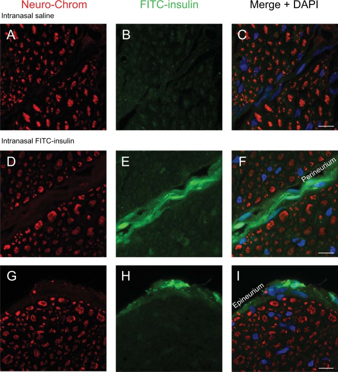
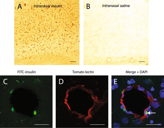
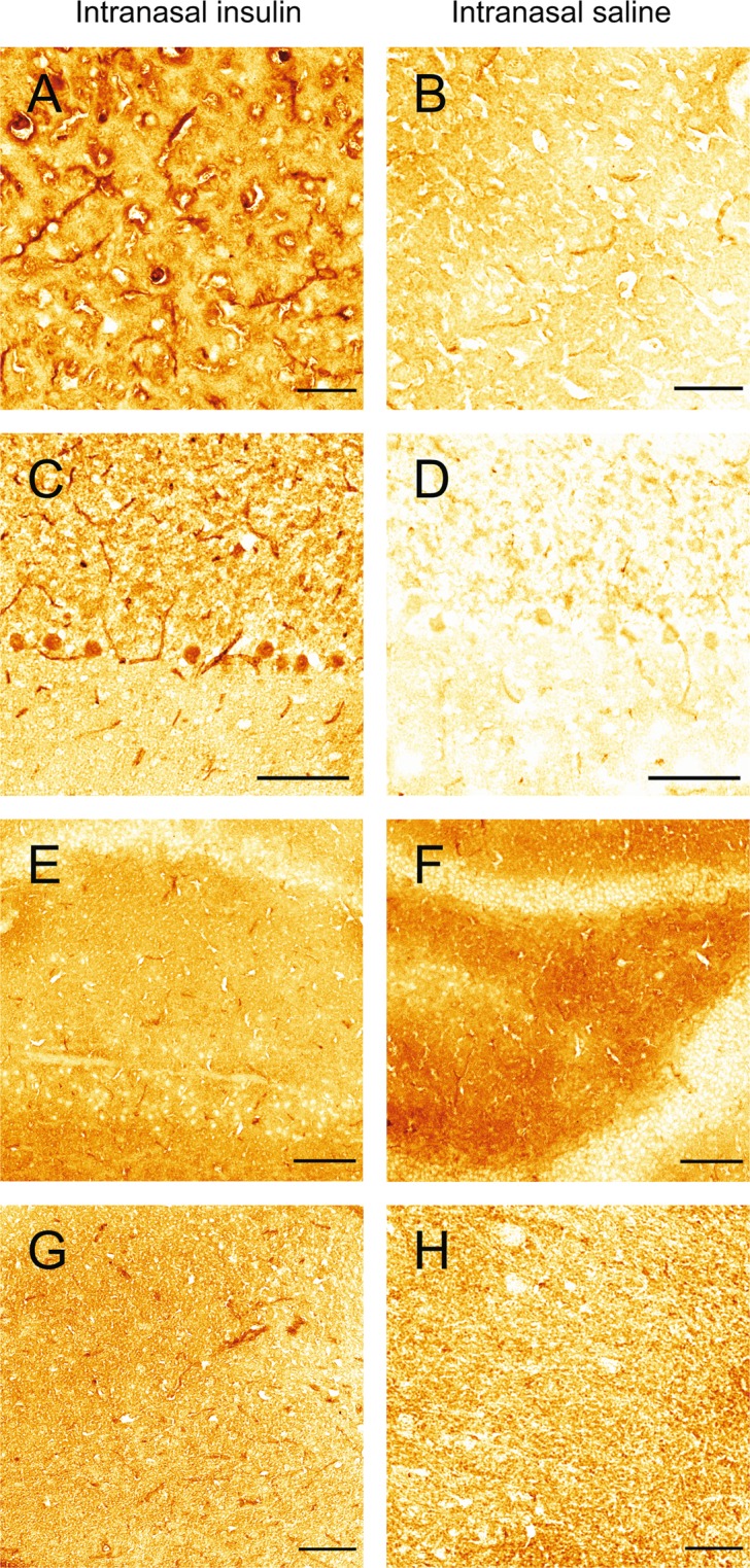
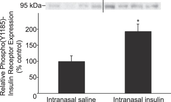
Verification
Claims the paper makes
The claims and statistics of this paper haven’t been checked yet.
Run the check — free, no account needed. It reads the open-access text and checks the sources it cites.
Claims other papers make about this one
Each is checked against this paper’s own text: does it say what the citing paper says it does?
In the citing paper
“In rats, transport along trigeminal nerve branches to brainstem regions has also been demonstrated.”
From Intranasal insulin (2021) · cites this paper as [34]
Self-check
Percentages, sums and reported differences re-computed from the paper’s own numbers.
The self-check needs the paper’s full text.
Citations
Cites
A Simple Practice Guide for Dose Conversion between Animals and Human
Nair, A., & Jacob, S. (2016) · Journal of Basic and Clinical Pharmacy · doi:10.4103/0976-0105.177703 · Cited by 6,174
Iliff, J. J., Wang, M., Liao, Y., Plogg, B. A., Peng, W., Gundersen, G. A., Benveniste, H., Vates, G. E., Deane, R., Goldman, S. A., et al. (2012) · Science Translational Medicine · doi:10.1126/scitranslmed.3003748 · Cited by 5,824
Brain insulin resistance in type 2 diabetes and Alzheimer disease: concepts and conundrums
Arnold, S. E., Arvanitakis, Z., Macauley-Rambach, S. L., Koenig, A. M., Wang, H.-Y., Ahima, R. S., Craft, S., Gandy, S., Buettner, C., Stoeckel, L. E., et al. (2018) · Nature Reviews Neurology · doi:10.1038/nrneurol.2017.185 · Cited by 1,597
Impairment of paravascular clearance pathways in the aging brain
Kress, B. T., Iliff, J. J., Xia, M., Wang, M., Wei, H. S., Zeppenfeld, D., Xie, L., Kang, H., Xu, Q., Liew, J. A., et al. (2014) · Annals of Neurology · doi:10.1002/ana.24271 · Cited by 1,521
Sniffing neuropeptides: A transnasal approach to the human brain
Born, J., Lange, T., Kern, W., McGregor, G. P., Bickel, U., & Fehm, H. L. (2002) · Nature Neuroscience · doi:10.1038/nn0602-849 · Cited by 1,356
Craft, S. (2012) · Archives of Neurology · doi:10.1001/archneurol.2011.233 · Cited by 1,253
Intranasal delivery of biologics to the central nervous system
Lochhead, J. J., & Thorne, R. G. (2012) · Advanced Drug Delivery Reviews · doi:10.1016/j.addr.2011.11.002 · Cited by 1,214
Self-citationImpairment of Glymphatic Pathway Function Promotes Tau Pathology after Traumatic Brain Injury
Iliff, J. J., Chen, M. J., Plog, B. A., Zeppenfeld, D. M., Soltero, M., Yang, L., Singh, I., Deane, R., & Nedergaard, M. (2014) · The Journal of Neuroscience · doi:10.1523/jneurosci.3020-14.2014 · Cited by 1,159
Thorne, R. G., Pronk, G. J., Padmanabhan, V., & Frey, W. H. (2004) · Neuroscience · doi:10.1016/j.neuroscience.2004.05.029 · Cited by 958
Intranasal insulin improves memory in humans
Benedict, C., Hallschmid, M., Hatke, A., Schultes, B., Fehm, H. L., Born, J., & Kern, W. (2004) · Psychoneuroendocrinology · doi:10.1016/j.psyneuen.2004.04.003 · Cited by 699
The role of insulin receptor signaling in the brain
Plum, L., Schubert, M., & Brüning, J. C. (2005) · Trends in Endocrinology & Metabolism · doi:10.1016/j.tem.2005.01.008 · Cited by 604
Insulin in the brain: There and back again
Banks, W. A., Owen, J. B., & Erickson, M. A. (2012) · Pharmacology & Therapeutics · doi:10.1016/j.pharmthera.2012.07.006 · Cited by 593
Suppression of glymphatic fluid transport in a mouse model of Alzheimer's disease
Peng, W., Achariyar, T. M., Li, B., Liao, Y., Mestre, H., Hitomi, E., Regan, S., Kasper, T., Peng, S., Ding, F., et al. (2016) · Neurobiology of Disease · doi:10.1016/j.nbd.2016.05.015 · Cited by 580
Freiherr, J., Hallschmid, M., Frey, W. H., Brünner, Y. F., Chapman, C. D., Hölscher, C., Craft, S., Felice, F. G. D., & Benedict, C. (2013) · CNS Drugs · doi:10.1007/s40263-013-0076-8 · Cited by 477
Aldridge, G. M., Podrebarac, D. M., Greenough, W. T., & Weiler, I. J. (2008) · Journal of Neuroscience Methods · doi:10.1016/j.jneumeth.2008.05.003 · Cited by 476
Insulin resistance and Parkinson’s disease: A new target for disease modification?
Athauda, D. S., & Foltynie, T. (2016) · Progress in Neurobiology · doi:10.1016/j.pneurobio.2016.10.001 · Cited by 340
Pizzo, M. E., Wolak, D. J., Kumar, N. N., Brunette, E., Brunnquell, C. L., Hannocks, M., Abbott, N. J., Meyerand, M. E., Sorokin, L., Stanimirovic, D. B., et al. (2018) · The Journal of Physiology · doi:10.1113/jp275105 · Cited by 272
Lochhead, J. J., Wolak, D. J., Pizzo, M. E., & Thorne, R. G. (2014) · Journal of Cerebral Blood Flow & Metabolism · doi:10.1038/jcbfm.2014.215 · Cited by 262
Self-citationImpaired insulin action in the human brain: causes and metabolic consequences
Heni, M., Kullmann, S., Preissl, H., Fritsche, A., & Häring, H.-U. (2015) · Nature Reviews Endocrinology · doi:10.1038/nrendo.2015.173 · Cited by 245
Insulin, cognition, and dementia
Cholerton, B., Baker, L. D., & Craft, S. (2013) · European Journal of Pharmacology · doi:10.1016/j.ejphar.2013.08.008 · Cited by 243
Banks, W. A., Jaspan, J. B., & Kastin, A. J. (1997) · Peptides · doi:10.1016/s0196-9781(97)00198-8 · Cited by 219
Wang, M., Ding, F., Deng, S., Guo, X., Wang, W., Iliff, J. J., & Nedergaard, M. (2017) · Journal of Neuroscience · doi:10.1523/jneurosci.2112-16.2017 · Cited by 199
Insulin transport across the blood–brain barrier can occur independently of the insulin receptor
Rhea, E. M., Rask‐Madsen, C., & Banks, W. A. (2018) · The Journal of Physiology · doi:10.1113/jp276149 · Cited by 153
Konishi, M., Sakaguchi, M., Lockhart, S. M., Cai, W., Li, M. E., Homan, E. P., Rask‐Madsen, C., & Kahn, C. R. (2017) · Proceedings of the National Academy of Sciences · doi:10.1073/pnas.1710625114 · Cited by 139
Central Nervous System Delivery of Intranasal Insulin: Mechanisms of Uptake and Effects on Cognition
Salameh, T. S., Bullock, K. M., Hujoel, I. A., Niehoff, M. L., Wolden‐Hanson, T., Kim, J., Morley, J. E., Farr, S. A., & Banks, W. A. (2015) · Journal of Alzheimer s Disease · doi:10.3233/jad-150307 · Cited by 136
Brain Insulin Dysregulation: Implication for Neurological and Neuropsychiatric Disorders
Ghasemi, R., Dargahi, L., Haeri, A., Moosavi, M., Mohamed, Z., & Ahmadiani, A. (2013) · Molecular Neurobiology · doi:10.1007/s12035-013-8404-z · Cited by 123
Intranasal delivery of insulin via the olfactory nerve pathway
Renner, D. B., Svitak, A. L., Gallus, N. J., Ericson, M. E., Frey, W. H., & Hanson, L. R. (2012) · Journal of Pharmacy and Pharmacology · doi:10.1111/j.2042-7158.2012.01555.x · Cited by 116
Intranasal Insulin and Insulin-Like Growth Factor 1 as Neuroprotectants in Acute Ischemic Stroke
Lioutas, V., Alfaro-Martinez, F., Bedoya, F. J., Chung, C., Maldonado, D. P., & Novak, V. (2015) · Translational Stroke Research · doi:10.1007/s12975-015-0409-7 · Cited by 115
Mao, Y., Guo, Z., Zheng, T., Jiang, Y., Yan, Y., Yin, X., Chen, Y., & Zhang, B. (2016) · Aging Cell · doi:10.1111/acel.12498 · Cited by 113
Safety of intranasal human insulin: A review
Schmid, V., Kullmann, S., Gfrörer, W., Hund, V., Hallschmid, M., Lipp, H., Häring, H., Preissl, H., Fritsche, A., & Heni, M. (2018) · Diabetes, Obesity and Metabolism · doi:10.1111/dom.13279 · Cited by 110
Unravelling the regulation of insulin transport across the brain endothelial cell
Gray, S. M., Aylor, K. W., & Barrett, E. J. (2017) · Diabetologia · doi:10.1007/s00125-017-4285-4 · Cited by 109
Quantitative Autoradiographic Evidence for Insulin Receptors in the Choroid Plexus of the Rat Brain
Baskin, D. G., Brewitt, B., Davidson, D. A., Paquette, T. L., Figlewicz, D. P., Lewellen, T. K., Graham, M. K., Woods, S. G., & Dorsa, D. M. (1986) · Diabetes · doi:10.2337/diab.35.2.246 · Cited by 104
Pang, Y., Lin, S., Wright, C., Shen, J., Carter, K., Bhatt, A., & Fan, L.-W. (2016) · Neuroscience · doi:10.1016/j.neuroscience.2016.01.020 · Cited by 99
Wang, C., Li, Y., Wible, B. A., Angelides, K. J., & Ishii, D. N. (1992) · Molecular Brain Research · doi:10.1016/0169-328x(92)90212-t · Cited by 91
Intranasal insulin treatment of an experimental model of moderate traumatic brain injury
Brabazon, F., Wilson, C. M., Jaiswal, S., Reed, J., Frey, W. H., & Byrnes, K. R. (2017) · Journal of Cerebral Blood Flow & Metabolism · doi:10.1177/0271678x16685106 · Cited by 88
Drejer, K., Vaag, A. A., Bech, K., Hansen, P. L., Sørensen, A. R., & Mygind, N. (1992) · Diabetic Medicine · doi:10.1111/j.1464-5491.1992.tb01792.x · Cited by 82
Insulin action in the brain: Roles in energy and glucose homeostasis
Dodd, G. T., & Tiganis, T. (2017) · Journal of Neuroendocrinology · doi:10.1111/jne.12513 · Cited by 77
Multiple insulin-responsive elements regulate transcription of the GAPDH gene
Alexander-Bridges, M., Dugast, I., Ercolani, L., Kong, X. F., Giere, L., & Nasrin, N. (1992) · Advances in Enzyme Regulation · doi:10.1016/0065-2571(92)90014-q · Cited by 63
Kirshner, Z. Z., & Gibbs, R. B. (2018) · Molecular and Cellular Endocrinology · doi:10.1016/j.mce.2018.01.015 · Cited by 57
Autofluorescent cells in rat brain can be convincing impostors in green fluorescent reporter studies
Spitzer, N., Sammons, G. S., & Price, E. M. (2011) · Journal of Neuroscience Methods · doi:10.1016/j.jneumeth.2011.01.029 · Cited by 54
Hjorth, C. F., Norrman, M., Wahlund, P., Benie, A. J., Petersen, B. O., Jessen, C. M., Pedersen, T. Å., Vestergaard, K., Steensgaard, D. B., Pedersen, J. S., et al. (2016) · Journal of Pharmaceutical Sciences · doi:10.1016/j.xphs.2016.01.003 · Cited by 49
Hersom, M., Helms, H. C. C., Schmalz, C., Pedersen, T. Å., Buckley, S. T., & Brodin, B. (2018) · American Journal of Physiology-Endocrinology and Metabolism · doi:10.1152/ajpendo.00350.2016 · Cited by 48
Pharmacokinetics of Intranasal versus Subcutaneous Insulin in the Mouse
Nedelcovych, M. T., Gadiano, A. J., Wu, Y., Manning, A. A., Thomas, A. G., Khuder, S. S., Yoo, S., Xu, J., McArthur, J. C., Haughey, N. J., et al. (2017) · ACS Chemical Neuroscience · doi:10.1021/acschemneuro.7b00434 · Cited by 46
Carpenter, M. C., & Wilcox, D. E. (2014) · Biochemistry · doi:10.1021/bi4016567 · Cited by 41
Oleic Acid Stimulates Glucose Uptake Into Adipocytes by Enhancing Insulin Receptor Signaling
Tsuchiya, A., Nagaya, H., Kanno, T., & Nishizaki, T. (2014) · Journal of Pharmacological Sciences · doi:10.1254/jphs.14182fp · Cited by 35
Mechanisms of action of brain insulin against neurodegenerative diseases
Ramalingam, M., & Kim, S. (2014) · Journal of Neural Transmission · doi:10.1007/s00702-013-1147-1 · Cited by 30
Rhea, E. M., Humann, S. R., Nirkhe, S., Farr, S. A., Morley, J. E., & Banks, W. A. (2017) · Journal of Alzheimer s Disease · doi:10.3233/jad-161095 · Cited by 29
Stearic Acid Serves as a Potent Inhibitor of Protein Tyrosine Phosphatase 1B
Tsuchiya, A., Kanno, T., & Nishizaki, T. (2013) · Cellular Physiology and Biochemistry · doi:10.1159/000356582 · Cited by 23
Regulation of β-actin gene transcription by insulin and phorbol esters
Messina, J. L., & Weinstock, R. S. (1992) · Experimental Cell Research · doi:10.1016/0014-4827(92)90206-n · Cited by 19
Temporal changes in islet polymerized and depolymerized tubulin during biphasic insulin release
McDaniel, M. L., Bry, C. G., HOMER, R. W., Fink, C., Ban, D., & Lacy, P. E. (1980) · Metabolism · doi:10.1016/0026-0495(80)90200-0 · Cited by 18
Cited by
Sorted by citations
Desforges, M., Le Coupanec, A., Dubeau, P., Bourgouin, A., Lajoie, L., Dubé, M., & Talbot, P. J. (2019) · Viruses · doi:10.3390/v12010014 · Cited by 1,048
“On the other hand, these viruses may also use other peripheral nerves like the trigeminal nerve, which possesses nociceptive neuronal cells present in the nasal cavity [64,65], or alternatively, the sensory fibers of the vagus nerve, which stems from the brainstem and innervates different organs of the respiratory tra…”
BackgroundKellar, D., & Craft, S. (2020) · The Lancet Neurology · doi:10.1016/s1474-4422(20)30231-3 · Cited by 832
Intranasal drug delivery: opportunities and toxicologic challenges during drug development
Keller, L.-A., Merkel, O. M., & Popp, A. (2021) · Drug Delivery and Translational Research · doi:10.1007/s13346-020-00891-5 · Cited by 636
Altered Insulin Signaling in Alzheimer’s Disease Brain – Special Emphasis on PI3K-Akt Pathway
Gabbouj, S., Ryhänen, S., Marttinen, M., Wittrahm, R., Takalo, M., Kemppainen, S., Martiskainen, H., Tanila, H., Haapasalo, A., Hiltunen, M., et al. (2019) · Frontiers in Neuroscience · doi:10.3389/fnins.2019.00629 · Cited by 401
Craft, S., Raman, R., Chow, T. W., Rafii, M. S., Sun, C.-K., Rissman, R. A., Donohue, M. C., Brewer, J. B., Jenkins, C., Harless, K., et al. (2020) · JAMA Neurology · doi:10.1001/jamaneurol.2020.1840 · Cited by 389
Metadata
- Authors
- Lochhead, J. J., Kellohen, K. L., Ronaldson, P. T., & Davis, T. P.
- Affiliations
- University of Arizona (US)
- Corresponding author
- Lochhead, Jeffrey J.
- Venue
- Scientific Reports
- Published
- 2019-02-22
- Volume
- 9
- Issue
- 1
- Pages
- 2621-2621
- ISSN
- 2045-2322 (ISSN-L)
- PMC
- PMC6385374
- OpenAlex
- W2917482585
- Cited by
- 146
- References
- 52
- Citations by year
- 2026: 17 · 2025: 24 · 2024: 19 · 2023: 18 · 2022: 21 · 2021: 20 · 2020: 21 · 2019: 6
- FWCI
- 7.26
- Type
- journal article
- Language
- en
- Open access
- gold
- doi.org
- Locations
- Scientific Reports (publishedVersion, cc-by)
- Europe PMC (PubMed Central) (publishedVersion, other-oa)
- PubMed Central (publishedVersion, cc-by)
- PubMed (publishedVersion)
- Funders
- American Heart Association (18CDA34110454)
- U.S. Department of Health & Human Services | NIH | National Institute of Neurological Disorders and Stroke (042652)
- Topics
- Life Sciences › Pharmacology, Toxicology and Pharmaceutics › Pharmaceutical Science › Advanced Drug Delivery Systems
- Life Sciences › Neuroscience › Neurology › Barrier Structure and Function Studies
- Life Sciences › Neuroscience › Cellular and Molecular Neuroscience › Alzheimer's disease research and treatments
- Keywords
- intranasal insulin, trigeminal nerve, insulin receptor activation, insulin receptor tyrosine kinase, neuroprotection, Alzheimer's disease, Parkinson's disease, traumatic brain injury, insulin signaling
- MeSH terms
- Administration, Intranasal, Animals, Brain, Female, Humans, Insulin, Receptor, Insulin, Tissue Distribution, Trigeminal Nerve, Fluorescein-5-isothiocyanate, Rats, Sprague-Dawley, Phosphotyrosine
About these checks
Registry, retraction, Cites and statistics results are computed, not judged, with one exception: a printed reference that no index lists is read once by a model before it counts as a citation error. Claims are extracted by a model and kept only when their quote appears verbatim in the text; each cited source is then checked, with a model’s help, for whether it says what the claim relies on. Those verdicts can be wrong — read the claim’s quote and the cited source before relying on one.
Statistics and arithmetic are recomputed, not judged.
Confidence bands are shown only for fields calibrated on our annotated test set. None is calibrated yet, so no band is shown.
What counts as a claim ›How we measure confidence ›How we check this ›
Are you an author, or do you see a problem with this page? Request a review
