

The DOI matches its Crossref record.
Nitric oxide and ROS mediate autophagy and regulate Alternaria alternata toxin-induced cell death in tobacco BY-2 cells
Abstract
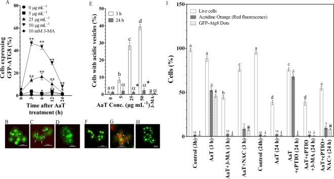
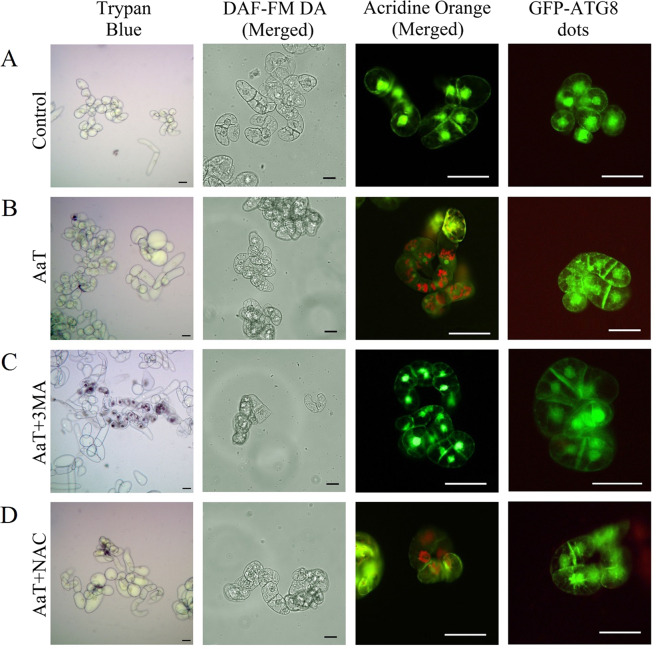
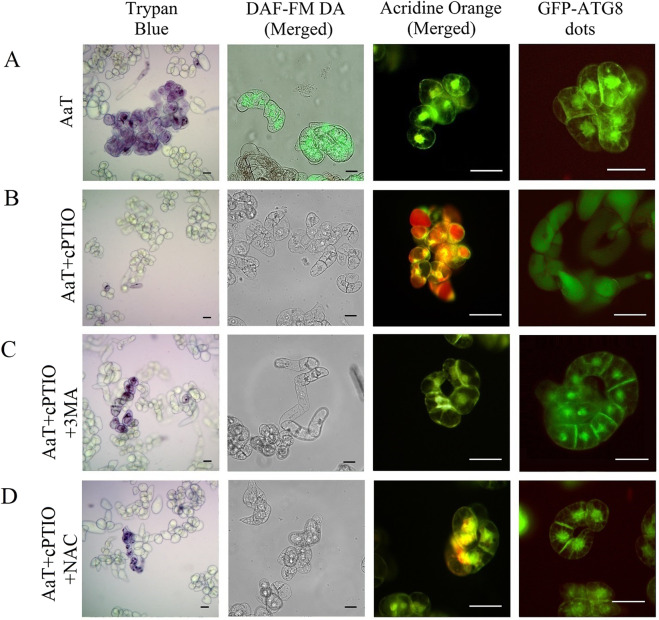
Synergistic interaction of nitric oxide (NO) and reactive oxygen species (ROS) is essential to initiate cell death mechanisms in plants. Though autophagy is salient in either restricting or promoting hypersensitivity response (HR)-related cell death, the crosstalk between the reactive intermediates and autophagy during hypersensitivity response is paradoxical. In this investigation, the consequences of Alternaria alternata toxin (AaT) in tobacco BY-2 cells were examined. At 3 h, AaT perturbed intracellular ROS homeostasis, altered antioxidant enzyme activities, triggered mitochondrial depolarization and induced autophagy. Suppression of autophagy by 3-Methyladenine caused a decline in cell viability in AaT treated cells, which indicated the vital role of autophagy in cell survival. After 24 h, AaT facilitated Ca 2+ influx with an accumulation of reactive oxidant intermediates and NO, to manifest necrotic cell death. Inhibition of NO accumulation by 2-(4-Carboxyphenyl)-4,4,5,5-tetramethylimidazoline-1-oxyl-3-oxide (cPTIO) decreased the level of necrotic cell death, and induced autophagy, which suggests NO accumulation represses autophagy and facilitates necrotic cell death at 24 h. Application of N-acetyl-L-cysteine at 3 h, confirmed ROS to be the key initiator of autophagy, and together with cPTIO for 24 h, revealed the combined effects of NO and ROS is required for necrotic HR cell death.
Overview
The claims and statistics of this paper haven’t been checked yet.
Run the check — free, no account needed. It reads the open-access text and checks the sources it cites.
Integrity signals
Registry
Cites
Statistics
Figures
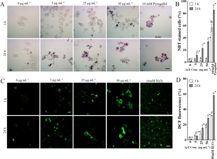
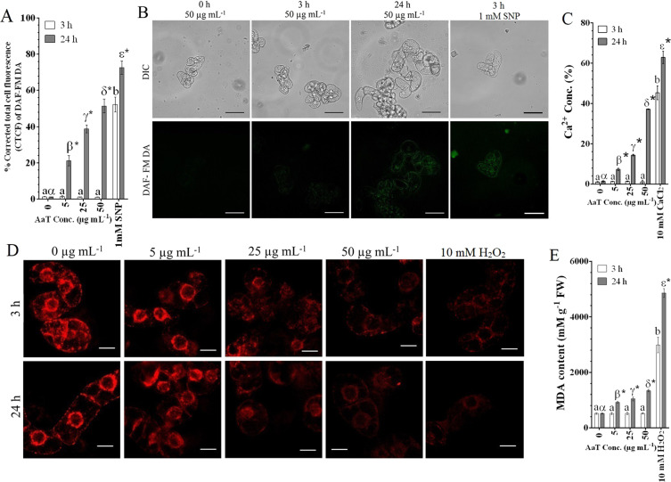
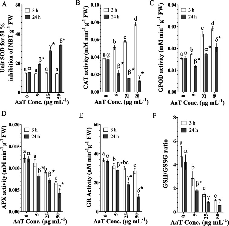
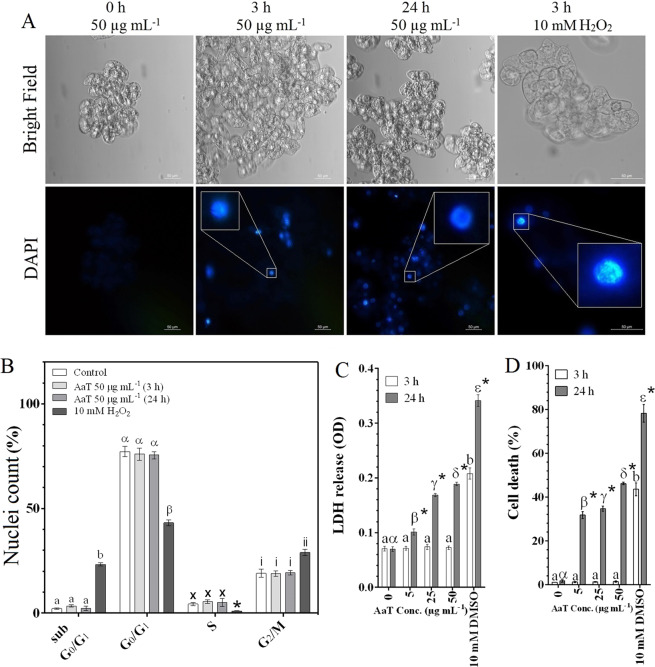
Verification
Claims the paper makes
The claims and statistics of this paper haven’t been checked yet.
Run the check — free, no account needed. It reads the open-access text and checks the sources it cites.
Claims other papers make about this one
Each is checked against this paper’s own text: does it say what the citing paper says it does?
In the citing paper
“Finally, N6-methyladenosine (m 6 A) mRNA modification plays an important role in regulating autophagy.”
From Guidelines for the use and interpretation of assays for monitoring autophagy (4th edition) 1 (2021) · cites this paper as [1010]
In the citing paper
“ULK1 mRNA undergoes m 6 A modification in the 3' UTR, and the m 6 Amarked ULK1 transcripts can further be targeted for degradation by YTHDF2 (YTH N6methyladenosine RNA binding protein 2).”
From Guidelines for the use and interpretation of assays for monitoring autophagy (4th edition) 1 (2021) · cites this paper as [1010]
In the citing paper
“Moreover, FTO (FTO alpha-ketoglutarate dependent dioxygenase) reverses the m 6 A mRNA modification of ULK1 transcripts, thereby promoting the initiation of autophagy [1010].”
From Guidelines for the use and interpretation of assays for monitoring autophagy (4th edition) 1 (2021) · cites this paper as [1010]
Self-check
Percentages, sums and reported differences re-computed from the paper’s own numbers.
The self-check needs the paper’s full text.
Citations
Cites
No references could be resolved yet.
Research works printed but not linked: 90
- Martin, G. B., Bogdanove, A. J. & Sessa, G. Understanding the functions of plant disease resistance proteins. Annu. Rev. Plant Biol. 54, 23–61 (2003). · DOI
- Nimchuk, Z., Eulgem, T., Holt, B. F. III & Dangl, J. L. Recognition and response in the plant immune system. Annu. Rev. Genet. 37, 579–609 (2003). · DOI
- Delledonne, M., Zeier, J., Marocco, A. & Lamb, C. Signal interactions between nitric oxide and reactive oxygen intermediates in the plant hypersensitive disease resistance response. P. Natl. Acad. Sci. USA 98, 13454–13459 (2001). · DOI
- Zaninotto, F., La Camera, S., Polverari, A. & Delledonne, M. Cross talk between reactive nitrogen and oxygen species during the hypersensitive disease resistance response. Plant Physiol. 141, 379–383 (2006). · DOI
- Pietrowska, E., Różalska, S., Kaźmierczak, A., Nawrocka, J. & Małolepsza, U. Reactive oxygen and nitrogen (ROS and RNS) species generation and cell death in tomato suspension cultures—Botrytis cinerea interaction. Protoplasma 252, 307–319 (2015). · DOI
- Doke, N. Involvement of superoxide anion generation in the hypersensitive response of potato tuber tissues to infection with an incompatible race of Phytophthora infestans and to the hyphal wall components. Physiol. Plant Pathol. 23, 345–357 (1983). · DOI
- Auh, C. K. & Murphy, T. M. Plasma membrane redox enzyme is involved in the synthesis of O2 − and H2O2 by Phytophthora elicitor-stimulated rose cells. Plant Physiol. 107, 1241–1247 (1995). · DOI
- Grant, M. et al. The RPM1 plant disease resistance gene facilitates a rapid and sustained increase in cytosolic calcium that is necessary for the oxidative burst and hypersensitive cell death. Plant J. 23, 441–450 (2000). · DOI
- McDowell, J. M. & Dangl, J. L. Signal transduction in the plant immune response. Trends Biochem. Sci. 25, 79–82 (2000). · DOI
- Liu, Y. Autophagy regulates programmed cell death during the plant innate immune response. Cell 121, 567–577 (2005). · DOI
- Wang, J. & Higgins, V. J. Nitric oxide has a regulatory effect in the germination of conidia of Colletotrichum coccodes. Fungal Genet. Biol. 42, 284–292 (2005). · DOI
- Moreau, M., Lindermayr, C., Durner, J. & Klessig, D. F. NO synthesis and signaling in plants–where do we stand? Physiol. Plant. 138, 372–383 (2010). · DOI
- Gas, E., Flores-Pérez, Ú., Sauret-Güeto, S. & Rodríguez-Concepción, M. Hunting for plant nitric oxide synthase provides new evidence of a central role for plastids in nitric oxide metabolism. Plant Cell 21, 18–23 (2009). · DOI
- Jeandroz, S. et al. Occurrence, structure, and evolution of nitric oxide synthase–like proteins in the plant kingdom. Sci. Signal. 9, 1–9 (2016). · DOI
- Santolini, J., André, F., Jeandroz, S. & Wendehenne, D. Nitric oxide synthase in plants: Where do we stand? Nitric Oxide 63, 30–38 (2017). · DOI
- Chamizo-Ampudia, A. et al. A dual system formed by the ARC and NR molybdoenzymes mediates nitrite‐dependent NO production in Chlamydomonas. Plant Cell Environ. 39, 2097–2107 (2016). · DOI
- Corpas, F. J., Palma, J. M., Del Río, L. A. & Barroso, J. B. Evidence supporting the existence of l-arginine-dependent nitric oxide synthase activity in plants. New Phytol. 184, 9–14 (2009). · DOI
- Rasul, S. et al. Nitric oxide production mediates oligogalacturonide‐triggered immunity and resistance to Botrytis cinerea in Arabidopsis thaliana. Plant Cell Environ. 35, 1483–1499 (2012). · DOI
- Howlett, B. J. Secondary metabolite toxins and nutrition of plant pathogenic fungi. Curr. Opin. Plant Biol. 9, 371–375 (2006). · DOI
- Koonin, E. V. & Aravind, L. Origin and evolution of eukaryotic apoptosis: the bacterial connection. Cell Death Differ. 9, 394–404 (2002). · DOI
- Van Doorn, W. G. et al. Morphological classification of plant cell deaths. Cell Death Differ. 18, 1241–1246 (2011). · DOI · Paper page
- Hofius, D. et al. Autophagic components contribute to hypersensitive cell death in Arabidopsis. Cell 137, 773–783 (2009). · DOI
- Komatsu, K. et al. Viral-induced systemic necrosis in plants involves both programmed cell death and the inhibition of viral multiplication, which are regulated by independent pathways. Mol. Plant Microbe In. 23, 283–293 (2010). · DOI
- Christofferson, D. E. & Yuan, J. Necroptosis as an alternative form of programmed cell death. Curr. Opin. Cell Biol. 22, 263–268 (2010). · DOI
- Patel, S. & Dinesh-Kumar, S. P. Arabidopsis ATG6 is required to limit the pathogen-associated cell death response. Autophagy 4, 20–27 (2008). · DOI
- Yoshimoto, K. et al. Autophagy negatively regulates cell death by controlling NPR1-dependent salicylic acid signaling during senescence and the innate immune response in Arabidopsis. Plant Cell 21, 2914–2927 (2009). · DOI
- Lai, Z., Wang, F., Zheng, Z., Fan, B. & Chen, Z. A critical role of autophagy in plant resistance to necrotrophic fungal pathogens. Plant J. 66, 953–968 (2011). · DOI
- Lenz, H. D. et al. Autophagy differentially controls plant basal immunity to biotrophic and necrotrophic pathogens. Plant J. 66, 818–830 (2011a). · DOI
- Lenz, H. D., Vierstra, R. D., Nürnberger, T. & Gust, A. A. ATG7 contributes to plant basal immunity towards fungal infection. Plant Signal Behav. 6, 1040–1042 (2011b). · DOI
- Duan, S. et al. Transcriptomic profile of tobacco in response to Alternaria longipes and Alternaria alternata infections. Sci. Rep, https://doi.org/10.1038/srep25635 (2016). · DOI
- Wang, H. C. et al. Phenotypic analysis of Alternaria alternata, the causal agent of tobacco brown spot. Plant Pathology J. 14, 79–85 (2015). · DOI
- Tsuge, T. et al. Host-selective toxins produced by the plant pathogenic fungus Alternaria alternata. FEMS Microbiol. Rev. 37, 44–66 (2013). · DOI
- Takao, K. The global regulator LaeA controls biosynthesis of host-specific toxins, pathogenicity and development of Alternaria alternata pathotypes. J. Gen. Plant Pathol. 82, 121–131 (2016). · DOI
- Saha, D. Identification of a polyketide synthase required for alternariol (AOH) and alternariol-9-methyl ether (AME) formation in Alternaria alternata. PLoS One, https://doi.org/10.1371/journal.pone.0040564 (2012). · DOI
- Oh, K. B., Tajima, Y., Saito, M., Chang, I. M. & Matsuoka, H. Cytotoxicity of extracts from Alternaria alternata against cultured tobacco BY-2 cells. Biocontrol Sci. 7, 127–130 (2002). · DOI
- Slavov, S., Mayama, S. & Atanassov, A. Toxin production of Alternaria alternata tobacco pathotype. Biotechnol. Biotec. Eq. 18, 90–95 (2004). · DOI
- Cheng, D. D. Characterization of the programmed cell death induced by metabolic products of Alternaria alternata in tobacco BY‐2 cells. Physiol. Plantarum 1, 117–129 (2011). · DOI
- McCabe, P. F. & Leaver, C. J. Programmed cell death in cell cultures in Programmed Cell Death in Higher Plants (eds Lam, E., Fukuda, H., Greenberg, J.) 115–124 (Springer, 2000). · DOI
- Gupta, N. S., Banerjee, M., Basu, S. K. & Acharya, K. Involvement of nitric oxide signal in Alternaria alternata toxin induced defense response in Rauvolfia serpentina Benth. ex Kurz calli. Plant Omics 6, 157–164 (2013).
- Hayward, A. P., Tsao, J. & Dinesh-Kumar, S. P. Autophagy and plant innate immunity: defense through degradation. Semin. Cell Dev. Biol. 20, 1041–1047 (2009). · DOI
- Minina, E. A., Bozhkov, P. V. & Hofius, D. Autophagy as initiator or executioner of cell death. Trends Plant Sci. 19, 692–697 (2014). · DOI
- Niu, L. & Liao, W. Hydrogen peroxide signaling in plant development and abiotic responses: crosstalk with nitric oxide and calcium. Front. Plant Sci. 7, 230 (2016).
- Hou, Y. et al. Comparative genomics of pathogens causing brown spot disease of tobacco: Alternaria longipes and Alternaria alternata. PloS one, https://doi.org/10.1371/journal.pone.0155258 (2016). · DOI
- Langhans, M. & Robinson, D. G. 1-Butanol targets the Golgi apparatus in tobacco BY-2 cells, but in a different way to Brefeldin A. J. Exp. Bot. 58, 3439–3447 (2007). · DOI
- Pérez-Pérez, M. E., Lemaire, S. D. & Crespo, J. L. Reactive oxygen species and autophagy in plants and algae. Plant physiol. 160, 156–164 (2012). · DOI
- Lamb, C. & Dixon, R. A. The oxidative burst in plant disease resistance. Annu. Rev. Plant Physiol. Plant Mol. Biol. 48, 251–275 (1997). · DOI
- Liu, H. B. et al. NADPH oxidase RBOHD contributes to autophagy and hypersensitive cell death during the plant defense response in Arabidopsis thaliana. Biol. Plantarum 59, 570–580 (2015). · DOI
- Jiménez-Quesada, M. J., Traverso, J. Á. & de Dios Alché, J. NADPH Oxidase-Dependent Superoxide Production in Plant Reproductive Tissues. Front. Plant Sci. 7, 1–13 (2016). · DOI
- Kadota, Y., Shirasu, K. & Zipfel, C. Regulation of the NADPH oxidase RBOHD during plant immunity. Plant Cell Physiol. 56, 1472–1480 (2015). · DOI
- Locato, V., Paradiso, A., Sabetta, W., De Gara, L. & de Pinto, M. C. Nitric oxide and reactive oxygen species in PCD signaling in Advances in Botanical Research: Nitric oxide and signaling in plants (eds Wendehenne, D., Jacquot, J. P., Gadal, P.) 165–192 (Academic Press, 2016). · DOI
- Mittler, R. Oxidative stress, antioxidants and stress tolerance. Trends Plant Sci. 7, 405–410 (2002). · DOI
- Kalogeris, T., Bao, Y. & Korthuis, R. J. Mitochondrial reactive oxygen species: a double edged sword in ischemia/reperfusion vs preconditioning. Redox Biol. 2, 702–714 (2014). · DOI
- Zorov, D. B., Juhaszova, M. & Sollott, S. J. Mitochondrial reactive oxygen species (ROS) and ROS-induced ROS release. Physiol. Rev. 94, 909–950 (2014). · DOI · Paper page
- Michaeli, S. & Galili, G. Degradation of organelles or specific organelle components via selective autophagy in plant cells. Int. J. Mol. Sci. 15, 7624–7638 (2014). · DOI
- Begara-Morales, J. C. et al. Differential molecular response of monodehydroascorbate reductase and glutathione reductase by nitration and S-nitrosylation. J. Exp. Bot. 66, 5983–5996 (2015). · DOI
- Clark, D., Durner, J., Navarre, D. A. & Klessig, D. F. Nitric oxide inhibition of tobacco catalase and ascorbate peroxidase. Mol. Plant. Microbe. In. 13, 1380–1384 (2000). · DOI
- Fares, A., Rossignol, M. & Peltier, J. B. Proteomics investigation of endogenous S-nitrosylation in Arabidopsis. Biochem. Bioph. Res. Co. 416, 331–336 (2011). · DOI
- Foyer, C. H. & Noctor, G. Ascorbate and glutathione: the heart of the redox hub. Plant Physiol. 155, 2–18 (2011). · DOI
- Keyster, M., Klein, A., Egbich, I., Jacobs, A. & Ludidi, N. Nitric oxide increases the enzymatic activity of three ascorbate peroxidase isoforms in soybean root nodules. Plant Signal Behav. 6, 956–961 (2011). · DOI
- Begara-Morales, J. C. et al. Dual regulation of cytosolic ascorbate peroxidase (APX) by tyrosine nitration and S-nitrosylation. J. Exp. Bot. 65, 527–538 (2014). · DOI
- Corpas, F. J., Alché, J. D. & Barroso, J. B. Current overview of S-nitrosoglutathione (GSNO) in higher plants. Front. Plant. Sci. 4, 126 (2013).
- de Pinto, M. C., Paradiso, A., Leonetti, P. & de Gara, L. Hydrogen peroxide, nitric oxide and cytosolic ascorbate peroxidase at the crossroad between defence and cell death. Plant J. 48, 784–795 (2006). · DOI
- Yakimova, E. T., Yordanova, Z., Slavov, S., Kapchina‐Toteva, V. M. & Woltering, E. J. Alternaria alternata AT toxin induces programmed cell death in tobacco. J. Phytopathol. 157, 592–601 (2009). · DOI
- Víteček, J. et al. Cell death induced by sodium nitroprusside and hydrogen peroxide in tobacco BY-2 cell suspension. Biol. Plantarum 51, 472–479 (2007). · DOI
- Hatsugai, N. et al. A plant vacuolar protease, VPE, mediates virus-induced hypersensitive cell death. Science 305, 855–858 (2004). · DOI
- Xiong, Y., Contento, A. L., Nguyen, P. Q. & Bassham, D. C. Degradation of oxidized proteins by autophagy during oxidative stress in Arabidopsis. Plant physiol. 143, 291–299 (2007). · DOI
- Delledonne, M., Xia, Y., Dixon, R. A. & Lamb, C. Nitric oxide functions as a signal in plant disease resistance. Nature 394, 85–88 (1998). · DOI
- Sarkar, S. et al. Complex inhibitory effects of nitric oxide on autophagy. Mol. Cell 43, 19–32 (2011). · DOI
- Shen, C., Yan, J., Erkocak, O. F., Zheng, X. F. & Chen, X. D. Nitric oxide inhibits autophagy via suppression of JNK in meniscal cells. Rheumatology 53, 1022–1033 (2014). · DOI
- Takatsuka, C. et al. Autophagy in tobacco BY-2 cells cultured under sucrose starvation conditions: isolation of the autolysosome and its characterization. Plant Cell Physiol. 52, 2074–2087 (2011). · DOI
- Santos, A. R. The impact of CdSe/ZnS quantum dots in cells of Medicago sativa in suspension culture. J. Nanobiotechnol. 8, 1–14 (2010). · DOI
- Huang, W., Xing, W., Li, D. & Liu, Y. Microcystin-RR induced apoptosis in tobacco BY-2 suspension cells is mediated by reactive oxygen species and mitochondrial permeability transition pore status. Toxicol. in vitro 22, 328–337 (2008). · DOI
- Morin, L. G. Direct colorimetric determination of serum calcium with o-cresolphthalein complexon. Am. J. Clin. Pathol. 61, 114–117 (1974). · DOI
- Sadhu, A., Ghosh, I., Moriyasu, Y., Mukherjee, A. & Bandyopadhyay, M. Role of cerium oxide nanoparticle-induced autophagy as a safeguard to exogenous H2O2-mediated DNA damage in tobacco BY-2 cells. Mutagenesis 33, 161–177 (2018). · DOI
- Ghosh, M., Bandyopadhyay, M. & Mukherjee, A. Genotoxicity of titanium dioxide (TiO2) nanoparticles at two trophic levels: plant and human lymphocytes. Chemosphere 81, 1253–1262 (2010). · DOI
- Bradford, M. M. A rapid and sensitive method for the quantitation of microgram quantities of protein utilizing the principle of protein-dye binding. Anal. Biochem. 72, 248–254 (1976). · DOI · Paper page
- Beauchamp, C. & Fridovich, I. Superoxide dismutase: improved assays and an assay applicable to acrylamide gels. Anal. Biochem. 44, 276–287 (1971). · DOI · Paper page
- Achary, V. M. M., Jena, S., Panda, K. K. & Panda, B. B. Aluminium induced oxidative stress and DNA damage in root cells of Allium cepa L. Ecotoxicol. Environ. Saf. 70, 300–310 (2008). · DOI
- Aebi, H. Catalase in vitro in Methods in enzymology, vol. 105 (ed. Packer, L.) 121–126 (Academic Press Inc,. 1984). · DOI · Paper page
- Chance, B. & Maehly, A. C. Assay of catalases and peroxidases in Methods in enzymology (ed. Glick, D.) 764–775 (Wiley, 1955). · DOI
- Gallego, S. M., Benavides, M. P. & Tomaro, M. L. Effect of heavy metal ion excess on sunflower leaves: evidence for involvement of oxidative stress. Plant Sci. 121, 151–159 (1996). · DOI
- Smith, I. K., Vierheller, T. L. & Thorne, C. A. Assay of glutathione reductase in crude tissue homogenates using 5, 5′-dithiobis (2-nitrobenzoic acid). Anal. Biochem. 175, 408–413 (1988). · DOI
- Anderson, M. E. Determination of glutathione and glutathione disulfide in biological samples in Methods in Enzymology, vol 113 (ed. Meister, A.) 548–555 (Elsevier, 1985). · DOI
- Sahoo, S., Awasthi J. P., Sunkar, R., Panda, S. K. Determining Glutathione Levels in Plants in Plant Stress Tolerance. Methods in Molecular Biology, vol 1631 (ed. Sunkar, R.) 273– 277 (Humana Press, 2017). · DOI
- Yamada, T., Takatsu, Y., Kasumi, M., Ichimura, K. & van Doorn, W. G. Nuclear fragmentation and DNA degradation during programmed cell death in petals of morning glory (Ipomoea nil). Planta 224, 1279–1290 (2006). · DOI
- Riccardi, C. & Nicoletti, I. Analysis of apoptosis by propidium iodide staining and flow cytometry. Nat. Protoc. 1, 1458–1461 (2006). · DOI · Paper page
- Chan, F. K. M., Moriwaki, K., De Rosa, M. J. Detection of necrosis by release of lactate dehydrogenase activity in Immune Homeostasis: Methods and Protocols (eds Snow, A. L. & Lenardo, M. J.) 65–70 (Springer, 2013). · DOI
- Ohno, R. et al. Cryptogein-Induced cell cycle arrest at G2 phase is associated with inhibition of cyclin-dependent kinases, suppression of expression of cell cycle-related genes and protein degradation in synchronized tobacco BY-2 cells. Plant Cell Physiol. 52, 922–932 (2011). · DOI
- Mitou, G., Budak, H. & Gozuacik, D. Techniques to study autophagy in plants. Int. J. Plant Genomics, https://doi.org/10.1155/2009/451357 (2009). · DOI
- Yano, K. et al. Dissection of autophagy in tobacco BY-2 cells under sucrose starvation conditions using the vacuolar H+-ATPase inhibitor concanamycin A and the autophagy-related protein Atg8. Plant Signal. Behav, https://doi.org/10.1080/15592324.2015.1082699 (2015). · DOI
Cited by
Sorted by citations
Guidelines for the use and interpretation of assays for monitoring autophagy (4th edition) 1
Klionsky, D. J., Abdel-Aziz, A. K., Abdelfatah, S., Abdellatif, M., Abdoli, A., Abel, S., Abeliovich, H., Abildgaard, M. H., Abudu, Y. P., Acevedo-Arozena, A., et al. (2021) · Autophagy · doi:10.1080/15548627.2020.1797280 · Cited by 2,897
Self-citation
Metadata
- Authors
- Sadhu, A., Moriyasu, Y., Acharya, K., & Bandyopadhyay, M.
- Venue
- Scientific Reports
- Published
- 2019-06-20
- Volume
- 9
- Issue
- 1
- ISSN
- 2045-2322
- PMC
- PMC6586778
- Cited by
- 48
- References
- 0
- Language
- en
- Update notices
- correction (2024-03-04)
- Funders
- Department of Science and Technology, Ministry of Science and Technology (DST/INT/JSPS/P-261/2017 dt. 16/10/17)
About these checks
Registry, retraction, Cites and statistics results are computed, not judged, with one exception: a printed reference that no index lists is read once by a model before it counts as a citation error. Claims are extracted by a model and kept only when their quote appears verbatim in the text; each cited source is then checked, with a model’s help, for whether it says what the claim relies on. Those verdicts can be wrong — read the claim’s quote and the cited source before relying on one.
Statistics and arithmetic are recomputed, not judged.
Confidence bands are shown only for fields calibrated on our annotated test set. None is calibrated yet, so no band is shown.
What counts as a claim ›How we measure confidence ›How we check this ›
Are you an author, or do you see a problem with this page? Request a review
