

Using diffusion tensor imaging to detect cortical changes in fronto-temporal dementia subtypes
Open accessThe DOI matches its Crossref record; 39 of the 39 references it cites were found in a registry or an index.
Abstract
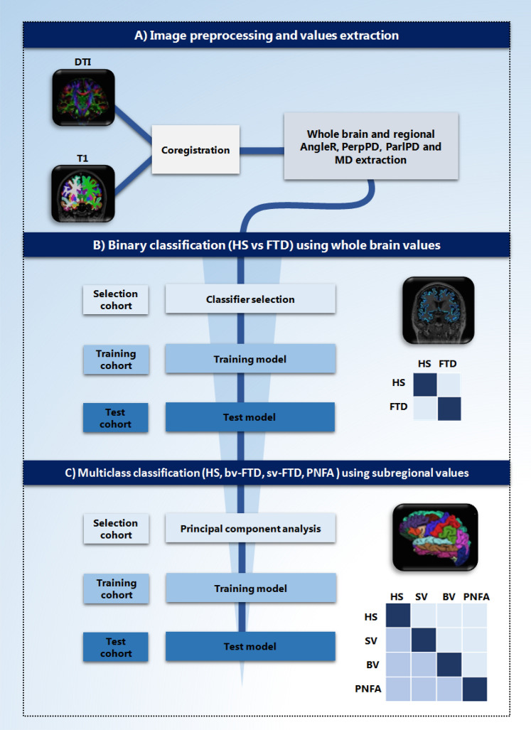
Fronto-temporal dementia (FTD) is a common type of presenile dementia, characterized by a heterogeneous clinical presentation that includes three main subtypes: behavioural-variant FTD, non-fluent/agrammatic variant primary progressive aphasia and semantic variant PPA. To better understand the FTD subtypes and develop more specific treatments, correct diagnosis is essential. This study aimed to test the discrimination power of a novel set of cortical Diffusion Tensor Imaging measures (DTI), on FTD subtypes. A total of 96 subjects with FTD and 84 healthy subjects (HS) were included in the study. A “selection cohort” was used to determine the set of features (measurements) and to use them to select the “best” machine learning classifier from a range of seven main models. The selected classifier was trained on a “training cohort” and tested on a third cohort (“test cohort”). The classifier was used to assess the classification power for binary (HS vs. FTD), and multiclass (HS and FTD subtypes) classification problems. In the binary classification, one of the new DTI features obtained the highest accuracy (85%) as a single feature, and when it was combined with other DTI features and two other common clinical measures (grey matter fraction and MMSE), obtained an accuracy of 88%. The new DTI features can distinguish between HS and FTD subgroups with an accuracy of 76%. These results suggest that DTI measures could support differential diagnosis in a clinical setting, potentially improve efficacy of new innovative drug treatments through effective patient selection, stratification and measurement of outcomes.
Overview
The claims and statistics of this paper haven’t been checked yet.
Free: it reads the open-access text and checks the first 5 claims that cite a source we can check. Create a free account to check every claim.
Integrity signals
Registry
Cites
Statistics
Figures
Verification
Claims the paper makes
The claims and statistics of this paper haven’t been checked yet.
Free: it reads the open-access text and checks the first 5 claims that cite a source we can check. Create a free account to check every claim.
Claims other papers make about this one
Each is checked against this paper’s own text: does it say what the citing paper says it does?
In the citing paper
“First, despite the larger sample size compared to similar previous studies in the literature (Kim et al., 2019; Torso et al., 2020), the sample was based on a unique database.”
From Multiclass characterization of frontotemporal dementia variants via multimodal brain network computational inference (2023) · cites this paper as Torso et al., 2020
Self-check
Percentages, sums and reported differences re-computed from the paper’s own numbers.
The self-check needs the paper’s full text.
Citations
Cites
The control of the false discovery rate in multiple testing under dependency
Benjamini, Y., & Yekutieli, D. (2001) · The Annals of Statistics · doi:10.1214/aos/1013699998 · Cited by 10,490
How do you feel — now? The anterior insula and human awareness
(Bud) Craig, A. D. (2009) · Nature Reviews Neuroscience · doi:10.1038/nrn2555 · Cited by 6,657
Sensitivity of revised diagnostic criteria for the behavioural variant of frontotemporal dementia
Rascovsky, K., Hodges, J. R., Knopman, D., Mendez, M. F., Kramer, J. H., Neuhaus, J., van Swieten, J. C., Seelaar, H., Dopper, E. G. P., Onyike, C. U., et al. (2011) · Brain · doi:10.1093/brain/awr179 · Cited by 5,221
Classification of primary progressive aphasia and its variants
Gorno-Tempini, M. L., Hillis, A. E., Weintraub, S., Kertesz, A., Mendez, M., Cappa, S. F., Ogar, J. M., Rohrer, J. D., Black, S., Boeve, B. F., et al. (2011) · Neurology · doi:10.1212/wnl.0b013e31821103e6 · Cited by 5,141
A common role of insula in feelings, empathy and uncertainty
Singer, T., Critchley, H. D., & Preuschoff, K. (2009) · Trends in Cognitive Sciences · doi:10.1016/j.tics.2009.05.001 · Cited by 1,270
Die Cytoarchitektonik der Hirnrinde des erwachsenen Menschen
Economo, C. V., & Koskinas, G. N. (1925) · Springer eBooks · Cited by 1,234
Bang, J., Spina, S., & Miller, B. L. (2015) · The Lancet · doi:10.1016/s0140-6736(15)00461-4 · Cited by 1,144
Maruyama, M., Shimada, H., Suhara, T., Shinotoh, H., Ji, B., Maeda, J., Zhang, M.-R., Trojanowski, J. Q., Lee, V. M.-Y., Ono, M., et al. (2013) · Neuron · doi:10.1016/j.neuron.2013.07.037 · Cited by 760
Patterns of temporal lobe atrophy in semantic dementia and Alzheimer's disease
Chan, D., Fox, N. C., Scahill, R. I., Crum, W. R., Whitwell, J. L., Leschziner, G., Rossor, A. M., Stevens, J. M., Cipolotti, L., & Rossor, M. N. (2001) · Annals of Neurology · doi:10.1002/ana.92 · Cited by 749
A role of right middle frontal gyrus in reorienting of attention: a case study
Japee, S., Holiday, K., Satyshur, M. D., Mukai, I., & Ungerleider, L. G. (2015) · Frontiers in Systems Neuroscience · doi:10.3389/fnsys.2015.00023 · Cited by 479
Cited by
Schwarz, A. J. (2021) · Neurotherapeutics · doi:10.1007/s13311-021-01027-4 · Cited by 42
Detection of Alzheimer's Disease using cortical diffusion tensor imaging
Torso, M., Bozzali, M., Zamboni, G., Jenkinson, M., Chance, S. A., & Initiative, A. D. N. (2020) · Human Brain Mapping · doi:10.1002/hbm.25271 · Cited by 40
Self-citationAdvanced Magnetic Resonance Imaging for Early Diagnosis and Monitoring of Movement Disorders
Ortega‐Robles, E., Alonso, B. D. C., Cantillo-Negrete, J., Carino-Escobar, R. I., & Arias-Carrión, Ó. (2025) · Brain Sciences · doi:10.3390/brainsci15010079 · Cited by 26
Torso, M., Ridgway, G. R., Jenkinson, M., Chance, S. A., & (4RTNI), T. F. L. D. N. I. A. T. 4.-R. T. N. I. (2021) · Alzheimer s Research & Therapy · doi:10.1186/s13195-021-00914-4 · Cited by 19
Self-citationTorso, M., Ridgway, G. R., Hardingham, I., Schwarz, A. J., & Chance, S. A. (2022) · The Journal of Prevention of Alzheimer s Disease · doi:10.14283/jpad.2022.59 · Cited by 19
Self-citation
Metadata
- Authors
- Torso, M., Bozzali, M., Cercignani, M., Jenkinson, M., & Chance, S. A.
- Affiliations
- University of Oxford (GB)
- Fondazione Santa Lucia (IT)
- Brighton and Sussex Medical School (GB)
- Corresponding author
- Torso, M.
- Venue
- Scientific Reports
- Published
- 2020-07-08
- Volume
- 10
- Issue
- 1
- Pages
- 11237-11237
- ISSN
- 2045-2322 (ISSN-L)
- PMC
- PMC7343779
- OpenAlex
- W3041423097
- Cited by
- 27
- References
- 39
- Citations by year
- 2026: 6 · 2025: 4 · 2024: 6 · 2023: 3 · 2022: 4 · 2021: 2 · 2020: 2
- FWCI
- 2.01
- Type
- journal article
- Language
- en
- Open access
- gold
- doi.org
- Licence
- cc-by
- creativecommons.org/licenses/by/4.0 (tdm)
- creativecommons.org/licenses/by/4.0 (vor)
- Locations
- Scientific Reports (publishedVersion, cc-by)
- PubMed Central (publishedVersion, cc-by)
- PubMed (publishedVersion)
- Figshare (submittedVersion, cc-by)
- Sussex Research Online (University of Sussex) (submittedVersion, cc-by)
- Funders
- Alzheimer's Research UK (ARUK-PPG2017A-19)
- Wellcome Trust (203139/Z/16/Z)
- Topics
- Health Sciences › Medicine › Psychiatry and Mental health › Dementia and Cognitive Impairment Research
- Health Sciences › Medicine › Radiology, Nuclear Medicine and Imaging › Advanced Neuroimaging Techniques and Applications
- Life Sciences › Neuroscience › Cognitive Neuroscience › Functional Brain Connectivity Studies
- Keywords
- frontotemporal dementia, diffusion tensor imaging, primary progressive aphasia, semantic variant PPA, differential diagnosis, machine learning classification, Mini-Mental State Examination
- MeSH terms
- Machine Learning, Aged, Cerebral Cortex, Diagnosis, Differential, Feasibility Studies, Female, Humans, Image Interpretation, Computer-Assisted, Male, Middle Aged, Cohort Studies, Case-Control Studies, Diffusion Tensor Imaging, Frontotemporal Dementia, Gray Matter
About these checks
Registry, retraction, Cites and statistics results are computed, not judged, with one exception: a printed reference that no index lists is read once by a model before it counts as a citation error. Claims are extracted by a model and kept only when their quote appears verbatim in the text; each cited source is then checked, with a model’s help, for whether it says what the claim relies on. Those verdicts can be wrong — read the claim’s quote and the cited source before relying on one.
Statistics and arithmetic are recomputed, not judged.
Confidence bands are shown only for fields calibrated on our annotated test set. None is calibrated yet, so no band is shown.
What counts as a claim ›How we measure confidence ›How we check this ›
Are you an author, or do you see a problem with this page? Request a review
How to cite
Cite CoChat for the checks on this page, and the paper for its own findings.
- This page
- CoChat. (2026). Citation check: Using diffusion tensor imaging to detect cortical changes in fronto-temporal dementia subtypes. https://app.cochat.ai/papers/10.1038/s41598-020-68118-8
- The paper
- Torso, M., Bozzali, M., Cercignani, M., Jenkinson, M., & Chance, S. A. (2020). Using diffusion tensor imaging to detect cortical changes in fronto-temporal dementia subtypes. Scientific Reports. https://doi.org/10.1038/s41598-020-68118-8
Data sources
Bibliographic data: Semantic Scholar, OpenAlex, Crossref.
Contains information from the Semantic Scholar Academic Graph Datasets, made available under the ODC Attribution License (ODC-BY 1.0).
Semantic Scholar’s data is described in: Kinney, R., et al. (2023). The Semantic Scholar Open Data Platform. arXiv:2301.10140.
