

The DOI matches its Crossref record; 62 of the 62 references it cites were found in a registry or an index; 33 of the 50 sources its claims cite were checked for support.
The relationship between resting-state functional connectivity, antidepressant discontinuation and depression relapse
Open accessPublished version of a preprint
Abstract
The risk of relapsing into depression after stopping antidepressants is high, but no established predictors exist. Resting-state functional magnetic resonance imaging (rsfMRI) measures may help predict relapse and identify the mechanisms by which relapses occur. rsfMRI data were acquired from healthy controls and from patients with remitted major depressive disorder on antidepressants. Patients were assessed a second time either before or after discontinuation of the antidepressant, and followed up for six months to assess relapse. A seed-based functional connectivity analysis was conducted focusing on the left subgenual anterior cingulate cortex and left posterior cingulate cortex. Seeds in the amygdala and dorsolateral prefrontal cortex were explored. 44 healthy controls (age: 33.8 (10.5), 73% female) and 84 patients (age: 34.23 (10.8), 80% female) were included in the analysis. 29 patients went on to relapse and 38 remained well. The seed-based analysis showed that discontinuation resulted in an increased functional connectivity between the right dorsolateral prefrontal cortex and the parietal cortex in non-relapsers. In an exploratory analysis, this functional connectivity predicted relapse risk with a balanced accuracy of 0.86. Further seed-based analyses, however, failed to reveal differences in functional connectivity between patients and controls, between relapsers and non-relapsers before discontinuation and changes due to discontinuation independent of relapse. In conclusion, changes in the connectivity between the dorsolateral prefrontal cortex and the posterior default mode network were associated with and predictive of relapse after open-label antidepressant discontinuation. This finding requires replication in a larger dataset.
Overview
CoChat Summary
TL;DR
In remitted medicated patients with major depressive disorder, resting-state functional connectivity did not differ from healthy controls, did not differ before discontinuation between future relapsers and non-relapsers, and did not change with antidepressant discontinuation for the main seeds tested. An exploratory interaction showed that right dorsolateral prefrontal cortex connectivity with posterior default mode regions increased in non-relapsers after discontinuation and predicted relapse with balanced accuracy 0.86, requiring replication.
Key findings
- No resting-state functional connectivity differences were found between 84 remitted medicated patients and 44 healthy controls for the left subgenual anterior cingulate cortex main seed or any exploratory seeds.
- No pre-discontinuation connectivity differences were found between 29 patients who later relapsed and 38 who remained well, and no discontinuation effects were found for the left posterior cingulate cortex main seed or exploratory seeds.
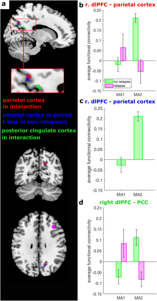
- A significant discontinuation-by-relapse interaction emerged for right dorsolateral prefrontal cortex connectivity with right parietal cortex (BA 7; peak MNI 14, −66, 38; p = 0.001) and posterior cingulate cortex (BA 23; peak MNI 12, −54, 14; p = 0.001); it was driven by increased right dlPFC–parietal connectivity in non-relapsers after discontinuation, while relapsers showed a numerical decrease that was not statistically significant.
- Post-hoc leave-one-out prediction using right dlPFC–parietal connectivity at MA2 or its change from MA1 to MA2 yielded a balanced accuracy of 0.86; the paper notes the change-based prediction is not independent evidence.
Methods
- The AIDA study was a two-centre observational randomized study of remitted patients on antidepressant medication who wanted to discontinue; patients were randomized to MA1-D-MA2 or MA1-MA2-D, assessed at main assessments MA1 and MA2, and followed for six months by telephone at weeks 1, 2, 4, 6, 8, 12, 16 and 21, with SCID-I if relapse signs appeared.
- Seed-based resting-state fMRI analysis used left sgACC as the main seed for patient-control and relapse comparisons and left PCC as the main seed for discontinuation analyses; exploratory seeds were right sgACC, left PCC, bilateral amygdala and bilateral dlPFC, with an affective mask including DMN, CEN, SN, amygdala and hippocampus; main results were FWE-corrected at p < 0.05 and exploratory results Bonferroni-corrected at p = 0.0083.
- The sample included 84 patients (mean age 34.23 [10.8], 80% female) and 44 controls (mean age 33.8 [10.5], 73% female); 29 patients relapsed and 38 remained well; post-hoc prediction used logistic regression nested in leave-one-out cross-validation.
Limitations
- The paper states the resting-state scan was only 5.5 min, the sgACC evidence base is heterogeneous and partly relies on overlapping samples, signal-to-noise ratio differed between sites, medication classes varied, motion-correction cut-offs are arbitrary, the naturalistic design cannot disentangle pharmacological from psychological effects of discontinuation, and the relapse sample was small.
- The paper states the prediction analysis was exploratory, focused on significant clusters, used a very small sample, and requires replication in an independent larger dataset; it also notes the change-based prediction is not orthogonal to the interaction effect.
- The text makes evident that the significant interaction did not remain significant when including participants with more than 1 mm framewise displacement and censoring scans with stick regressors, and that the study’s power was sufficient mainly for moderate-to-large effects, limiting interpretation of negative findings.
Model-written summary. Check it against the paper before relying on it.
Integrity signals
Registry
Cites
Statistics
Figures


Verification
Claims the paper makes
16 Valid · 2 Not valid · 7 Not checked · 55 Own results and interpretations
Every source the paper cites for this claim was checked and supports it.
In the paper
“but no established predictors exist”
§ The relationship between resting-state functional connectivity, antidepressant discontinuation and depression relapse > Abstract
What the sources say
- Supportedcites [5] Berwian, I. M., Walter, H., Seifritz, E. & Huys, Q. J. Predicting relapse after antidepressant discontinuation a systematic review. Psychol. Med. 47, 426–437 (2017). source
The Abstract states that although several predictors are promising, the evidence is weak and there are no established, validated markers of individual relapse risk after antidepressant cessation. This supports the claim in its stated context of relapse after discontinuation. (LLM, conf=0.94)
plain- Asserted: passes — The authors put it forward as true.
- Checkable: passes — It can be checked against evidence.
- Load-bearing: passes — The paper’s conclusions depend on it.
- Based: passes — The paper gives a reason why it holds.
- Supported
The cited sources support only part of this claim — read each source’s verdict below.
In the paper
“standard demographic and clinical variables appear to have very limited predictive power5”
§ The relationship between resting-state functional connectivity, antidepressant discontinuation and depression relapse > Introduction
What the sources say
- Partly supportedcites [5] Berwian, I. M., Walter, H., Seifritz, E. & Huys, Q. J. Predicting relapse after antidepressant discontinuation a systematic review. Psychol. Med. 47, 426–437 (2017). sourceExperimental — accuracy not yet measured
The Conclusion says few predictors of overall relapse risk appear to distinguish relapse risk after continuing versus discontinuing antidepressants. But the Discussion describes some clinical factors, including prior episodes and residual symptoms, as robust predictors of overall relapse risk. The claim omits that distinction and overstates the finding for relapse generally; page numbers are unavailable in our copy. (LLM, conf=0.85)
hedged- Asserted: passes — The authors put it forward as true.
- Checkable: passes — It can be checked against evidence.
- Load-bearing: passes — The paper’s conclusions depend on it.
- Based: passes — The paper gives a reason why it holds.
- Partly supported
The cited sources support only part of this claim — read each source’s verdict below.
In the paper
“the subgenual anterior cingulate cortex (sgACC) appears to have a particularly central role in relation to treatment response, remitted depression and relapse.”
§ The relationship between resting-state functional connectivity, antidepressant discontinuation and depression relapse > Introduction
What the sources say
- Not checked — no full text of the sourcecites [13] Mayberg, H. S. et al. Cingulate function in depression: a potential predictor of treatment response. NeuroReport 8, 1057–1061 (1997). source
- Partly supportedcites [14] McGrath, C. L. et al. Pretreatment brain states identify likely nonresponse to standard treatments for depression. Biol. Psychiatry 76, 527–535 (2014). sourceExperimental — accuracy not yet measured
The Results and Discussion (page not provided) link higher pretreatment subcallosal cingulate activity to treatment nonresponse and compare patients who did and did not remit. They do not establish a role in relapse or examine the brain state of people with remitted depression, so the broader claim is only partly supported. (LLM, conf=0.86)
- Supportedcites [26] Workman, C. I. et al. A novel resting-state functional magnetic resonance imaging signature of resilience to recurrent depression. Psychol. Med. 47, 597–607 (2017). source
The Introduction describes the sgACC as central to depression and reports connectivity findings in remitted patients that differed according to subsequent recurrence. The Discussion reports that increased sgACC connectivity in current depression normalizes with treatment. Together, these passages support the claim's qualified description of its role. (LLM, conf=0.90)
- Partly supportedcites [27] Langenecker, S. A. et al. Cognitive control neuroimaging measures differentiate between those with and without future recurrence of depression. Neuroimage Clin. 20, 1001–1009 (2018). sourceExperimental — accuracy not yet measured
The Results (Left subgenual anterior cingulate connectivity; page unavailable) report greater sgACC connectivity in participants with a history of depression who later had a recurrence than in resilient participants and healthy controls. This supports a link to remission and relapse, but the paper does not establish a central role for sgACC in treatment response. (LLM, conf=0.86)
hedgedstates an association- Asserted: passes — The authors put it forward as true.
- Checkable: passes — It can be checked against evidence.
- Load-bearing: passes — The paper’s conclusions depend on it.
- Based: passes — The paper gives a reason why it holds.
The cited sources support only part of this claim — read each source’s verdict below.
In the paper
“One of the more replicated findings is a decrease in connectivity between PCC and various brain regions (including amygdala31, right inferior temporal gyrus36, right inferior frontal gyrus33)”
§ The relationship between resting-state functional connectivity, antidepressant discontinuation and depression relapse > Introduction
What the sources say
- Partly supportedcites [31] Cullen, K. R. et al. Neural correlates of antidepressant treatment response in adolescents with major depressive disorder. J. Child Adolesc. Psychopharmacol. 26, 705–712 (2016). sourceExperimental — accuracy not yet measured
The Results and Discussion report decreased amygdala–posterior cingulate resting-state connectivity associated with clinical improvement after SSRI treatment, supporting the amygdala example cited in the sentence. The Discussion also says the study found no overall pre–post connectivity change meeting its statistical criteria, so it does not establish a general effect of antidepressant initiation or that this finding is replicated. (LLM, conf=0.87)
- Not checked — no full text of the sourcecites [36] Qin, J. et al. Predicting clinical responses in major depression using intrinsic functional connectivity. NeuroReport 26, 675–680 (2015). source
- Supportedcites [33] Karim, H. T. et al. Intrinsic functional connectivity in late-life depression: trajectories over the course of pharmacotherapy in remitters and non-remitters. Mol. Psychiatry 22, 450–457 (2017). source
The citation to this paper is attached to the right inferior frontal gyrus example, not the other regions listed. The Results (Default mode network; page unavailable) report decreased connectivity with that region over time in remitters, and the Discussion identifies the connection as PCC–IFG. The paper does not need to establish that the finding is replicated across studies to support this example. (LLM, conf=0.90)
plainstates a cause- Asserted: passes — The authors put it forward as true.
- Checkable: passes — It can be checked against evidence.
- Load-bearing: unclear — It is unclear whether the paper’s conclusions depend on it.
- Based: passes — The paper gives a reason why it holds.
- Partly supported
Every source the paper cites for this claim was checked and supports it.
In the paper
“increased connectivity between the sgACC and regions in the CEN differentiates patients who will experience a relapse and those who will not27”
§ The relationship between resting-state functional connectivity, antidepressant discontinuation and depression relapse > Introduction
What the sources say
- Supportedcites [27] Langenecker, S. A. et al. Cognitive control neuroimaging measures differentiate between those with and without future recurrence of depression. Neuroimage Clin. 20, 1001–1009 (2018). source
The Results section (Left subgenual anterior cingulate connectivity; page not provided) reports greater SGAC connectivity to cognitive-control network regions in the group with future MDD recurrence than in the resilient group. The Discussion likewise describes heightened SGAC-to-CCN connectivity in those with recurrence within one year. (LLM, conf=0.93)
plainstates an association- Asserted: passes — The authors put it forward as true.
- Checkable: passes — It can be checked against evidence.
- Load-bearing: passes — The paper’s conclusions depend on it.
- Based: passes — The paper gives a reason why it holds.
- Supported
32 more claims · 2 with their cited sources checked
Create a free account to see all 80 claims and every source verdict.
- Probably not a substantial claim. 27
- It may state two separate claims as one. 23
- The quote only partly states this claim. 2
Claims other papers make about this one
Each is checked against this paper’s own text: does it say what the citing paper says it does?
The cited source says what the claim attributes to it.
In the citing paper
“So far, findings of abnormal RSFC in remitted MDD (rMDD) have been inconsistent: both increased, decreased, or no abnormal connectivity have been found (Berwian et al., 2020; Bhaumik et al., 2017; Dong et al., 2019; Jenkins et al., 2017; Langenecker et al., 2018; Peters et al., 2016a; Vega et al., 2019).”
From Understanding and predicting future relapse in depression from resting state functional connectivity and self-referential processing (2023) · cites this paper as Berwian et al., 2020
The cited source says what the claim attributes to it.
In the citing paper
“This is in line with some of the scarce earlier studies in (young or medicated) rMDD patients (Berwian et al., 2020).”
From Understanding and predicting future relapse in depression from resting state functional connectivity and self-referential processing (2023) · cites this paper as Berwian et al., 2020
Statistics recomputed
Every reported test result, recomputed from its test statistic and degrees of freedom. A result shows as not adding up only once an independent check has confirmed it.
- Consistentt(126) = 7.76reported p < .001 · recomputed p < .001
“t(126) = 7.76, p < .001”
Self-check
Percentages, sums and reported differences re-computed from the paper’s own numbers.
Each contradiction between two passages listed here was confirmed by an independent check.
Rated minor: it would not touch a result — a value no result rests on, a p-value that stays on the same side of the significance level, or a conflict that is clerical, resolved elsewhere in the paper, or outside its own findings.
In the paper
§ The relationship between resting-state functional connectivity, antidepressant discontinuation and depression relapse > Discussion
“This interaction was driven by those who remained well during the follow-up period showing a significant increase in RSFC between the dlPFC and two areas in the posterior DMN, namely the parietal cortex and the PCC”
§ The relationship between resting-state functional connectivity, antidepressant discontinuation and depression relapse > Results > Interaction of discontinuation and relapse
“None of the post-hoc tests on the connectivity between right dlPFC and PCC reached significance, although a similar pattern of results is visible as for the connectivity of right dlPFC and parietal cortex”
Rated minor: it would not touch a result — a value no result rests on, a p-value that stays on the same side of the significance level, or a conflict that is clerical, resolved elsewhere in the paper, or outside its own findings.
In the paper
§ The relationship between resting-state functional connectivity, antidepressant discontinuation and depression relapse > Results > Interaction of discontinuation and relapse
“However, examining the exploratory seeds a significant interaction between discontinuation and relapse emerged for RSFC between the right dlPFC and two peaks in the right posterior DMN”
§ The relationship between resting-state functional connectivity, antidepressant discontinuation and depression relapse > Results > Exploratory analyses without correction for multiple comparisons
“No significant effects emerged for any of the exploratory seeds in any of the analyses when we dropped the correction for multiple comparison for number of seeds and set the required significance level to 0.05 FWE-corrected.”
Citations
Cites
Statistical power analyses using G∗Power 3.1: tests for correlation and regression analyses
Faul, F., Erdfelder, E., Buchner, A., & Lang, A.-G. (2009) · Behavior Research Methods · doi:10.3758/brm.41.4.1149 · Cited by 37,615
The human brain is intrinsically organized into dynamic, anticorrelated functional networks
Fox, M. D., Snyder, A. Z., Vincent, J. L., Corbetta, M., Van Essen, D. C., & Raichle, M. E. (2005) · Proceedings of the National Academy of Sciences · doi:10.1073/pnas.0504136102 · Cited by 8,553
Power, J. D., Barnes, K. A., Snyder, A. Z., Schlaggar, B. L., & Petersen, S. E. (2012) · NeuroImage · doi:10.1016/j.neuroimage.2011.10.018 · Cited by 7,429 · cited 1×
“The subgenual anterior cingulate cortex (sgACC) appears to have a particularly central role in the illness and treatment of depression(7; 8): neuroimaging measures of metabolic activity within this region have been shown to have predictive power for response to antidepressant and other treatments(9; 10) and RSFC of th…”
BackgroundEmotion-regulation strategies across psychopathology: A meta-analytic review
Aldao, A., Nolen-Hoeksema, S., & Schweizer, S. (2010) · Clinical Psychology Review · doi:10.1016/j.cpr.2009.11.004 · Cited by 6,686
Conn : A Functional Connectivity Toolbox for Correlated and Anticorrelated Brain Networks
Whitfield-Gabrieli, S., & Nieto-Castanon, A. (2012) · Brain Connectivity · doi:10.1089/brain.2012.0073 · Cited by 4,885
A reproducible evaluation of ANTs similarity metric performance in brain image registration
Avants, B. B., Tustison, N. J., Song, G., Cook, P. A., Klein, A., & Gee, J. C. (2011) · NeuroImage · doi:10.1016/j.neuroimage.2010.09.025 · Cited by 4,796
The cognitive control of emotion
OCHSNER, K., & GROSS, J. (2005) · Trends in Cognitive Sciences · doi:10.1016/j.tics.2005.03.010 · Cited by 4,495
A new SPM toolbox for combining probabilistic cytoarchitectonic maps and functional imaging data
Eickhoff, S. B., Stephan, K. E., Mohlberg, H., Grefkes, C., Fink, G. R., Amunts, K., & Zilles, K. (2005) · NeuroImage · doi:10.1016/j.neuroimage.2004.12.034 · Cited by 4,033
Self-citationRumination reconsidered: a psychometric analysis.
Treynor, W., Gonzalez, R., & Nolen-Hoeksema, S. (2003) · Cognitive Therapy and Research · doi:10.1023/a:1023910315561 · Cited by 3,808
SUSAN—a new approach to low level image processing
Smith, S. M., & Brady, J. M. (1997) · International Journal of Computer Vision · doi:10.1023/a:1007963824710 · Cited by 3,333
Research works printed but not linked: 2
- Wittchen, H.-U. & Fydrich, T. Strukturiertes klinisches Interview für DSM-IV. Manual zum SKID-I und SKID-II (Hofgrefe, Göttingen, 1997).
- Cohen, J. Statistical power analysis for the behavioural sciences (Academic Press, San Diego, 1969).
Cited by
Gut microbiota-derived metabolites in CRC progression and causation
Dalal, N., Jalandra, R., Bayal, N., Yadav, A. K., Harshulika, Sharma, M., Makharia, G. K., Kumar, P., Singh, R. K., Solanki, P. R., et al. (2021) · Journal of Cancer Research and Clinical Oncology · doi:10.1007/s00432-021-03729-w · Cited by 105
Dunlop, K., Grosenick, L., Downar, J. H., Vila‐Rodriguez, F., Gunning, F. M., Daskalakis, Z. J., Blumberger, D. M., & Liston, C. M. (2024) · Biological Psychiatry · doi:10.1016/j.biopsych.2024.01.012 · Cited by 37
Sun, J., Chen, L., He, J., Wang, Z., Guo, C., Ma, Y., Luo, Y., Gao, D.-Q., Yang, H., Fang, J., et al. (2022) · Frontiers in Psychology · doi:10.3389/fpsyg.2022.849847 · Cited by 26
Amygdala Reactivity, Antidepressant Discontinuation, and Relapse
Erdmann, T., Berwian, I. M., Stephan, K. E., Seifritz, E., Walter, H., & Huys, Q. J. M. (2024) · JAMA Psychiatry · doi:10.1001/jamapsychiatry.2024.2136 · Cited by 18
Self-citationFarb, N. A. S., Desormeau, P., Anderson, A. K., & Segal, Z. V. (2022) · NeuroImage: Clinical · doi:10.1016/j.nicl.2022.102969 · Cited by 16
“Functional connectivity studies support this view, as cognitive control networks including the LPFC are observed less frequently in patients remitted from MDD (58), whereas non-relapsers tend to show increased connectivity of the LPFC with executive control regions following antidepressant discontinuation (59).”
Metadata
- Authors
- Berwian, I. M., Wenzel, J. G., Kuehn, L., Schnuerer, I., Kasper, L., Veer, I. M., Seifritz, E., Stephan, K. E., Walter, H., & Huys, Q. J. M.
- Affiliations
- Princeton University (US)
- Charité – Universitätsmedizin Berlin (DE)
- University of Zurich (CH)
- Wellcome Centre for Human Neuroimaging (GB)
- Corresponding author
- Berwian, Isabel M.
- ORCID
- Venue
- Scientific Reports
- Published
- 2020-12-18
- Volume
- 10
- Issue
- 1
- Pages
- 22346-22346
- ISSN
- 2045-2322 (ISSN-L)
- PMC
- PMC7749105
- OpenAlex
- W3114734180
- Cited by
- 29
- References
- 55
- Citations by year
- 2026: 3 · 2025: 3 · 2024: 11 · 2023: 6 · 2022: 4 · 2021: 2
- FWCI
- 1.61
- Type
- journal article
- Language
- en
- Open access
- gold
- doi.org
- Licence
- cc-by
- creativecommons.org/licenses/by/4.0 (tdm)
- creativecommons.org/licenses/by/4.0 (vor)
- Locations
- Scientific Reports (publishedVersion, cc-by)
- bioRxiv (Cold Spring Harbor Laboratory) (acceptedVersion, cc-by-nc-nd)
- PubMed Central (publishedVersion, cc-by)
- PubMed (publishedVersion)
- Zurich Open Repository and Archive (University of Zurich) (acceptedVersion, cc-by)
- DOAJ (DOAJ: Directory of Open Access Journals) (submittedVersion, cc-by-sa)
- UCL Discovery (University College London) (submittedVersion)
- MPG.PuRe (Max Planck Society) (submittedVersion)
- Repository for Publications and Research Data (ETH Zurich) (submittedVersion, cc-by)
- Zurich Open Repository and Archive (University of Zurich) (submittedVersion, cc-by)
- ETH Zürich Research Collection (cc-by)
- Zurich Open Repository and Archive (University of Zurich)
- Funders
- Clinical Research Priority Program "Molecular Imaging" at the University of Zurich
- René and Susanne Braginsky Foundation
- Deutsche Forschungsgemeinschaft (WA 1539/5-1)
- Schweizerischer Nationalfonds zur Förderung der Wissenschaftlichen Forschung (320030L_153449 / 1)
- Topics
- Life Sciences › Neuroscience › Cognitive Neuroscience › Functional Brain Connectivity Studies
- Health Sciences › Medicine › Pharmacology (medical) › Treatment of Major Depression
- Social Sciences › Psychology › Experimental and Cognitive Psychology › Mental Health Research Topics
- Keywords
- depression relapse, antidepressant discontinuation, resting-state functional connectivity, seed-based functional connectivity, dorsolateral prefrontal cortex, posterior default mode network, subgenual anterior cingulate cortex, posterior cingulate cortex, amygdala, remitted depression, recurrence prediction
- MeSH terms
- Adult, Amygdala, Antidepressive Agents, Brain Mapping, Depression, Female, Gyrus Cinguli, Humans, Magnetic Resonance Imaging, Male, Middle Aged, Neural Pathways, Recurrence, Prefrontal Cortex, Secondary Prevention
About these checks
Registry, retraction, Cites and statistics results are computed, not judged, with one exception: a printed reference that no index lists is read once by a model before it counts as a citation error. Claims are extracted by a model and kept only when their quote appears verbatim in the text; each cited source is then checked, with a model’s help, for whether it says what the claim relies on. Those verdicts can be wrong — read the claim’s quote and the cited source before relying on one.
The claim verdicts this report counts are experimental: their accuracy is not yet measured.
A language model drew the claims from the paper on 2026-10-10, under version 3 of the claim definition (model deepseek-v4.1-flash, prompt extract-v3).
A second model labelled each claim on 2026-10-10 (model jev-1.13-20260917, definition revision gold-guideline-v0.2).
Cited sources are checked with a language model’s help (gpt-6-sol).
Statistics and arithmetic are recomputed, not judged.
Confidence bands are shown only for fields calibrated on our annotated test set. None is calibrated yet, so no band is shown.
What counts as a claim ›How we measure confidence ›How we check this ›
Are you an author, or do you see a problem with this page? Request a review
How to cite
Cite CoChat for the checks on this page, and the paper for its own findings.
- This page
- CoChat. (2026). Citation check: The relationship between resting-state functional connectivity, antidepressant discontinuation and depression relapse. https://app.cochat.ai/papers/10.1038/s41598-020-79170-9
- The paper
- Berwian, I. M., Wenzel, J. G., Kuehn, L., Schnuerer, I., Kasper, L., Veer, I. M., Seifritz, E., Stephan, K. E., Walter, H., & Huys, Q. J. M. (2020). The relationship between resting-state functional connectivity, antidepressant discontinuation and depression relapse. Scientific Reports. https://doi.org/10.1038/s41598-020-79170-9
Data sources
Bibliographic data: Semantic Scholar, OpenAlex, Crossref.
Contains information from the Semantic Scholar Academic Graph Datasets, made available under the ODC Attribution License (ODC-BY 1.0).
Semantic Scholar’s data is described in: Kinney, R., et al. (2023). The Semantic Scholar Open Data Platform. arXiv:2301.10140.
