

The DOI matches its Crossref record; 48 of the 48 references it cites were found in a registry or an index.
A comparative analysis of lipoprotein transport proteins: LolA and LolB from Vibrio cholerae and LolA from Porphyromonas gingivalis
Open accessPublished version of a preprint
Abstract
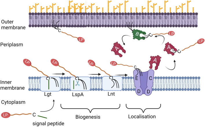
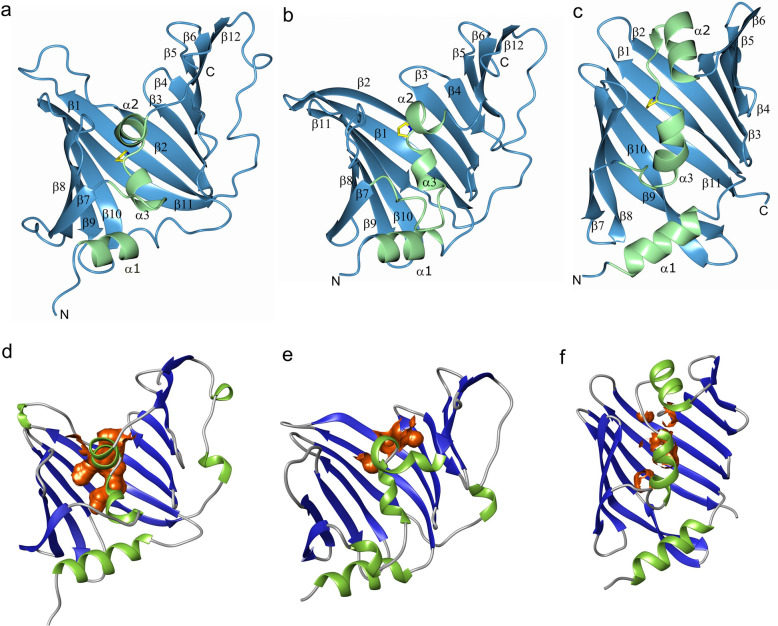
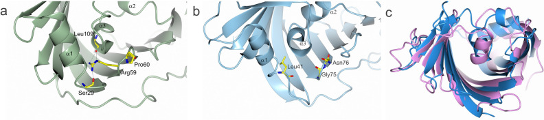
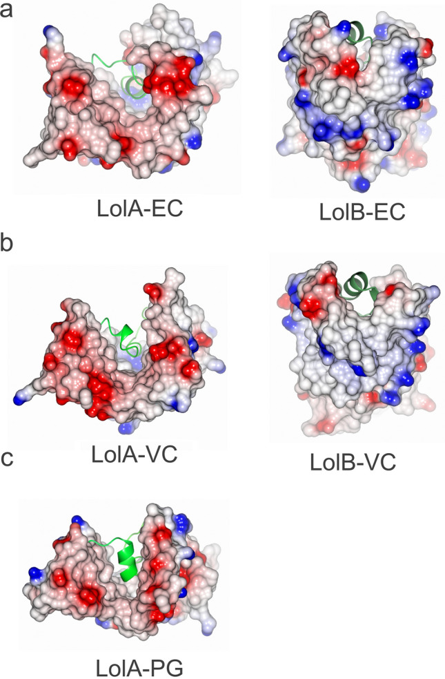
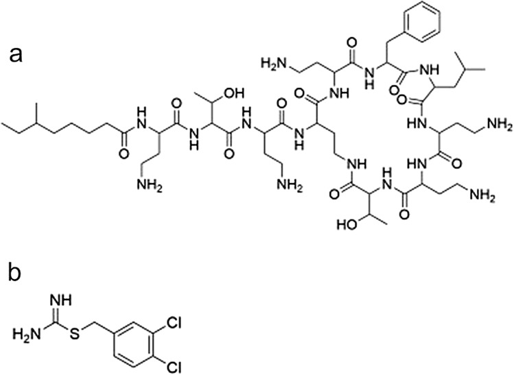
In Gram-negative bacteria, N-terminal lipidation is a signal for protein trafficking from the inner membrane (IM) to the outer membrane (OM). The IM complex LolCDE extracts lipoproteins from the membrane and moves them to the chaperone LolA. The LolA-lipoprotein complex crosses the periplasm after which the lipoprotein is anchored to the OM. In γ-proteobacteria anchoring is assisted by the receptor LolB, while a corresponding protein has not been identified in other phyla. In light of the low sequence similarity between Lol-systems from different phyla and that they may use different Lol components, it is crucial to compare representative proteins from several species. Here we present a structure–function study of LolA and LolB from two phyla: LolA from Porphyromonas gingivalis (phylum bacteroidota), and LolA and LolB from Vibrio cholerae (phylum proteobacteria). Despite large sequence differences, the LolA structures are very similar, hence structure and function have been conserved throughout evolution. However, an Arg-Pro motif crucial for function in γ-proteobacteria has no counterpart in bacteroidota. We also show that LolA from both phyla bind the antibiotic polymyxin B whereas LolB does not. Collectively, these studies will facilitate the development of antibiotics as they provide awareness of both differences and similarities across phyla.
Overview
The claims and statistics of this paper haven’t been checked yet.
Run the check — free, no account needed. It reads the open-access text and checks the sources it cites.
Integrity signals
Registry
Cites
Statistics
Figures
Verification
Claims the paper makes
The claims and statistics of this paper haven’t been checked yet.
Run the check — free, no account needed. It reads the open-access text and checks the sources it cites.
Self-check
Percentages, sums and reported differences re-computed from the paper’s own numbers.
The self-check needs the paper’s full text.
Citations
Cites
Highly accurate protein structure prediction with AlphaFold
Jumper, J., Evans, R., Pritzel, A., Green, T., Figurnov, M., Ronneberger, O., Tunyasuvunakool, K., Bates, R., Žídek, A., Potapenko, A., et al. (2021) · Nature · doi:10.1038/s41586-021-03819-2 · Cited by 43,764
Features and development of Coot
Emsley, P., Lohkamp, B., Scott, W. G., & Cowtan, K. (2010) · Acta Crystallographica Section D Biological Crystallography · doi:10.1107/s0907444910007493 · Cited by 29,915
Kabsch, W. (2010) · Acta Crystallographica Section D Biological Crystallography · doi:10.1107/s0907444909047337 · Cited by 16,463
Overview of the CCP 4 suite and current developments
Winn, M. D., Ballard, C. C., Cowtan, K. D., Dodson, E. J., Emsley, P., Evans, P. R., Keegan, R. M., Krissinel, E. B., Leslie, A. G. W., McCoy, A., et al. (2011) · Acta Crystallographica Section D Biological Crystallography · doi:10.1107/s0907444910045749 · Cited by 12,353
Towards automated crystallographic structure refinement with phenix.refine
Afonine, P. V., Grosse-Kunstleve, R. W., Echols, N., Headd, J. J., Moriarty, N. W., Mustyakimov, M., Terwilliger, T. C., Urzhumtsev, A., Zwart, P. H., & Adams, P. D. (2012) · Acta Crystallographica Section D Biological Crystallography · doi:10.1107/s0907444912001308 · Cited by 5,511
How good are my data and what is the resolution?
Evans, P. R., & Murshudov, G. N. (2013) · Acta Crystallographica Section D Biological Crystallography · doi:10.1107/s0907444913000061 · Cited by 4,697
Dali server: conservation mapping in 3D
Holm, L., & Rosenstr�m, P. (2010) · Nucleic Acids Research · doi:10.1093/nar/gkq366 · Cited by 3,640
CASTp 3.0: computed atlas of surface topography of proteins
Tian, W., Chen, C., Lei, X., Zhao, J., & Liang, J. (2018) · Nucleic Acids Research · doi:10.1093/nar/gky473 · Cited by 2,389
CASTp: Computed Atlas of Surface Topography of proteins
Binkowski, T. A. (2003) · Nucleic Acids Research · doi:10.1093/nar/gkg512 · Cited by 1,693
Automated macromolecular model building for X-ray crystallography using ARP/wARP version 7
Langer, G., Cohen, S. X., Lamzin, V. S., & Perrakis, A. (2008) · Nature Protocols · doi:10.1038/nprot.2008.91 · Cited by 1,606
Metadata
- Authors
- Jaiman, D., Nagampalli, R., & Persson, K.
- Affiliations
- Umeå Centre for Microbial Research (SE)
- Corresponding author
- Persson, Karina
- Venue
- Scientific Reports
- Published
- 2023-04-24
- Volume
- 13
- Issue
- 1
- Pages
- 6605-6605
- ISSN
- 2045-2322 (ISSN-L)
- PMC
- PMC10126205
- OpenAlex
- W4366829709
- Cited by
- 8
- References
- 48
- Citations by year
- 2026: 4 · 2024: 3 · 2023: 1
- FWCI
- 1.33
- Type
- journal article
- Language
- en
- Open access
- gold
- doi.org
- Licence
- cc-by
- creativecommons.org/licenses/by/4.0 (tdm)
- creativecommons.org/licenses/by/4.0 (vor)
- Locations
- Scientific Reports (publishedVersion, cc-by)
- Research Square (acceptedVersion, cc-by)
- PubMed Central (publishedVersion, cc-by)
- PubMed (publishedVersion)
- KTH Publication Database DiVA (KTH Royal Institute of Technology) (submittedVersion, other-oa)
- Desy publication database (The Deutsches Elektronen-Synchrotron) (submittedVersion, other-oa)
- DOAJ (DOAJ: Directory of Open Access Journals) (submittedVersion, cc-by-sa)
- DESY Publication Database (PUBDB) (Deutsches Elektronen-Synchrotron)
- Funders
- Vetenskapsrådet (2007-08673, 2016-05009)
- Stiftelsen Lars Hiertas Minne
- Medical faculty, Umeå University
- Umea University
- Topics
- Life Sciences › Biochemistry, Genetics and Molecular Biology › Genetics › Bacterial Genetics and Biotechnology
- Life Sciences › Biochemistry, Genetics and Molecular Biology › Molecular Biology › Lipid Membrane Structure and Behavior
- Life Sciences › Immunology and Microbiology › Microbiology › Bacterial Infections and Vaccines
- Keywords
- lipoprotein transport, LOLA, Gram-negative bacteria, Vibrio cholerae, Porphyromonas gingivalis, periplasmic chaperone
- MeSH terms
- Bacterial Outer Membrane Proteins, Carrier Proteins, Cell Membrane, Escherichia coli, Lipoproteins, Vibrio cholerae, Porphyromonas gingivalis, Protein Transport, Escherichia coli Proteins, Periplasmic Binding Proteins
About these checks
Registry, retraction, Cites and statistics results are computed, not judged, with one exception: a printed reference that no index lists is read once by a model before it counts as a citation error. Claims are extracted by a model and kept only when their quote appears verbatim in the text; each cited source is then checked, with a model’s help, for whether it says what the claim relies on. Those verdicts can be wrong — read the claim’s quote and the cited source before relying on one.
Statistics and arithmetic are recomputed, not judged.
Confidence bands are shown only for fields calibrated on our annotated test set. None is calibrated yet, so no band is shown.
What counts as a claim ›How we measure confidence ›How we check this ›
Are you an author, or do you see a problem with this page? Request a review
How to cite
Cite CoChat for the checks on this page, and the paper for its own findings.
- This page
- CoChat. (2026). Citation check: A comparative analysis of lipoprotein transport proteins: LolA and LolB from Vibrio cholerae and LolA from Porphyromonas gingivalis. https://app.cochat.ai/papers/10.1038/s41598-023-33705-y
- The paper
- Jaiman, D., Nagampalli, R., & Persson, K. (2023). A comparative analysis of lipoprotein transport proteins: LolA and LolB from Vibrio cholerae and LolA from Porphyromonas gingivalis. Scientific Reports. https://doi.org/10.1038/s41598-023-33705-y
Data sources
Bibliographic data: Semantic Scholar, OpenAlex, Crossref.
Contains information from the Semantic Scholar Academic Graph Datasets, made available under the ODC Attribution License (ODC-BY 1.0).
Semantic Scholar’s data is described in: Kinney, R., et al. (2023). The Semantic Scholar Open Data Platform. arXiv:2301.10140.
