

The DOI matches its Crossref record.
Effects of tumor-infiltrating lymphocytes on nonresponse rate of neoadjuvant chemotherapy in patients with invasive breast cancer
Open accessAbstract
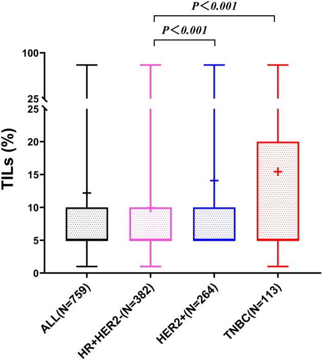
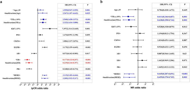
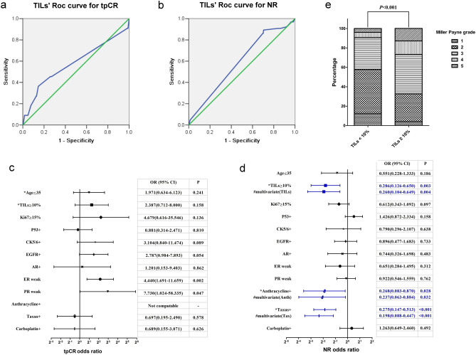
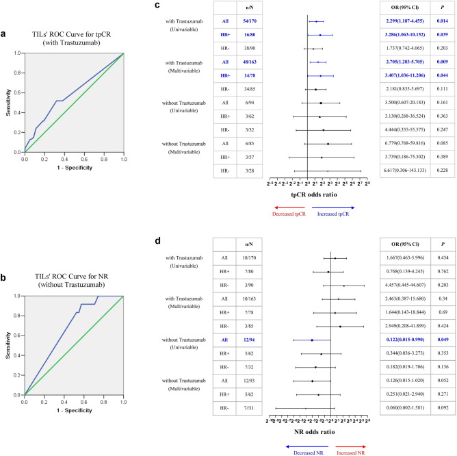
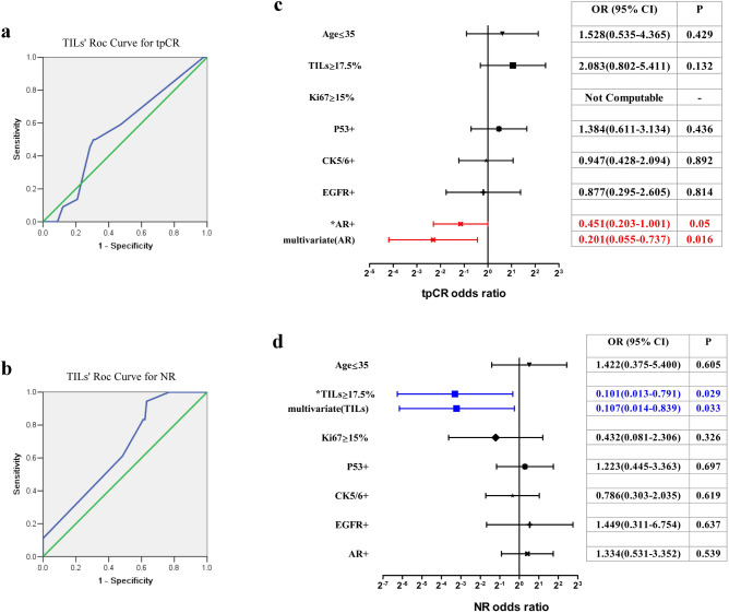
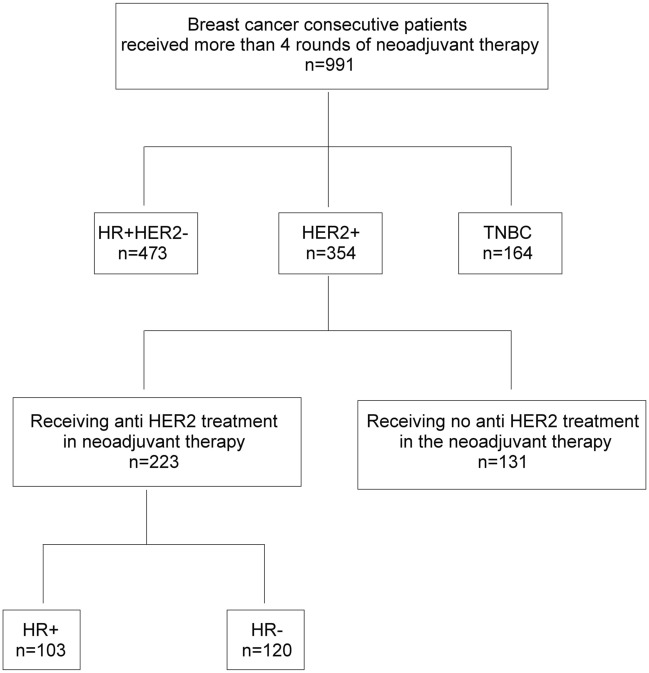
High level of tumor-infiltrating lymphocytes (TILs) can predict the rate of total pathological complete remission (tpCR) of breast cancer patients who receive neoadjuvant chemotherapy (NACT). This study focused on evaluating the data of patients whose primary tumor and/or lymph node metastasis show nonresponse (NR) to NACT, trying to provide a basis for the clinical decision which patients will develop NACT resistance. The study included breast cancers from 991 patients who received NACT. ROC curve analysis confirmed that TILs showed significant predictive value for NR of hormone receptor (HR)+HER2− and triple-negative breast cancer (TNBC). Among HR+HER2− breast cancer, TILs ≥ 10% was an independent predictor for low NR rate. Furthermore, positive correlation of TILs with Ki67 index and Miller-Payne grade, and negative correlation with ER and PR H-scores were only identified in this subgroup. In TNBC, TILs ≥ 17.5% was an independent predictor for low NR rate. The predictive value of low TILs on NR may facilitate to screen patients with HR+HER2− or TNBC who may not benefit from NACT. HR+HER2− breast cancer with low levels of TILs should be carefully treated with neoadjuvant chemotherapy, and other alternatives such as neoadjuvant endocrine therapy can be considered.
Overview
The claims and statistics of this paper haven’t been checked yet.
Run the check — free, no account needed. It reads the open-access text and checks the sources it cites.
Integrity signals
Registry
Cites
Statistics
Figures
Verification
Claims the paper makes
The claims and statistics of this paper haven’t been checked yet.
Run the check — free, no account needed. It reads the open-access text and checks the sources it cites.
Claims other papers make about this one
Each is checked against this paper’s own text: does it say what the citing paper says it does?
The source backs part of the claim, not all of it — or backs it for a different population, condition or comparison.
In the citing paper
“These results are in line with previously published studies [58–66].”
From Digital image analysis and machine learning-assisted prediction of neoadjuvant chemotherapy response in triple-negative breast cancer (2024) · cites this paper as [62]
Experimental — accuracy not yet measured
Self-check
Percentages, sums and reported differences re-computed from the paper’s own numbers.
The self-check needs the paper’s full text.
Citations
Cites
No references could be resolved yet.
Research works printed but not linked: 30
- Fisher, E. R., Wang, J., Bryant, J., Fisher, B. & Wolmark, N. Pathobiology of preoperative chemotherapy: Findings from the national surgical adjuvant breast and bowel (NSABP) protocol B-18. Cancer 95, 681–695. https://doi.org/10.1002/cncr.10741 (2002). · DOI
- Hortobagyi, G. N. Comprehensive management of locally advanced breast cancer. Cancer 66, 1387. https://doi.org/10.1002/1097-0142(19900915)66:14+3.0.CO;2-I (1990). · DOI
- von Minckwitz, G. et al. Trastuzumab emtansine for residual invasive her2-positive breast cancer. New Engl. J. Med. 380, 617–628. https://doi.org/10.1056/NEJMoa1814017 (2018). · DOI
- Kuerer, H. M. et al. Clinical course of breast cancer patients with complete pathologic primary tumor and axillary lymph node response to doxorubicin-based neoadjuvant chemotherapy. J. Clin. Oncol. 17, 460–469. https://doi.org/10.1200/JCO.1999.17.2.460 (1999). · DOI
- Rosenberg, S., Spiess, P. & Lafreniere, R. A new approach to the adoptive immunotherapy of cancer with tumor-infiltrating lymphocytes. Science 233, 1318–1321. https://doi.org/10.1126/science.3489291 (1986). · DOI
- Tsuta, K. et al. Primary lung adenocarcinoma with massive lymphocyte infiltration. Am. J. Clin. Pathol. 123, 547–552. https://doi.org/10.1309/APKQ-4Q9D-52GN-LR8W (2005). · DOI
- Stanton, S. E. & Disis, M. L. Clinical significance of tumor-infiltrating lymphocytes in breast cancer. J. Immunother. Cancer 4, 59. https://doi.org/10.1186/s40425-016-0165-6 (2016). · DOI
- Cortazar, P. et al. Pathological complete response and long-term clinical benefit in breast cancer: The CTNeoBC pooled analysis. Lancet 384, 164–172. https://doi.org/10.1016/S0140-6736(13)62422-8 (2014). · DOI
- Minckwitz, G. V., Untch, M., Blohmer, J. U., Costa, S. D. & Loibl, S. Definition and impact of pathologic complete response on prognosis after neoadjuvant chemotherapy in various intrinsic breast cancer subtypes. J. Clin. Oncol. 30, 1796–1804. https://doi.org/10.1200/JCO.2011.38.8595 (2012). · DOI · Paper page
- Ogston, K. N. et al. A new histological grading system to assess response of breast cancers to primary chemotherapy: Prognostic significance and survival. Breast 12, 320–327. https://doi.org/10.1016/S0960-9776(03)00106-1 (2003). · DOI
- Sataloff, D. M. et al. Pathologic response to induction chemotherapy in locally advanced carcinoma of the breast: A determinant of outcome. J. Am. Coll. Surg. 180, 297–306 (1995).
- Karagiannis, G. S. et al. Neoadjuvant chemotherapy induces breast cancer metastasis through a TMEM-mediated mechanism. Sci. Transl. Med. 9, eaan0026. https://doi.org/10.1126/scitranslmed.aan0026 (2017). · DOI
- Keklikoglou, I. et al. Chemotherapy elicits pro-metastatic extracellular vesicles in breast cancer models. Nat. Cell Biol. 21, 190–202. https://doi.org/10.1038/s41556-018-0256-3 (2019). · DOI
- von Minckwitz, G. et al. Response-guided neoadjuvant chemotherapy for breast cancer. J. Clin. Oncol. 31, 3623–3630. https://doi.org/10.1200/JCO.2012.45.0940 (2013). · DOI
- Stanton, S. E., Adams, S. & Disis, M. L. Variation in the incidence and magnitude of tumor-infiltrating lymphocytes in breast cancer subtypes: A systematic review. JAMA Oncol. 2, 1354–1360. https://doi.org/10.1001/jamaoncol.2016.1061 (2016). · DOI
- Salgado, R. et al. Tumor-infiltrating lymphocytes and associations with pathological complete response and event-free survival in HER2-positive early-stage breast cancer treated with lapatinib and trastuzumab: a secondary analysis of the NeoALTTO trial. JAMA Oncol. 1, 448–454. https://doi.org/10.1001/jamaoncol.2015.0830 (2015). · DOI
- Loi, S. et al. Tumor infiltrating lymphocytes are prognostic in triple negative breast cancer and predictive for trastuzumab benefit in early breast cancer: results from the finher trial. Eur. Phys. J. D. 41, 485–492. https://doi.org/10.1140/epjd/e2007-00013-1 (2014). · DOI
- Luen, S. J. et al. Tumour-infiltrating lymphocytes in advanced HER2-positive breast cancer treated with pertuzumab or placebo in addition to trastuzumab and docetaxel: A retrospective analysis of the cleopatra study. Lancet Oncol. 18, 52–62. https://doi.org/10.1016/S1470-2045(16)30631-3 (2017). · DOI
- Hwang, H. W. et al. A nomogram to predict pathologic complete response (pCR) and the value of tumor-infiltrating lymphocytes (TILs) for prediction of response to neoadjuvant chemotherapy (NAC) in breast cancer patients. Breast Cancer Res. Treat. 173, 255–256. https://doi.org/10.1007/s10549-018-4981-x (2019). · DOI
- Luen, S. J., Salgado, R., Dieci, M. V., Vingiani, A. & Loi, S. Prognostic implications of residual disease tumor-infiltrating lymphocytes and residual cancer burden in triple-negative breast cancer patients after neoadjuvant chemotherapy. Ann. Oncol. 30, 236–242. https://doi.org/10.1093/annonc/mdy547 (2019). · DOI
- Criscitiello, C., Vingiani, A., Maisonneuve, P., Viale, G. & Curigliano, G. Tumor-infiltrating lymphocytes (tils) in ER+/HER2 breast cancer. Breast Cancer Res. Treat. 183, 347–354. https://doi.org/10.1007/s10549-020-05771-7 (2020). · DOI
- Denkert, C. et al. Tumour-infiltrating lymphocytes and prognosis in different subtypes of breast cancer: A pooled analysis of 3771 patients treated with neoadjuvant therapy. Lancet Oncol. 19, 40–50. https://doi.org/10.1016/S1470-2045(17)30904-X (2018). · DOI · Paper page
- Rugo, H. et al. Performance of PD-L1 immunohistochemistry assays in unresectable locally advanced or metastatic triple-negative breast cancer: Post hoc analysis of IMpassion130. Ann. Oncol. 30, v858–v859. https://doi.org/10.1093/annonc/mdz394.009 (2019). · DOI · Paper page
- Dieci, M. V., Miglietta, F. & Guarneri, V. Immune infiltrates in breast cancer: Recent updates and clinical implications. Cells 10, 223. https://doi.org/10.3390/cells10020223 (2021). · DOI
- Prat, A. et al. HER2-enriched subtype and ERBB2 expression in HER2-positive breast cancer treated with dual HER2 blockade. J. Natl. Cancer I(112), 46–54. https://doi.org/10.1093/jnci/djz042 (2020). · DOI
- Salgado, R. et al. International TILs Working Group 2014 The evaluation of tumor-infiltrating lymphocytes (TILs) in breast cancer: Recommendations by an International TILs Working Group 2014. Ann. Oncol. 26, 259–271. https://doi.org/10.1093/annonc/mdu450 (2015). · DOI
- Qian, X. L. et al. Syndecan binding protein (SDCBP) is over expressed in estrogen recaptor negative breast cancers, and is a potential promoter for tumor proliferation. PLoS ONE 8, e60046. https://doi.org/10.1371/journal.pone.0060046 (2013). · DOI
- Hammond, M. E. et al. American Society of Clinical Oncology/College of American Pathologists guideline recommendations for immunohistochemical testing of estrogen and progesterone receptors in breast cancer. J. Clin. Oncol. 28, 2784–2795. https://doi.org/10.1200/JCO.2009.25.6529 (2010). · DOI
- Mccarty, K. S., Szabo, E., Flowers, J. L., Cox, E. B. & Creasman, W. T. Use of a monoclonal anti-estrogen receptor antibody in the immunohistochemical evaluation of human tumors. Cancer Res. 46, 4244s–4248s. https://doi.org/10.1016/0304-3835(86)90123-0 (1986). · DOI
- Wolff, A. C. et al. Human epidermal growth factor receptor 2 testing in breast cancer: American Society of Clinical Oncology/College of American Pathologists Clinical Practice Guideline Focused Update. J. Clin. Oncol. 36, 2105–2122. https://doi.org/10.1200/JCO.2018.77.8738 (2018). · DOI
Cited by
Sorted by citations
Fisher, T. B., Saini, G., Rekha, T. S., Krishnamurthy, J., Bhattarai, S., Callagy, G., Webber, M., Janssen, E. A. M., Kong, J., & Aneja, R. (2024) · Breast Cancer Research · doi:10.1186/s13058-023-01752-y · Cited by 42
Metadata
- Authors
- Qian, X.-L., Xia, X.-Q., Li, Y.-Q., Jia, Y.-M., Sun, Y.-Y., Song, Y.-M., Xue, H.-Q., Hao, Y.-F., Wang, J., Wang, X.-Z., Liu, C.-Y., Zhang, X.-M., Zhang, L.-N., & Guo, X.-J.
- Affiliations
- Tianjin Medical University Cancer Institute and Hospital (CN)
- ORCID
- Liu, Chen-Ying
- Venue
- Scientific Reports
- Published
- 2023-06-07
- Volume
- 13
- Issue
- 1
- Pages
- 9256-9256
- ISSN
- 2045-2322 (ISSN-L)
- PMC
- PMC10247751
- OpenAlex
- W4379767997
- Cited by
- 8
- References
- 0
- Citations by year
- 2026: 1 · 2025: 4 · 2024: 2 · 2023: 1
- FWCI
- 1.02
- Type
- journal article
- Language
- en
- Open access
- gold
- doi.org
- Locations
- Scientific Reports (publishedVersion, cc-by)
- PubMed Central (publishedVersion, cc-by)
- PubMed (publishedVersion)
- DOAJ (DOAJ: Directory of Open Access Journals) (submittedVersion, cc-by-sa)
- Funders
- National Natural Science Foundation of China (81772840)
- Topics
- Life Sciences › Biochemistry, Genetics and Molecular Biology › Cancer Research › Breast Cancer Treatment Studies
- Health Sciences › Medicine › Pathology and Forensic Medicine › Breast Lesions and Carcinomas
- Health Sciences › Medicine › Radiology, Nuclear Medicine and Imaging › MRI in cancer diagnosis
- Keywords
- tumor-infiltrating lymphocytes, neoadjuvant chemotherapy, breast cancer, treatment non-response, predictive biomarkers, triple-negative breast cancer, hormone receptor-positive HER2-negative breast cancer, Ki-67, endocrine therapy, lymph node metastasis
- MeSH terms
- Antineoplastic Combined Chemotherapy Protocols, Breast Neoplasms, Female, Humans, Prognosis, Lymphocytes, Tumor-Infiltrating, Receptor, ErbB-2, Erb-b2 Receptor Tyrosine Kinases, Neoadjuvant Therapy, Triple Negative Breast Neoplasms
About these checks
Registry, retraction, Cites and statistics results are computed, not judged, with one exception: a printed reference that no index lists is read once by a model before it counts as a citation error. Claims are extracted by a model and kept only when their quote appears verbatim in the text; each cited source is then checked, with a model’s help, for whether it says what the claim relies on. Those verdicts can be wrong — read the claim’s quote and the cited source before relying on one.
Statistics and arithmetic are recomputed, not judged.
Confidence bands are shown only for fields calibrated on our annotated test set. None is calibrated yet, so no band is shown.
What counts as a claim ›How we measure confidence ›How we check this ›
Are you an author, or do you see a problem with this page? Request a review
How to cite
Cite CoChat for the checks on this page, and the paper for its own findings.
- This page
- CoChat. (2026). Citation check: Effects of tumor-infiltrating lymphocytes on nonresponse rate of neoadjuvant chemotherapy in patients with invasive breast cancer. https://app.cochat.ai/papers/10.1038/s41598-023-36517-2
- The paper
- Qian, X.-L., Xia, X.-Q., Li, Y.-Q., Jia, Y.-M., Sun, Y.-Y., Song, Y.-M., Xue, H.-Q., Hao, Y.-F., Wang, J., Wang, X.-Z., Liu, C.-Y., Zhang, X.-M., Zhang, L.-N., & Guo, X.-J. (2023). Effects of tumor-infiltrating lymphocytes on nonresponse rate of neoadjuvant chemotherapy in patients with invasive breast cancer. Scientific Reports. https://doi.org/10.1038/s41598-023-36517-2
