

The DOI matches its Crossref record; 2 of the 2 references it cites were found in a registry or an index.
Lusca: FIJI (ImageJ) based tool for automated morphological analysis of cellular and subcellular structures
Abstract
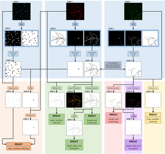
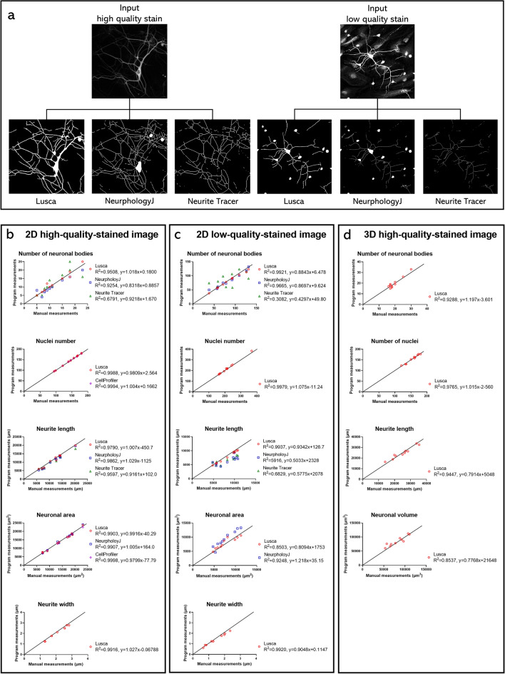
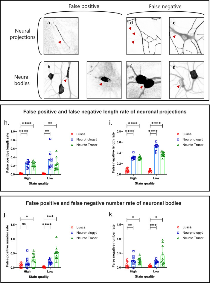
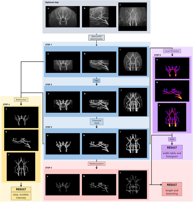
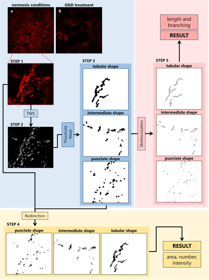
The human body consists of diverse subcellular, cellular and supracellular structures. Neurons possess varying-sized projections that interact with different cellular structures leading to the development of highly complex morphologies. Aiming to enhance image analysis of complex biological forms including neurons using available FIJI (ImageJ) plugins, Lusca, an advanced open-source tool, was developed. Lusca utilizes machine learning for image segmentation with intensity and size thresholds. It performs particle analysis to ascertain parameters such as area/volume, quantity, and intensity, in addition to skeletonization for determining length, branching, and width. Moreover, in conjunction with colocalization measurements, it provides an extensive set of 29 morphometric parameters for both 2D and 3D analysis. This is a significant enhancement compared to other scripts that offer only 5–15 parameters. Consequently, it ensures quicker and more precise quantification by effectively eliminating noise and discerning subtle details. With three times larger execution speed, fewer false positive and negative results, and the capacity to measure various parameters, Lusca surpasses other existing open-source solutions. Its implementation of machine learning-based segmentation facilitates versatile applications for different cell types and biological structures, including mitochondria, fibres, and vessels. Lusca’s automated and precise measurement capability makes it an ideal choice for diverse biological image analyses.
Overview
The claims and statistics of this paper haven’t been checked yet.
Run the check — free, no account needed. It reads the open-access text and checks the sources it cites.
Integrity signals
Registry
Cites
Statistics
Figures
Verification
Claims the paper makes
The claims and statistics of this paper haven’t been checked yet.
Run the check — free, no account needed. It reads the open-access text and checks the sources it cites.
Claims other papers make about this one
Each is checked against this paper’s own text: does it say what the citing paper says it does?
In the citing paper
“Weka, an open-source software tool that enables users to perform ML-based image segmentation, object classification, and feature extraction with minimal programming experience,33-35 has been widely used in biological and clinical research to quantitatively analyze various types of microorganisms, cells, and tissues.36-40”
From SEMTWIST Quantification of Biofilm Infection in Human Chronic Wound Using Scanning Electron Microscopy and Machine Learning (2025) · cites this paper as [37]
Self-check
Percentages, sums and reported differences re-computed from the paper’s own numbers.
The self-check needs the paper’s full text.
Citations
Cites
Image analysis and machine learning in digital pathology: challenges and opportunities
Madabhushi, A., & Lee, G. (2016) · Medical Image Analysis · doi:10.1016/j.media.2016.06.037 · Cited by 1,093
Computational classification of mitochondrial shapes reflects stress and redox state
Ahmad, T., Aggarwal, K., Pattnaik, B., Mukherjee, S., Sethi, T., Tiwari, B. K., Kumar, M., Micheal, A., Mabalirajan, U., Ghosh, B., et al. (2013) · Cell Death & Disease · doi:10.1038/cddis.2012.213 · Cited by 233
Research works printed but not linked: 30
- Titus, A. D. J. et al. Hypobaric hypoxia-induced dendritic atrophy of hippocampal neurons is associated with cognitive impairment in adult rats. Neuroscience 145, 265–278 (2007). · DOI
- York, E. M., LeDue, J. M., Bernier, L.-P. & MacVicar, B. A. 3DMorph automatic analysis of microglial morphology in three dimensions from ex vivo and in vivo imaging. eNeuro 5(6), ENEURO.0266–18.2018. (2018). · DOI
- Abramoff, M. D., Magalhaes, P. J. & Ram, S. J. Image processing with ImageJ. Biophoton. Int. 11, 36–42 (2004).
- Carpenter, A. E. et al. Cell Profiler: Image analysis software for identifying and quantifying cell phenotypes. Genome Biol. 7, R100 (2006). · DOI
- Meijering, E. et al. Design and validation of a tool for neurite tracing and analysis in fluorescence microscopy images. Cytometry. A 58, 167–176 (2004). · DOI
- Myatt, D. R., Hadlington, T., Ascoli, G. A. & Nasuto, S. J. Neuromantic—from semi-manual to semi-automatic reconstruction of neuron morphology. Front. Neuroinform. 6, 4 (2012). · DOI
- Peng, H., Ruan, Z., Long, F., Simpson, J. H. & Myers, E. W. V3D enables real-time 3D visualization and quantitative analysis of large-scale biological image data sets. Nat. Biotechnol. 28, 348–353 (2010). · DOI · Paper page
- Peng, H., Long, F., Zhao, T. & Myers, E. Proof-editing is the bottleneck of 3D neuron reconstruction: The problem and solutions. Neuroinformatics 9, 103–105 (2011). · DOI
- Rodriguez, A., Ehlenberger, D. B., Hof, P. R. & Wearne, S. L. Rayburst sampling, an algorithm for automated three-dimensional shape analysis from laser scanning microscopy images. Nat. Protoc. 1, 2152–2161 (2006). · DOI
- Pool, M., Thiemann, J., Bar-Or, A. & Fournier, A. E. NeuriteTracer: A novel ImageJ plugin for automated quantification of neurite outgrowth. J. Neurosci. Methods 168, 134–139 (2008). · DOI
- Ho, S.-Y. et al. NeurphologyJ: An automatic neuronal morphology quantification method and its application in pharmacological discovery. BMC Bioinform. 12, 230 (2011). · DOI
- Boulan, B. et al. AutoNeuriteJ: An ImageJ plugin for measurement and classification of neuritic extensions. PLoS ONE 15, e0234529 (2020). · DOI
- Arganda-Carreras, I. et al. Trainable weka segmentation: A machine learning tool for microscopy pixel classification. Bioinformatics 33, 2424–2426 (2017). · DOI · Paper page
- Bolte, S. & Cordelières, F. P. A guided tour into subcellular colocalization analysis in light microscopy. J. Microsc. 224, 213–232 (2006). · DOI
- Cruz-Matías, I. et al. Sphericity and roundness computation for particles using the extreme vertices model. J. Comput. Sci. 30, 28–40 (2019). · DOI
- Arganda-Carreras, I., Fernández-González, R., Muñoz-Barrutia, A. & Ortiz-De-Solorzano, C. 3D reconstruction of histological sections: Application to mammary gland tissue. Microsc. Res. Tech. 73, 1019–1029 (2010). · DOI
- Dougherty, R. & Kunzelmann, K. Computing local thickness of 3D structures with ImageJ. Microsc. Microanal. 13, 1678–1679 (2007). · DOI
- Dunn, K. W., Kamocka, M. M. & McDonald, J. H. A practical guide to evaluating colocalization in biological microscopy. Am. J. Physiol. Cell Physiol. 300, C723–C742 (2011). · DOI
- Encarnacion-Rivera, L., Foltz, S., Hartzell, H. C. & Choo, H. Myosoft: An automated muscle histology analysis tool using machine learning algorithm utilizing FIJI/ImageJ software. PLoS ONE 15, e0229041 (2020). · DOI
- Justić, H. et al. Redefining the Koizumi model of mouse cerebral ischemia: A comparative longitudinal study of cerebral and retinal ischemia in the Koizumi and Longa middle cerebral artery occlusion models. J. Cereb. Blood Flow Metab Off. J. Int. Soc. Cereb. Blood Flow Metab. 42, 2080–2094 (2022). · DOI
- Jagečić, D., Petrović, D. J., Šimunić, I., Isaković, J. & Mitrečić, D. The oxygen and glucose deprivation of immature cells of the nervous system exerts distinct effects on mitochondria, mitophagy, and autophagy, depending on the cells’ differentiation stage differentiation stage. Brain Sci. 13, 910 (2023). · DOI
- Schindelin, J. et al. Fiji: An open-source platform for biological-image analysis. Nat. Methods 9, 676–682 (2012). · DOI · Paper page
- van Ginneken, B. Fifty years of computer analysis in chest imaging: Rule-based, machine learning, deep learning. Radiol. Phys. Technol. 10, 23–32 (2017). · DOI
- Deo, R. C. Machine learning in medicine. Circulation 132, 1920–1930 (2015). · DOI · Paper page
- Gurcan, M. N. et al. Histopathological image analysis: A review. IEEE Rev. Biomed. Eng. 2, 147–171 (2009). · DOI · Paper page
- Patel, L., Shukla, T., Huang, X., Ussery, D. W. & Wang, S. Machine learning methods in drug discovery. Molecules 25, 5277 (2020). · DOI · Paper page
- Koizumi, J. et al. Experimental studies of ischemic brain edema. Jpn. J. Stroke 8, 1–8 (1986). · DOI · Paper page
- Longa, E. Z., Weinstein, P. R., Carlson, S. & Cummins, R. Reversible middle cerebral artery occlusion without craniectomy in rats. Stroke 20, 84–91 (1989). · DOI · Paper page
- Stirling, D. R. et al. Cell Profiler 4: Improvements in speed, utility and usability. BMC Bioinform. 22, 433 (2021). · DOI
- Pani, G. et al. MorphoNeuroNet: An automated method for dense neurite network analysis. Cytom. Part A J. Int. Soc. Anal. Cytol. 85, 188–199 (2014). · DOI
Cited by
Sorted by citations
Singh, S., Muniz De Oliveira, F., Wang, C., Kumar, M., Xuan, Y., DeMazumder, D., Sen, C. K., & Roy, S. (2025) · Advances in Wound Care · doi:10.1089/wound.2024.0291 · Cited by 6
Metadata
- Authors
- Šimunić, I., Jagečić, D., Isaković, J., Dobrivojević Radmilović, M., & Mitrečić, D.
- Venue
- Scientific Reports
- Published
- 2024-03-28
- Volume
- 14
- Issue
- 1
- ISSN
- 2045-2322
- PMC
- PMC10978859
- Cited by
- 18
- References
- 2
- Language
- en
- Update notices
- correction (2024-04-19)
- Funders
- Hrvatska Zaklada za Znanost (UIP-2017-05-8082, IP-2022-10-4656)
About these checks
Registry, retraction, Cites and statistics results are computed, not judged, with one exception: a printed reference that no index lists is read once by a model before it counts as a citation error. Claims are extracted by a model and kept only when their quote appears verbatim in the text; each cited source is then checked, with a model’s help, for whether it says what the claim relies on. Those verdicts can be wrong — read the claim’s quote and the cited source before relying on one.
Statistics and arithmetic are recomputed, not judged.
Confidence bands are shown only for fields calibrated on our annotated test set. None is calibrated yet, so no band is shown.
What counts as a claim ›How we measure confidence ›How we check this ›
Are you an author, or do you see a problem with this page? Request a review
How to cite
Cite CoChat for the checks on this page, and the paper for its own findings.
- This page
- CoChat. (2026). Citation check: Lusca: FIJI (ImageJ) based tool for automated morphological analysis of cellular and subcellular structures. https://app.cochat.ai/papers/10.1038/s41598-024-57650-6
- The paper
- Šimunić, I., Jagečić, D., Isaković, J., Dobrivojević Radmilović, M., & Mitrečić, D. (2024). Lusca: FIJI (ImageJ) based tool for automated morphological analysis of cellular and subcellular structures. Scientific Reports. https://doi.org/10.1038/s41598-024-57650-6
