

The DOI matches its Crossref record; 210 of the 210 references it cites were found in a registry or an index.
Real world perspectives on endometriosis disease phenotyping through surgery, omics, health data, and artificial intelligence
Open accessAbstract
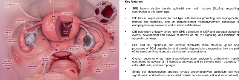
Endometriosis is an enigmatic disease whose diagnosis and management are being transformed through innovative surgical, molecular, and computational technologies. Integrating single-cell and other omic disease data with clinical and surgical metadata can identify multiple disease subtypes with translation to novel diagnostics and therapeutics. Herein, we present real-world perspectives on endometriosis and the importance of multidisciplinary collaboration in informing molecular, epidemiologic, and cell-specific data in the clinical and surgical contexts.
Overview
The claims and statistics of this paper haven’t been checked yet.
Run the check — free, no account needed. It reads the open-access text and checks the sources it cites.
Integrity signals
Registry
Cites
Statistics
Figures
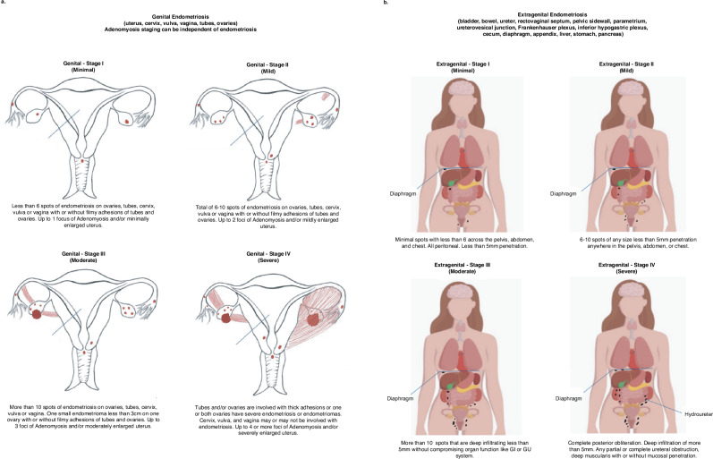
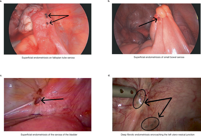
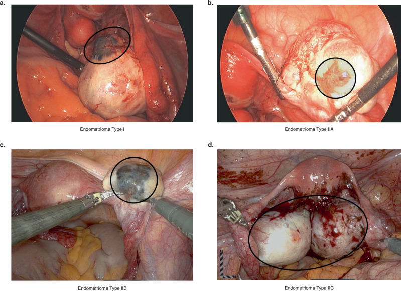
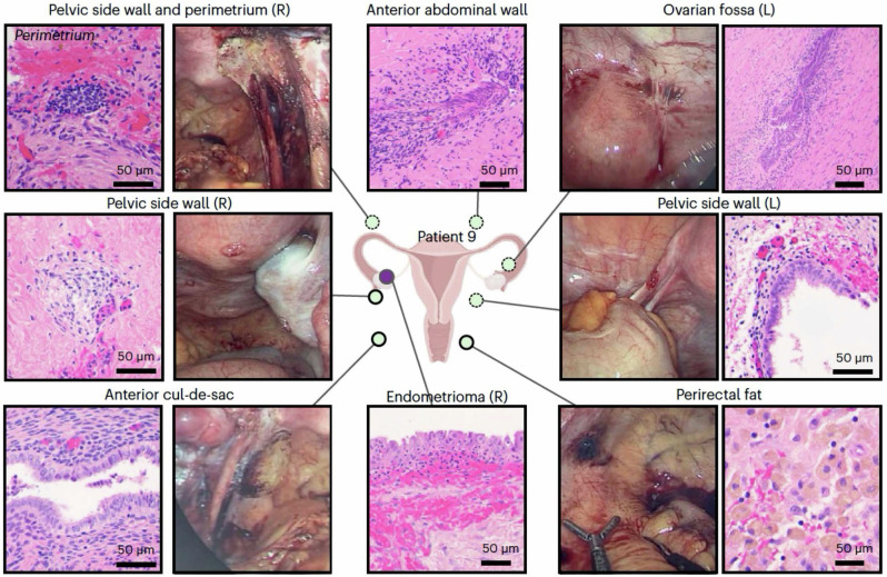
Verification
Claims the paper makes
The claims and statistics of this paper haven’t been checked yet.
Run the check — free, no account needed. It reads the open-access text and checks the sources it cites.
Claims other papers make about this one
Each is checked against this paper’s own text: does it say what the citing paper says it does?
The check wasn’t confident either way, so it gives no verdict — never disputed.
In the citing paper
“9 patients (3.81%) were classified as Stage I, 28 (11.86%) as Stage II, 58 (24.58%) as Stage III, and 141 (59.75%) as Stage IV”
From Selective Appendectomy in Patients Undergoing Minimally Invasive Surgery for Endometriosis: A Retrospective Cohort Study (2025) · cites this paper as [17]
Experimental — accuracy not yet measured
Self-check
Percentages, sums and reported differences re-computed from the paper’s own numbers.
The self-check needs the paper’s full text.
Citations
Cites
Items 201–210 of 210
Phosphoproteomics Analysis of Endometrium in Women with or without Endometriosis
Xu, H., Deng, H., Liu, C., Chen, Y., & Zhang, Z. (2015) · Chinese Medical Journal · doi:10.4103/0366-6999.166022 · Cited by 6
Nezhat, F. R., Kavic, M. S., Nezhat, C., & NEZHAT, C. R. (2023) · JSLS Journal of the Society of Laparoscopic & Robotic Surgeons · doi:10.4293/jsls.2022.00073 · Cited by 6
Self-citationColgrave, E. M., Keast, J. R., Nowell, C. J., Healey, M., Rogers, P. A. W., Holdsworth‐Carson, S. J., & Girling, J. E. (2023) · Reproductive BioMedicine Online · doi:10.1016/j.rbmo.2023.103610 · Cited by 6
Bui, B. N., Ardisasmita, A. I., Kuijk, E., Altmäe, S., Steba, G. S., Mackens, S., Fuchs, S. A., Broekmans, F. J., & Nieuwenhuis, E. E. S. (2023) · Human Reproduction · doi:10.1093/humrep/dead257 · Cited by 5
Almonte, A. L., Wang, W., Flynn, E., Houshdaran, S., Vallvé-Juanico, J., Irwin, J. C., Kushnoor, D., Combes, A. J., Fragiadakis, G. K., Sirota, M., et al. (2023) · Fertility and Sterility · doi:10.1016/j.fertnstert.2023.08.204 · Cited by 4
Self-citationChen, C. V., Orlando, M. S., Abel, M. K., Opoku‐Anane, J., & Rabban, J. T. (2024) · International Journal of Gynecological Pathology · doi:10.1097/pgp.0000000000001071 · Cited by 4
Towards a more biologically informative system of endometriosis classification
Mijatovic, V. (2020) · Human Reproduction · doi:10.1093/humrep/deaa265 · Cited by 3
Maekawa, R., Ota, Y., Ota, I., Mihara, Y., Takasaki, H., Sato, S., Tamura, I., Shirafuta, Y., Shinagawa, M., Fujimura, T., et al. (2023) · Reproductive Medicine and Biology · doi:10.1002/rmb2.12548 · Cited by 2
Nezhat, C., & Schipper, E. (2011) · Jaypee Brothers Medical Publishers (P) Ltd. eBooks · doi:10.5005/jp/books/12770_4 · Cited by 1
Self-citationMetabolomics as a Candidate for Endometriosis Biomarker: A Systematic Review
Meidyana, S., & Fedora, K. (2024) · Fertility & Reproduction · doi:10.1142/s2661318224500075 · Cited by 1
Research works printed but not linked: 5
- Burney, R. O. & Giudice, L. C. In Degroot’s Endocrinology (eds. Burney, R. O. et al.) Vol. 18, 2079–2092 (Elesiver, 2022).
- Giudice, L. C. Advances in approaches to diagnose endometriosis. Glob Reprod. Health 9, e0074 (2024).
- Köhler, G. & Lorenz, G. Zur Korrelation von endoskopischem und histologischem Bild der Endometriose. Endometriose 4, 56–60 (1991).
- Nezhat, F., Nezhat, C., Nezhat, C. & Admon, D. A fresh look at ovarian endometriomas. Contemp. Ob. Gyn. 39, 81–94 (1994).
- Nezhat, C. R., Nezhat, F. & Nezhat, C. H. Nezhat’s Video-Assisted and Robotic-Assisted Laparoscopy and Hysteroscopy (Cambridge University Press, 2013).
Cited by
Sorted by citations
Non-Hormonal Strategies in Endometriosis: Targets with Future Clinical Potential
Ramos‐Niño, M. E. (2025) · Journal of Clinical Medicine · doi:10.3390/jcm14145091 · Cited by 16
Graciano-España, M. D. C., Barnhart, K. T., Gonzáléz-Monfort, M., Arenas-Barrero, M., Legro, R. S., Thomas, T. R., Rush, M., Vilella, F., Sánchez, M. F., Simón, C., et al. (2025) · Fertility and Sterility · doi:10.1016/j.fertnstert.2025.06.035 · Cited by 6
The Nexus of Iron, Senescence, and Fibrosis in Endometriosis: A Narrative Review
Patel, R., Kalthur, G., Datta, R., Shah, S., & Dutta, R. (2025) · Reproductive Sciences · doi:10.1007/s43032-025-01999-0 · Cited by 6
Adamyan, L. V., Михалева, Л. М., Сереженков, В. А., Tkachev, N. А., Pivazyan, L. G., Кузнецова, М., Mailova, K. S., Stepanyan, A. A., & Полеткина, А. А. (2025) · Russian Journal of Human Reproduction · doi:10.17116/repro20253104126 · Cited by 4
Manu, A., Poenaru, E., Duică, F., Stoleru, S., Bauşic, A. I. G., Coroleuca, B.-C., Coroleucă, C. A., Iacob, C. M., & Brătilă, E. (2025) · Journal of Clinical Medicine · doi:10.3390/jcm14207256 · Cited by 4
Metadata
- Authors
- Nezhat, C. R., Oskotsky, T. T., Robinson, J. F., Fisher, S. J., Tsuei, A., Liu, B., Irwin, J. C., Gaudilliere, B., Sirota, M., Stevenson, D. K., & Giudice, L. C.
- Affiliations
- University of California, San Francisco (US)
- Center for Special Minimally Invasive and Robotic Surgery (US)
- Stanford Medicine (US)
- Stanford University (US)
- Corresponding author
- Nezhat, Camran R.
- Giudice, Linda C.
- ORCID
- Venue
- npj Women s Health
- Published
- 2025-02-06
- Volume
- 3
- Issue
- 1
- Pages
- 8-8
- ISSN
- 2948-1716 (ISSN-L)
- PMC
- PMC11802455
- OpenAlex
- W4407167810
- Cited by
- 25
- References
- 209
- Citations by year
- 2026: 15 · 2025: 10
- FWCI
- 3.44
- Type
- journal article
- Language
- en
- Open access
- gold
- doi.org
- Licence
- Locations
- npj Women s Health (publishedVersion, cc-by-nc-nd)
- PubMed Central (publishedVersion, cc-by-nc-nd)
- PubMed (publishedVersion)
- eScholarship (California Digital Library) (submittedVersion, cc-by-nc-nd)
- DOAJ (DOAJ: Directory of Open Access Journals) (submittedVersion)
- Funders
- National Institutes of Health (HD P01 106414)
- Topics
- Health Sciences › Medicine › Reproductive Medicine › Endometriosis Research and Treatment
- Health Sciences › Medicine › Obstetrics and Gynecology › Endometrial and Cervical Cancer Treatments
- Health Sciences › Medicine › Obstetrics and Gynecology › Gynecological conditions and treatments
- Keywords
- endometriosis, disease phenotyping, endometriosis subtypes, single-cell omics, surgical data, health data, artificial intelligence, endometriosis diagnosis, multidisciplinary collaboration
About these checks
Registry, retraction, Cites and statistics results are computed, not judged, with one exception: a printed reference that no index lists is read once by a model before it counts as a citation error. Claims are extracted by a model and kept only when their quote appears verbatim in the text; each cited source is then checked, with a model’s help, for whether it says what the claim relies on. Those verdicts can be wrong — read the claim’s quote and the cited source before relying on one.
Statistics and arithmetic are recomputed, not judged.
Confidence bands are shown only for fields calibrated on our annotated test set. None is calibrated yet, so no band is shown.
What counts as a claim ›How we measure confidence ›How we check this ›
Are you an author, or do you see a problem with this page? Request a review
