Student or educator? Sign up with your school email and get 6 months of Pro free.
Cited by 44

The DOI matches its Crossref record; 57 of the 57 references it cites were found in a registry or an index.

HnRNP L and hnRNP LL antagonistically modulate PTB-mediated splicing suppression of CHRNA1 pre-mRNA

Rahman, Mohammad Alinoor, Masuda, Akio, Ohe, Kenji, Ito, Mikako, Hutchinson, David O., Mayeda, Akila, Engel, Andrew G., Ohno, Kinji·2013·Scientific Reports

Abstract

CHRNA1 gene, encoding the muscle nicotinic acetylcholine receptor alpha subunit, harbors an inframe exon P3A. Inclusion of exon P3A disables assembly of the acetylcholine receptor subunits. A single nucleotide mutation in exon P3A identified in congenital myasthenic syndrome causes exclusive inclusion of exon P3A. The mutation gains a de novo binding affinity for a splicing enhancing RNA-binding protein, hnRNP LL and displaces binding of a splicing suppressing RNA-binding protein, hnRNP L. The hnRNP L binds to another splicing repressor PTB through the proline-rich region and promotes PTB binding to the polypyrimidine tract upstream of exon P3A, whereas hnRNP LL lacking the proline-rich region cannot bind to PTB. Interaction of hnRNP L with PTB inhibits association of U2AF 65 and U1 snRNP with the upstream and downstream of P3A, respectively, which causes a defect in exon P3A definition. HnRNP L and hnRNP LL thus antagonistically modulate PTB-mediated splicing suppression of exon P3A.

Overview

The claims and statistics of this paper haven’t been checked yet.

Run the check — free, no account needed. It reads the open-access text and checks the sources it cites.

A few free checks a day · papers already verified open instantly · results are public

Integrity signals

Registry

Crossref · 10.1038/srep02931

Cites

57 of 57 references verified0 citation errors · 0 not checkable

Statistics

Figures

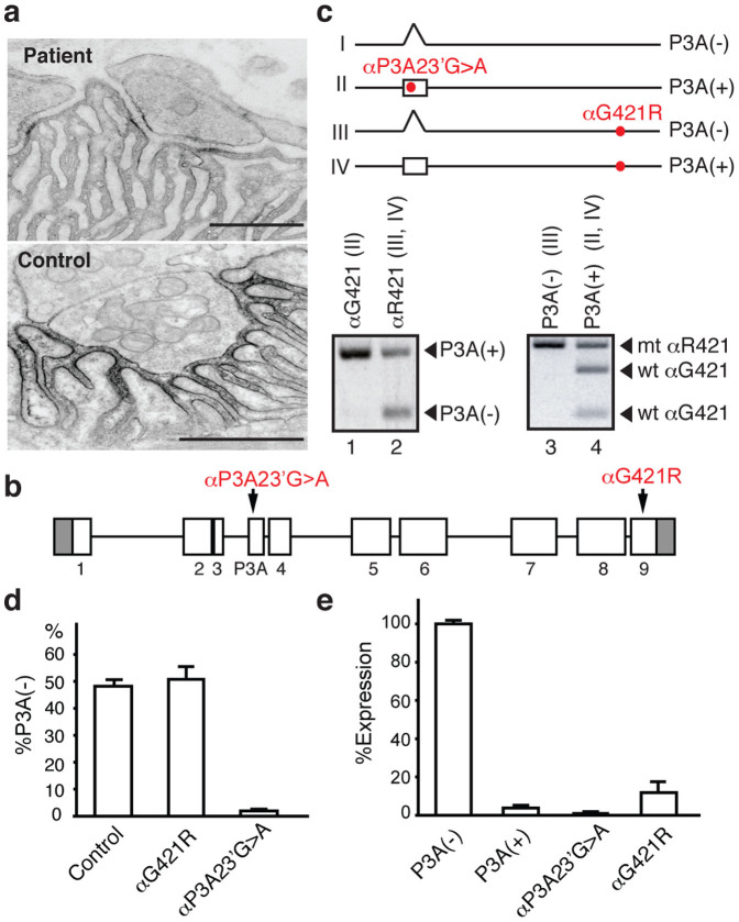
Figure 1 Ultrastructure of the patient endplate, identified mutations, and their functional consequences. (a) AChR expression at the patient and control endplate visualized with peroxidase-labeled α-bungarotoxin. Note restricted distribution and attenuated expression of AChR at the patient endplate. Bars = 1 μm. (b) Structure of CHRNA1 gene and two identified mutations. (c) Four possible transcripts in muscle of the patient. Only transcript I can make a normal α subunit. A segment spanning exon P3A of transcripts from αG421- and αR421-alleles is specifically amplified by allele-specific RT-PCR using patient muscle. Transcript I is not detectable (lanes 1 and 2). P3A(−) and P3A(+) transcripts are specifically amplified by allele-specific RT-PCR of patient muscle. Nested RT-PCR products spanning αG421R are digested by NlaIV, which cuts only the wild-type αG421 fragment (lane 4) and leaves the mutant αR421 fragment undigested (lanes 3 and 4). The P3A(−) transcript almost exclusively arises from an allele with αR421 (transcript III) (lane 3), whereas P3A(+) transcripts arise from both alleles (transcripts II and IV) (lane 4). Again, transcript I is not detectable (lanes 3 and 4). (d) Allele-specific real-time RT-PCR of patient muscle. The αP3A23′G > A allele barely generates P3A(−) transcript. (e) Expression of AChR on the HEK293 cell surface introduced with the indicated α cDNAs along with the wild-type β, δ, and ε cDNAs. The expression level of αG421R-AChR is 14.7 ± 5.1% of normal (mean ± SD, n = 3).
Figure 1 Ultrastructure of the patient endplate, identified mutations, and their functional consequences. (a) AChR expression at the patient and control endplate visualized with peroxidase-labeled α-bungarotoxin. Note restricted distribution and attenuated expression of AChR at the patient endplate. Bars = 1 μm. (b) Structure of CHRNA1 gene and two identified mutations. (c) Four possible transcripts in muscle of the patient. Only transcript I can make a normal α subunit. A segment spanning exon P3A of transcripts from αG421- and αR421-alleles is specifically amplified by allele-specific RT-PCR using patient muscle. Transcript I is not detectable (lanes 1 and 2). P3A(−) and P3A(+) transcripts are specifically amplified by allele-specific RT-PCR of patient muscle. Nested RT-PCR products spanning αG421R are digested by NlaIV, which cuts only the wild-type αG421 fragment (lane 4) and leaves the mutant αR421 fragment undigested (lanes 3 and 4). The P3A(−) transcript almost exclusively arises from an allele with αR421 (transcript III) (lane 3), whereas P3A(+) transcripts arise from both alleles (transcripts II and IV) (lane 4). Again, transcript I is not detectable (lanes 3 and 4). (d) Allele-specific real-time RT-PCR of patient muscle. The αP3A23′G > A allele barely generates P3A(−) transcript. (e) Expression of AChR on the HEK293 cell surface introduced with the indicated α cDNAs along with the wild-type β, δ, and ε cDNAs. The expression level of αG421R-AChR is 14.7 ± 5.1% of normal (mean ± SD, n = 3).CC BY · PubMed Central
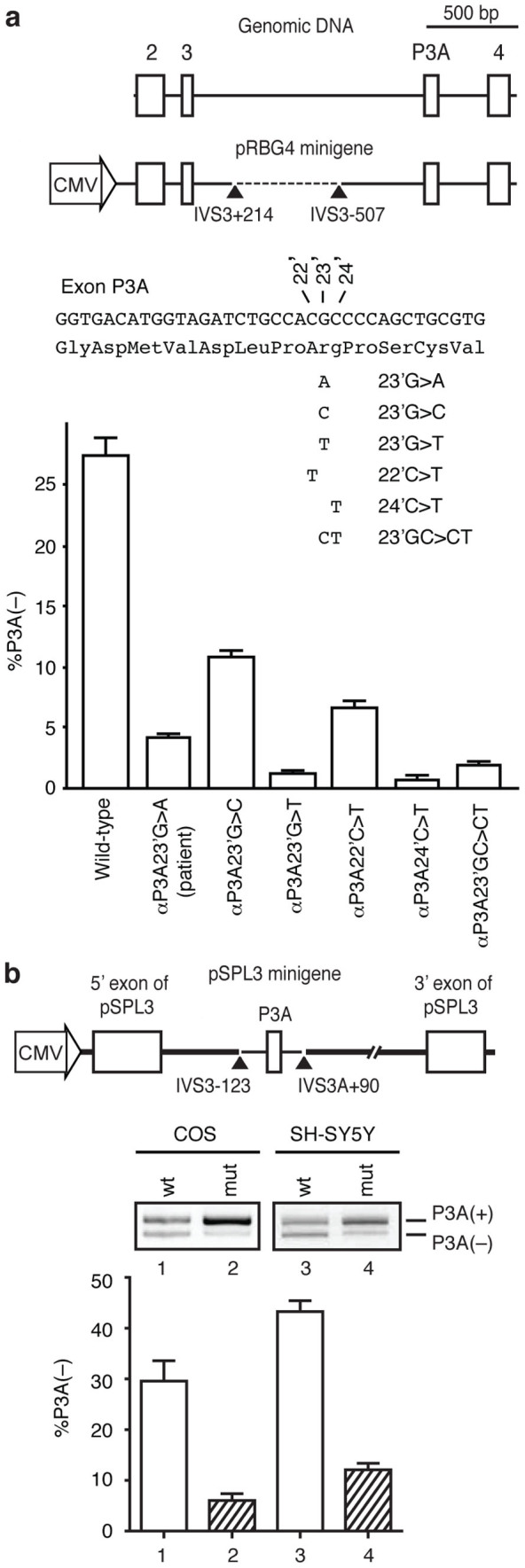
Figure 2 Construction of minigenes and splicing assays. (a) Structure of CHRNA1 gene and pRBG4 minigene. A 589-bp segment (broken line) in intron 3 is deleted in pRBG4 minigene. The patient's mutation and five artificial mutations are introduced into pRBG4 minigene. Ratios of exon P3A skipping are quantified by real-time RT-PCR of COS cells transfected with pRBG4 minigenes. (b) pSPL3 minigene harboring CHRNA1 exon P3A and flanking introns. Arrowheads point to the boundaries of CHRNA1 and pSPL3 vector. Ratios of exon skipping are analyzed by RT-PCR of COS cells and SH-SY5Y cells transfected with pSPL3 minigenes. Bars and lines represent mean and standard deviation (SD), respectively, of three independent experiments for both (a) and (b).
Figure 2 Construction of minigenes and splicing assays. (a) Structure of CHRNA1 gene and pRBG4 minigene. A 589-bp segment (broken line) in intron 3 is deleted in pRBG4 minigene. The patient's mutation and five artificial mutations are introduced into pRBG4 minigene. Ratios of exon P3A skipping are quantified by real-time RT-PCR of COS cells transfected with pRBG4 minigenes. (b) pSPL3 minigene harboring CHRNA1 exon P3A and flanking introns. Arrowheads point to the boundaries of CHRNA1 and pSPL3 vector. Ratios of exon skipping are analyzed by RT-PCR of COS cells and SH-SY5Y cells transfected with pSPL3 minigenes. Bars and lines represent mean and standard deviation (SD), respectively, of three independent experiments for both (a) and (b).CC BY · PubMed Central
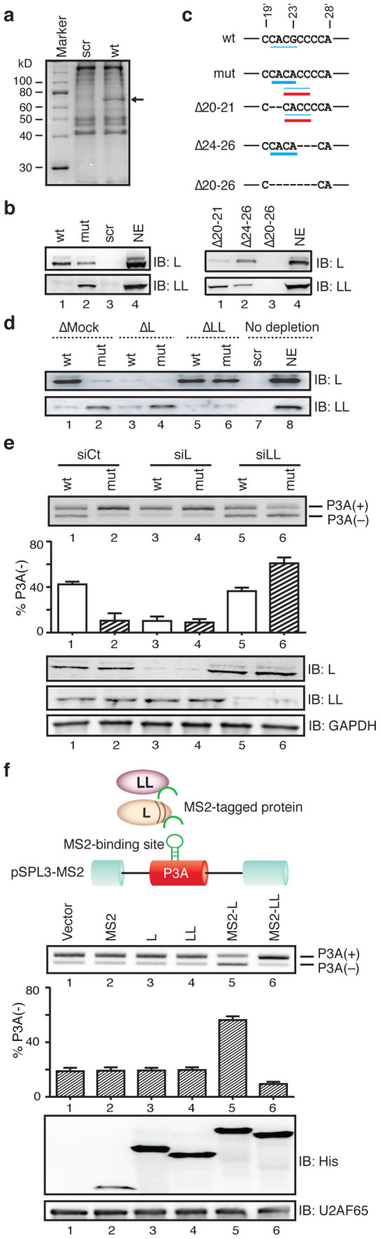
Figure 3 αP3A23′G>A compromises binding affinity for a splicing-suppressing hnRNP L and gains binding affinity for a splicing-enhancing hnRNP LL. (a) Coomassie blue staining of RNA affinity-purified products using SH-SY5Y nuclear extract with biotinylated RNA probes. A ∼70-kDa protein (arrow) is detected only with the wild-type (wt) probe but not with the scrambled (scr) probe. (b) Immunoblots (IB) probed with candidate splicing trans-factors. The wild-type exon P3A (wt) binds to hnRNP L (L) and the αP3A23′G>A (mut) disrupts its binding. The mutation gains de novo binding to hnRNP LL (LL). NE indicates 10% of nuclear extract used. (c) Deletion mutagenesis to map the binding sequences of hnRNPs L and LL in exon P3A. Underlines indicate putative binding motifs for hnRNP LL (red lines) and hnRNP L (thin and thick blue lines for weak and strong motifs, respectively)27. Note that the G-to-A mutation de novo generates a binding motif of CACC for hnRNPs L and LL. Immunoblots of RNA affinity-purified products are detected with antibodies against hnRNPs L and LL. (d) Mock-, hnRNP L-, and hnRNP LL-depleted SH-SY5Y nuclear extracts were affinity-purified with RNA probes and resolved by immunoblotting. (e) RT-PCR of wild-type (wt) and mutant (mut) pSPL3 minigenes in SH-SY5Y cells treated with siRNA against control (siCt), hnRNP L (siL), and hnRNP LL (siLL). Immunoblotting shows down regulation of hnRNPs L and LL. (f) Schematic presentation of a reporter minigene (pSPL3-MS2) and trans-acting effectors. HnRNPs L and LL (ovals) are fused to the artificial MS2 coat protein (inverted U shape). MS2 coat protein-binding hairpin RNA is substituted for the splicing regulatory site of exon P3A to directly tether MS2 coat protein-fused hnRNPs L and LL. RT-PCR of pSPL3-MS2 minigenes in SH-SY5Y cells that are co-transfected with the indicated effectors. Immunoblotting shows expression of effectors in the nuclear lysate of SH-SY5Y cells. The MS2-L construct has 16 additional codons arising from the multiple cloning site compared to MS2-LL. Bars and lines represent mean and SD, respectively, of three independent experiments for panels (e) and (f).
Figure 3 αP3A23′G>A compromises binding affinity for a splicing-suppressing hnRNP L and gains binding affinity for a splicing-enhancing hnRNP LL. (a) Coomassie blue staining of RNA affinity-purified products using SH-SY5Y nuclear extract with biotinylated RNA probes. A ∼70-kDa protein (arrow) is detected only with the wild-type (wt) probe but not with the scrambled (scr) probe. (b) Immunoblots (IB) probed with candidate splicing trans-factors. The wild-type exon P3A (wt) binds to hnRNP L (L) and the αP3A23′G>A (mut) disrupts its binding. The mutation gains de novo binding to hnRNP LL (LL). NE indicates 10% of nuclear extract used. (c) Deletion mutagenesis to map the binding sequences of hnRNPs L and LL in exon P3A. Underlines indicate putative binding motifs for hnRNP LL (red lines) and hnRNP L (thin and thick blue lines for weak and strong motifs, respectively)27. Note that the G-to-A mutation de novo generates a binding motif of CACC for hnRNPs L and LL. Immunoblots of RNA affinity-purified products are detected with antibodies against hnRNPs L and LL. (d) Mock-, hnRNP L-, and hnRNP LL-depleted SH-SY5Y nuclear extracts were affinity-purified with RNA probes and resolved by immunoblotting. (e) RT-PCR of wild-type (wt) and mutant (mut) pSPL3 minigenes in SH-SY5Y cells treated with siRNA against control (siCt), hnRNP L (siL), and hnRNP LL (siLL). Immunoblotting shows down regulation of hnRNPs L and LL. (f) Schematic presentation of a reporter minigene (pSPL3-MS2) and trans-acting effectors. HnRNPs L and LL (ovals) are fused to the artificial MS2 coat protein (inverted U shape). MS2 coat protein-binding hairpin RNA is substituted for the splicing regulatory site of exon P3A to directly tether MS2 coat protein-fused hnRNPs L and LL. RT-PCR of pSPL3-MS2 minigenes in SH-SY5Y cells that are co-transfected with the indicated effectors. Immunoblotting shows expression of effectors in the nuclear lysate of SH-SY5Y cells. The MS2-L construct has 16 additional codons arising from the multiple cloning site compared to MS2-LL. Bars and lines represent mean and SD, respectively, of three independent experiments for panels (e) and (f).CC BY · PubMed Central
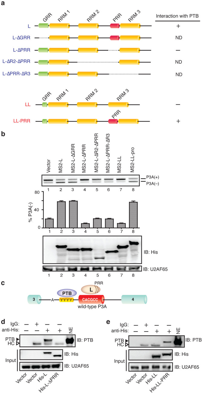
Figure 4 HnRNP L interacts with PTB through proline-rich region (PRR) to synergistically repress inclusion of exon P3A. (a) Schematic domain-structures of hnRNPs L and LL and their mutant derivatives. Interaction of each protein product with PTB is indicated on right according to the results from panels (d) and (e). RRM, GRR and ND indicate RNA recognition motif, glycine-rich region and not detected respectively. (b) RT-PCR of pSPL3-MS2 products after the indicated MS2-tagged trans-acting effectors are introduced into SH-SY5Y cells. Bars and lines represent mean and SD, respectively, of three independent experiments. Immunoblotting shows expression of effectors in the nuclear lysate of SH-SY5Y cells. (c) Schematic binding sites of hnRNP L and PTB in exon P3A and upstream PPT (YYYY) respectively. (d) Interaction of PTB with His-tagged hnRNP L and its indicated mutant. Nuclear extract of transfected SH-SY5Y cells is immunoprecipitated with anti-His antibody and assayed for interaction with endogenous PTB by immunoblotting (IB). Open arrowheads point to IgG heavy chain (HC) that was non-specifically precipitated. (e) Interaction of PTB with His-tagged hnRNP LL and its indicated mutant. See (d) for the procedures and other labels.
Figure 4 HnRNP L interacts with PTB through proline-rich region (PRR) to synergistically repress inclusion of exon P3A. (a) Schematic domain-structures of hnRNPs L and LL and their mutant derivatives. Interaction of each protein product with PTB is indicated on right according to the results from panels (d) and (e). RRM, GRR and ND indicate RNA recognition motif, glycine-rich region and not detected respectively. (b) RT-PCR of pSPL3-MS2 products after the indicated MS2-tagged trans-acting effectors are introduced into SH-SY5Y cells. Bars and lines represent mean and SD, respectively, of three independent experiments. Immunoblotting shows expression of effectors in the nuclear lysate of SH-SY5Y cells. (c) Schematic binding sites of hnRNP L and PTB in exon P3A and upstream PPT (YYYY) respectively. (d) Interaction of PTB with His-tagged hnRNP L and its indicated mutant. Nuclear extract of transfected SH-SY5Y cells is immunoprecipitated with anti-His antibody and assayed for interaction with endogenous PTB by immunoblotting (IB). Open arrowheads point to IgG heavy chain (HC) that was non-specifically precipitated. (e) Interaction of PTB with His-tagged hnRNP LL and its indicated mutant. See (d) for the procedures and other labels.CC BY · PubMed Central
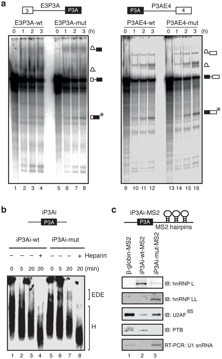
Figure 5 Skipping of exon P3A is promoted by impairing the formation of exon-defined E (EDE) complex in the wild-type pre-mRNA. (a) Time-course data obtained from in vitro splicing of the 32P-labeled pre-mRNAs from E3P3A (wt and mut) and P3AE4 (wt and mut) minigenes. The splicing products are shown schematically on the right. The spliced mRNA (asterisk) is increased in E3P3A-mut compared to E3P3A-wt. Although intron lariats are apparently increased in E3P3A-wt, the intron lariat and high molecular weight RNAs are not clearly resolved for E3P3A, and the increase of the intron lariats cannot be precisely estimated. Poor resolution of splicing products of E3P3A compared to P3AE4 is likely due to binding of a splicing repressing PTB. (b) Time-course analysis of early exon-defined spliceosome (EDE complex) that assembles across the P3A exon of 32P-labeled substrates (iP3Ai-wt and iP3A-mut) in the absence of ATP. Native agarose gel electrophoresis resolves the indicated nonspecific complex H (H) and the exon-defined E complex (EDE). (c) Schematic structures of MS2-attached wild-type (wt) and mutant (mut) substrates used for isolation of EDE complex. Immunoblotting (IB) and RT-PCR analyses of purified E complex assembled on indicated substrates. PTB was likely bound to a CUCUCUCU sequence in intron 1 of β-globin-MS2 pre-mRNA.
Figure 5 Skipping of exon P3A is promoted by impairing the formation of exon-defined E (EDE) complex in the wild-type pre-mRNA. (a) Time-course data obtained from in vitro splicing of the 32P-labeled pre-mRNAs from E3P3A (wt and mut) and P3AE4 (wt and mut) minigenes. The splicing products are shown schematically on the right. The spliced mRNA (asterisk) is increased in E3P3A-mut compared to E3P3A-wt. Although intron lariats are apparently increased in E3P3A-wt, the intron lariat and high molecular weight RNAs are not clearly resolved for E3P3A, and the increase of the intron lariats cannot be precisely estimated. Poor resolution of splicing products of E3P3A compared to P3AE4 is likely due to binding of a splicing repressing PTB. (b) Time-course analysis of early exon-defined spliceosome (EDE complex) that assembles across the P3A exon of 32P-labeled substrates (iP3Ai-wt and iP3A-mut) in the absence of ATP. Native agarose gel electrophoresis resolves the indicated nonspecific complex H (H) and the exon-defined E complex (EDE). (c) Schematic structures of MS2-attached wild-type (wt) and mutant (mut) substrates used for isolation of EDE complex. Immunoblotting (IB) and RT-PCR analyses of purified E complex assembled on indicated substrates. PTB was likely bound to a CUCUCUCU sequence in intron 1 of β-globin-MS2 pre-mRNA.CC BY · PubMed Central
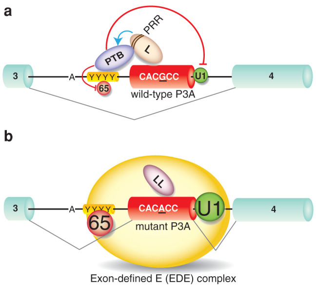
Figure 6 Model of pathogenic mutation (αP3A23′G>A)-induced aberrant exon P3A inclusion that is antagonistically regulated by hnRNPs L and LL. Early spliceosome complex formation on CHRNA1 pre-mRNA with alternative exon P3A are schematically shown. Large letters indicate functional binding of splicing factors, whereas small letters represent compromised binding of splicing factors. The sequence of point mutation in exon P3A (αP3A23′G>A) is underlined. (a) HnRNP L (L) binds to wild-type exon P3A and interacts with PTB through the proline-rich region (PRR), which stabilizes PTB binding to the upstream PPT (YYYY). The hnRNP L–PTB interaction prevents association of U2AF65 (65) to PPT and U1 snRNP (U1) to the 5′ splice site. The formation of exon-defined E (EDE) complex is thus impaired, which leads to skipping of exon P3A. (b) The αP3A23′G>A-mutation switches binding of hnRNP L to hnRNP LL (LL). Lack of PRR in hnRNP LL fails to stabilize PTB binding to the upstream PPT, which allows binding of U1 snRNP (U1) and U2AF65 (65) on pre-mRNA. The formation of the exon-defined E (EDE) complex facilitates inclusion of exon P3A.
Figure 6 Model of pathogenic mutation (αP3A23′G>A)-induced aberrant exon P3A inclusion that is antagonistically regulated by hnRNPs L and LL. Early spliceosome complex formation on CHRNA1 pre-mRNA with alternative exon P3A are schematically shown. Large letters indicate functional binding of splicing factors, whereas small letters represent compromised binding of splicing factors. The sequence of point mutation in exon P3A (αP3A23′G>A) is underlined. (a) HnRNP L (L) binds to wild-type exon P3A and interacts with PTB through the proline-rich region (PRR), which stabilizes PTB binding to the upstream PPT (YYYY). The hnRNP L–PTB interaction prevents association of U2AF65 (65) to PPT and U1 snRNP (U1) to the 5′ splice site. The formation of exon-defined E (EDE) complex is thus impaired, which leads to skipping of exon P3A. (b) The αP3A23′G>A-mutation switches binding of hnRNP L to hnRNP LL (LL). Lack of PRR in hnRNP LL fails to stabilize PTB binding to the upstream PPT, which allows binding of U1 snRNP (U1) and U2AF65 (65) on pre-mRNA. The formation of the exon-defined E (EDE) complex facilitates inclusion of exon P3A.CC BY · PubMed Central

Verification

Claims the paper makes

The claims and statistics of this paper haven’t been checked yet.

Run the check — free, no account needed. It reads the open-access text and checks the sources it cites.

A few free checks a day · papers already verified open instantly · results are public

Claims other papers make about this one

Each is checked against this paper’s own text: does it say what the citing paper says it does?

Self-check

Percentages, sums and reported differences re-computed from the paper’s own numbers.

The self-check needs the paper’s full text.

Citations

Cites

Show all 57 references

Research works printed but not linked: 3

  1. Newland, C. F., Beeson, D., Vincent, A. & Newsom-Davis, J. Functional and non-functional isoforms of the human muscle acetylcholine receptor. J Physiol 489, 767–778 (1995). · DOI
  2. Preussner, M. et al. HnRNP L and L-like cooperate in multiple-exon regulation of CD45 alternative splicing. Nucleic Acids Res 40, 5666–5678 (2012). · DOI
  3. Krull, M., Petrusma, M., Makalowski, W., Brosius, J. & Schmitz, J. Functional persistence of exonized mammalian-wide interspersed repeat elements (MIRs). Genome Res 17, 1139–1145 (2007). · DOI

Cited by

See all 44

Metadata

Authors
Rahman, M. A., Masuda, A., Ohe, K., Ito, M., Hutchinson, D. O., Mayeda, A., Engel, A. G., & Ohno, K.
Venue
Scientific Reports
Published
2013-10-14
Volume
3
Issue
1
ISSN
2045-2322
Cited by
44
References
57
Language
en
Licence

About these checks

Registry, retraction, Cites and statistics results are computed, not judged, with one exception: a printed reference that no index lists is read once by a model before it counts as a citation error. Claims are extracted by a model and kept only when their quote appears verbatim in the text; each cited source is then checked, with a model’s help, for whether it says what the claim relies on. Those verdicts can be wrong — read the claim’s quote and the cited source before relying on one.

Statistics and arithmetic are recomputed, not judged.

Confidence bands are shown only for fields calibrated on our annotated test set. None is calibrated yet, so no band is shown.

What counts as a claim ›How we measure confidence ›How we check this ›

Are you an author, or do you see a problem with this page? Request a review

How to cite

Cite CoChat for the checks on this page, and the paper for its own findings.

This page
CoChat. (2026). Citation check: HnRNP L and hnRNP LL antagonistically modulate PTB-mediated splicing suppression of CHRNA1 pre-mRNA. https://app.cochat.ai/papers/10.1038/srep02931
The paper
Rahman, M. A., Masuda, A., Ohe, K., Ito, M., Hutchinson, D. O., Mayeda, A., Engel, A. G., & Ohno, K. (2013). HnRNP L and hnRNP LL antagonistically modulate PTB-mediated splicing suppression of CHRNA1 pre-mRNA. Scientific Reports. https://doi.org/10.1038/srep02931

Data sources

Bibliographic data: Semantic Scholar, OpenAlex, Crossref.

Contains information from the Semantic Scholar Academic Graph Datasets, made available under the ODC Attribution License (ODC-BY 1.0).

Semantic Scholar’s data is described in: Kinney, R., et al. (2023). The Semantic Scholar Open Data Platform. arXiv:2301.10140.