

The DOI matches its Crossref record.
The scanning electron microscope in microbiology and diagnosis of infectious disease
Abstract
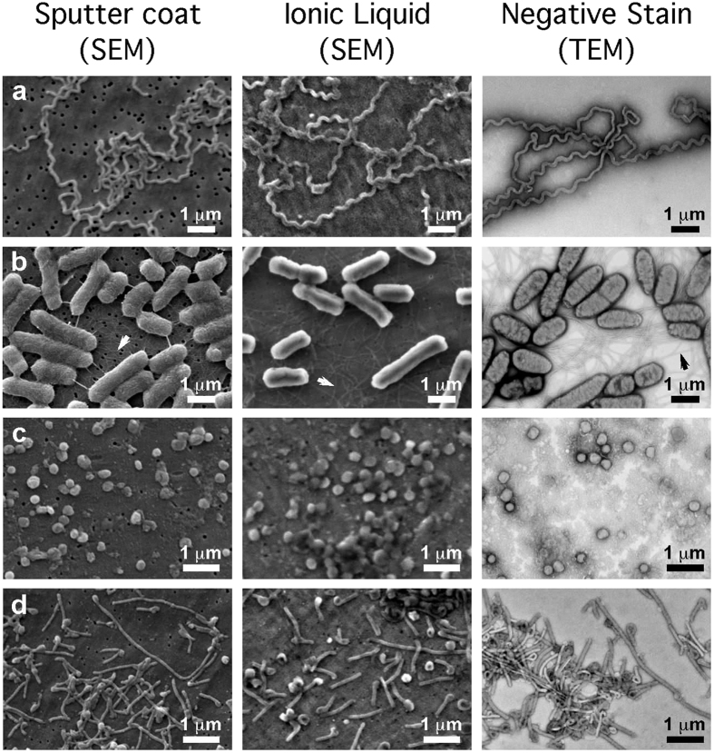
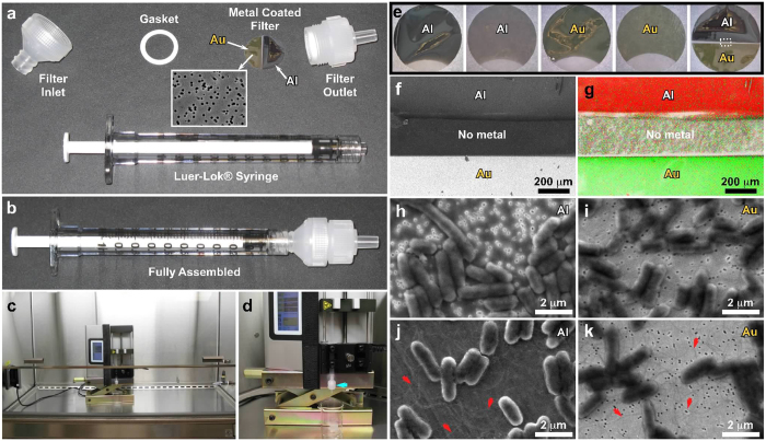
Despite being an excellent tool for investigating ultrastructure, scanning electron microscopy (SEM) is less frequently used than transmission electron microscopy for microbes such as viruses or bacteria. Here we describe rapid methods that allow SEM imaging of fully hydrated, unfixed microbes without using conventional sample preparation methods. We demonstrate improved ultrastructural preservation, with greatly reduced dehydration and shrinkage, for specimens including bacteria and viruses such as Ebola virus using infiltration with ionic liquid on conducting filter substrates for SEM.
Overview
The claims and statistics of this paper haven’t been checked yet.
Run the check — free, no account needed. It reads the open-access text and checks the sources it cites.
Integrity signals
Registry
Cites
Statistics
Figures
Verification
Claims the paper makes
The claims and statistics of this paper haven’t been checked yet.
Run the check — free, no account needed. It reads the open-access text and checks the sources it cites.
Claims other papers make about this one
Each is checked against this paper’s own text: does it say what the citing paper says it does?
In the citing paper
“Of various methods available for qualitative structural BFI detection on abiotic or biotic surfaces, scanning electron microscopy (SEM)-based detection of BFI structure is considered the gold standard.20-22”
From SEMTWIST Quantification of Biofilm Infection in Human Chronic Wound Using Scanning Electron Microscopy and Machine Learning (2025) · cites this paper as [22]
Self-check
Percentages, sums and reported differences re-computed from the paper’s own numbers.
The self-check needs the paper’s full text.
Citations
Cites
No references could be resolved yet.
Research works printed but not linked: 34
- van Rooyen, C. E. & SCOTT, G. D. Smallpox diagnosis with special reference to electron microscopy. Can. J. Public Health 39, 467–477 (1948).
- Goldsmith, C. S. & Miller, S. E. Modern uses of electron microscopy for detection of viruses. Clin. Microbiol. Rev. 22, 552–563, 10.1128/CMR.00027-09 (2009). · DOI
- Hazelton, P. R. & Gelderblom, H. R. Electron microscopy for rapid diagnosis of infectious agents in emergent situations. Emerg. Infect. Dis. 9, 294–303 (2003). · DOI
- Nitsche, A., Gelderblom, H. R., Eisendle, K., Romani, N. & Pauli, G. Pitfalls in diagnosing human poxvirus infections. J. Clin. Virol. 38, 165–168 (2007). · DOI
- Biel, S. S. et al. Detection of human polyomaviruses in urine from bone marrow transplant patients: comparison of electron microscopy with PCR. Clin. Chem. 50, 306–312 (2004). · DOI
- Biel, S. S. & Gelderblom, H. R. Diagnostic electron microscopy is still a timely and rewarding method. J. Clin. Virol. 13, 105–119 (1999). · DOI
- Beniac, D. R., Siemens, C. G., Wright, C. J. & Booth, T. F. A filtration based technique for simultaneous SEM and TEM sample preparation for the rapid detection of pathogens. Viruses. 6, 3458–3471, 10.3390/v6093458 (2014). · DOI
- Beniac, D. R., Hiebert, S. L., Siemens, C. G., Corbett, C. R. & Booth, T. F. A mobile biosafety microanalysis system for infectious agents. Sci. Rep. 5, 9505, 10.1038/srep09505 (2015). · DOI
- Thiberge, S. et al. Scanning electron microscopy of cells and tissues under fully hydrated conditions. Proc. Natl. Acad. Sci. USA 101, 3346–3351 (2004). · DOI
- de Jonge, N., Peckys, D. B., Kremers, G. J. & Piston, D. W. Electron microscopy of whole cells in liquid with nanometer resolution. Proc. Natl. Acad. Sci. USA 106, 2159–2164, 10.1073/pnas.0809567106 (2009). · DOI
- Ogura, T. Nanoscale analysis of unstained biological specimens in water without radiation damage using high-resolution frequency transmission electric-field system based on FE-SEM. Biochem. Biophys. Res. Commun. 459, 521–528, 10.1016/j.bbrc.2015.02.140 (2015). · DOI
- Bergmans, L., Moisiadis, P., Van, M. B., Quirynen, M. & Lambrechts, P. Microscopic observation of bacteria: review highlighting the use of environmental SEM. Int. Endod. J. 38, 775–788 (2005). · DOI
- Walther, P. & Muller, M. Biological ultrastructure as revealed by high resolution cryo-SEM of block faces after cryo-sectioning. J. Microsc. 196, 279–287 (1999). · DOI
- Hayles, M. F., Stokes, D. J., Phifer, D. & Findlay, K. C. A technique for improved focused ion beam milling of cryo-prepared life science specimens. J. Microsc. 226, 263–269 (2007). · DOI
- Katsen-Globa, A., Puetz, N., Gepp, M. M., Neubauer, J. C. & Zimmermann, H. Study of SEM preparation artefacts with correlative microscopy: Cell shrinkage of adherent cells by HMDS-drying. Scanning, 10.1002/sca.21310 (2016). · DOI
- Beniac, D. R. et al. The organisation of Ebola virus reveals a capacity for extensive, modular polyploidy. PLoS. One. 7, e29608, 10.1371/journal.pone.0029608 (2012). · DOI
- Cotelesage, J. J. et al. Hepatitis B Virus Genotype G forms core-like particles with unique structural properties. J. Viral Hepat. 18, 443–448, 10.1111/j.1365-2893.2010.01330.x (2010). · DOI
- Frank, J. et al. SPIDER and WEB: processing and visualization of images in 3D electron microscopy and related fields. J. Struct. Biol. 116, 190–199 (1996). · DOI
- Kuwabata, S., Kongkanand, A., Oyamatsu, D. & Torimoto, T. Observation of ionic liquid by scanning electron microscope. Chemistry Letters 35, 600–601, 10.1246/cl.2006.600 (2006). · DOI
- Dwiranti, A. et al. Chromosome observation by scanning electron microscopy using ionic liquid. Microsc. Res. Tech. 75, 1113–1118, 10.1002/jemt.22038 (2012). · DOI
- Takahashi, C., Shirai, T. & Fuji, M. FE-SEM observation of swelled seaweed using hydrophilic ionic liquid; 1-butyl-3-methylimidazolium tetrafluoroborate. Microsc. Res. Tech. 76, 66–71, 10.1002/jemt.22137 (2013). · DOI
- Ishigaki, Y. et al. Ionic liquid enables simple and rapid sample preparation of human culturing cells for scanning electron microscope analysis. Microsc. Res. Tech. 74, 415–420, 10.1002/jemt.20924 (2011). · DOI
- Kawai, K., Kaneko, K., Kawakami, H., Narushima, T. & Yonezawa, T. Simple pretreatment of non-conductive small hydrous bio-samples with choline-type ionic liquid and membrane filter for microsample mounting. Colloids Surf. B Biointerfaces. 102, 9–12, 10.1016/j.colsurfb.2012.08.019 (2013). · DOI
- Torimoto, T., Tsuda, T., Okazaki, K. & Kuwabata, S. New frontiers in materials science opened by ionic liquids. Adv. Mater. 22, 1196–1221, 10.1002/adma.200902184 (2010). · DOI
- Arimoto, S., Oyamatsu, D., Torimoto, T. & Kuwabata, S. Development of in situ electrochemical scanning electron microscopy with ionic liquids as electrolytes. Chemphyschem. 9, 763–767, 10.1002/cphc.200700758 (2008). · DOI
- Klungthong, C. et al. The impact of primer and probe-template mismatches on the sensitivity of pandemic influenza A/H1N1/2009 virus detection by real-time RT-PCR. J Clin. Virol. 48, 91–95, 10.1016/j.jcv.2010.03.012 (2010). · DOI
- Pyne, M. T., Brown, K. L. & Hillyard, D. R. Evaluation of the Roche Cobas AmpliPrep/Cobas TaqMan HIV-1 test and identification of rare polymorphisms potentially affecting assay performance. J Clin. Microbiol. 48, 2852–2858, 10.1128/JCM.00776-10 (2010). · DOI
- Mahony, J. B. & Richardson, S. Molecular diagnosis of severe acute respiratory syndrome: the state of the art. J Mol. Diagn. 7, 551–559 (2005). · DOI
- Gelderblom, H. R. & Hazelton, P. R. Specimen collection for electron microscopy. Emerg. Infect. Dis. 6, 433–434 (2000). · DOI
- Goldstein, J. I. et al. Scanning Electron Microscopy and X-ray Microanalysis (Springer, New York 2003). · DOI
- Levett, P. N. Leptospirosis. Clin. Microbiol. Rev. 14, 296–326 (2001). · DOI
- Fàbrega, A. & Vila, J. Salmonella enterica serovar Typhimurium skills to succeed in the host: virulence and regulation. Clin. Microbiol. Rev. 26, 308–341, 10.1128/CMR.00066-12 (2013). · DOI
- Halfmann, P. et al. Replication-deficient ebolavirus as a vaccine candidate. J. Virol. 83, 3810–3815, 10.1128/JVI.00074-09 (2009). · DOI
- Abramoff, M. D., Magelhaes, P. J. & Ram, S. J. Image Processing with ImageJ. Biophotonics International 11, 36–42 (2004).
Cited by
Sorted by citations
Singh, S., Muniz De Oliveira, F., Wang, C., Kumar, M., Xuan, Y., DeMazumder, D., Sen, C. K., & Roy, S. (2025) · Advances in Wound Care · doi:10.1089/wound.2024.0291 · Cited by 6
Metadata
- Authors
- Golding, C. G., Lamboo, L. L., Beniac, D. R., & Booth, T. F.
- Venue
- Scientific Reports
- Published
- 2016-05-23
- Volume
- 6
- Issue
- 1
- ISSN
- 2045-2322
- PMC
- PMC4876401
- Cited by
- 236
- References
- 0
- Language
- en
About these checks
Registry, retraction, Cites and statistics results are computed, not judged, with one exception: a printed reference that no index lists is read once by a model before it counts as a citation error. Claims are extracted by a model and kept only when their quote appears verbatim in the text; each cited source is then checked, with a model’s help, for whether it says what the claim relies on. Those verdicts can be wrong — read the claim’s quote and the cited source before relying on one.
Statistics and arithmetic are recomputed, not judged.
Confidence bands are shown only for fields calibrated on our annotated test set. None is calibrated yet, so no band is shown.
What counts as a claim ›How we measure confidence ›How we check this ›
Are you an author, or do you see a problem with this page? Request a review
How to cite
Cite CoChat for the checks on this page, and the paper for its own findings.
- This page
- CoChat. (2026). Citation check: The scanning electron microscope in microbiology and diagnosis of infectious disease. https://app.cochat.ai/papers/10.1038/srep26516
- The paper
- Golding, C. G., Lamboo, L. L., Beniac, D. R., & Booth, T. F. (2016). The scanning electron microscope in microbiology and diagnosis of infectious disease. Scientific Reports. https://doi.org/10.1038/srep26516
