Cited by 661Metabolism, Diabetes, and CancerMitochondrial Function and PathologyATP Synthase and ATPases Research

The DOI matches its Crossref record; 78 of the 79 references it cites were found in a registry or an index.

Effects of metformin and other biguanides on oxidative phosphorylation in mitochondria

Open access
Bridges, Hannah R., Jones, Andrew J. Y., Pollak, Michael N., Hirst, Judy·2014·Biochemical Journal

Abstract

The biguanide metformin is widely prescribed for Type II diabetes and has anti-neoplastic activity in laboratory models. Despite evidence that inhibition of mitochondrial respiratory complex I by metformin is the primary cause of its cell-lineage-specific actions and therapeutic effects, the molecular interaction(s) between metformin and complex I remain uncharacterized. In the present paper, we describe the effects of five pharmacologically relevant biguanides on oxidative phosphorylation in mammalian mitochondria. We report that biguanides inhibit complex I by inhibiting ubiquinone reduction (but not competitively) and, independently, stimulate reactive oxygen species production by the complex I flavin. Biguanides also inhibit mitochondrial ATP synthase, and two of them inhibit only ATP hydrolysis, not synthesis. Thus we identify biguanides as a new class of complex I and ATP synthase inhibitor. By comparing biguanide effects on isolated complex I and cultured cells, we distinguish three anti-diabetic and potentially anti-neoplastic biguanides (metformin, buformin and phenformin) from two anti-malarial biguanides (cycloguanil and proguanil): the former are accumulated into mammalian mitochondria and affect oxidative phosphorylation, whereas the latter are excluded so act only on the parasite. Our mechanistic and pharmacokinetic insights are relevant to understanding and developing the role of biguanides in new and existing therapeutic applications, including cancer, diabetes and malaria.

Overview

The claims and statistics of this paper haven’t been checked yet.

Run the check — free, no account needed. It reads the open-access text and checks the sources it cites.

A few free checks a day · papers already verified open instantly · results are public

Integrity signals

Registry

Crossref · 10.1042/bj20140620

Cites

78 of 78 references verified0 citation errors · 0 not checkable

Statistics

Figures

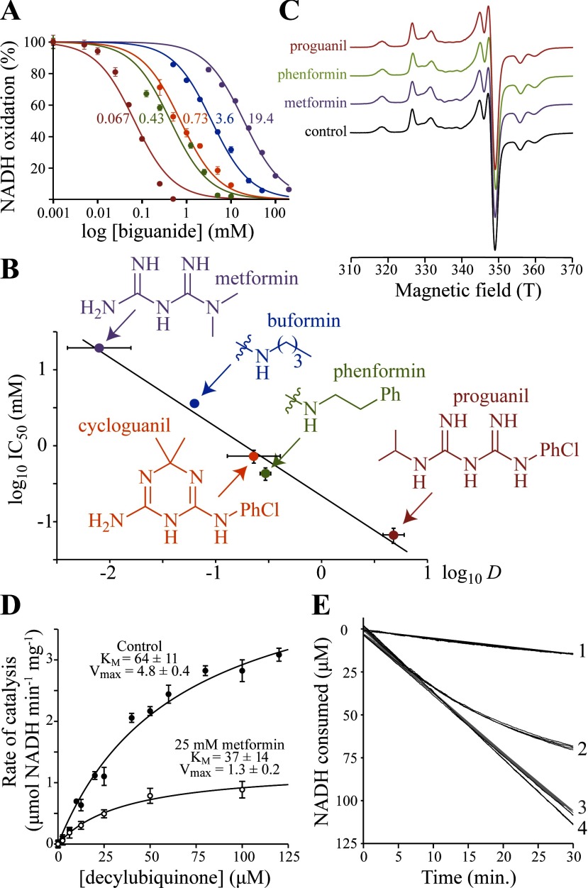
Figure 1 Effects of biguanides on isolated bovine complex I (A) Dependence of NADH oxidation on biguanide concentration, relative to a biguanide-free control. Colours are as in (B), and the IC50 values (in mM) are noted. Data points are means±S.E.M. (n=3–5). (B) Relationship between the inhibition IC50 values and octanol:PBS distribution coefficient (D) values of the biguanides. IC50 values are in mM with 95% confidence intervals; log D values are means±S.E.M. (n=3). Linear fit with R2=0.963. Biguanide structures shown are for the neutral forms. (C) 12 K EPR spectra of the FeS clusters in complex I in the presence and absence of biguanides. Ph, phenyl; PhCl, para-chlorophenyl. (D) The effect of 25 mM metformin on NADH:decylubiquinone oxidoreduction, presented as the measurement of KM for decylubiquinone. The data (means±S.E.M.; n=3) were fit using the Michaelis–Menten equation. (E) Metformin inhibition of NADH oxidation by SMPs (4.5 μg protein/ml). Trace 1, 100 mM metformin added before initiation of catalysis by 200 μM NADH at t=0. Trace 2, catalysis initiated by 200 μM NADH 10 min before the addition of 100 mM metformin at t=0. Traces 3 and 4, controls for 1 and 2, with NaCl instead of metformin. Four traces are overlaid for each condition.
Figure 1 Effects of biguanides on isolated bovine complex I (A) Dependence of NADH oxidation on biguanide concentration, relative to a biguanide-free control. Colours are as in (B), and the IC50 values (in mM) are noted. Data points are means±S.E.M. (n=3–5). (B) Relationship between the inhibition IC50 values and octanol:PBS distribution coefficient (D) values of the biguanides. IC50 values are in mM with 95% confidence intervals; log D values are means±S.E.M. (n=3). Linear fit with R2=0.963. Biguanide structures shown are for the neutral forms. (C) 12 K EPR spectra of the FeS clusters in complex I in the presence and absence of biguanides. Ph, phenyl; PhCl, para-chlorophenyl. (D) The effect of 25 mM metformin on NADH:decylubiquinone oxidoreduction, presented as the measurement of KM for decylubiquinone. The data (means±S.E.M.; n=3) were fit using the Michaelis–Menten equation. (E) Metformin inhibition of NADH oxidation by SMPs (4.5 μg protein/ml). Trace 1, 100 mM metformin added before initiation of catalysis by 200 μM NADH at t=0. Trace 2, catalysis initiated by 200 μM NADH 10 min before the addition of 100 mM metformin at t=0. Traces 3 and 4, controls for 1 and 2, with NaCl instead of metformin. Four traces are overlaid for each condition.CC BY · PubMed Central
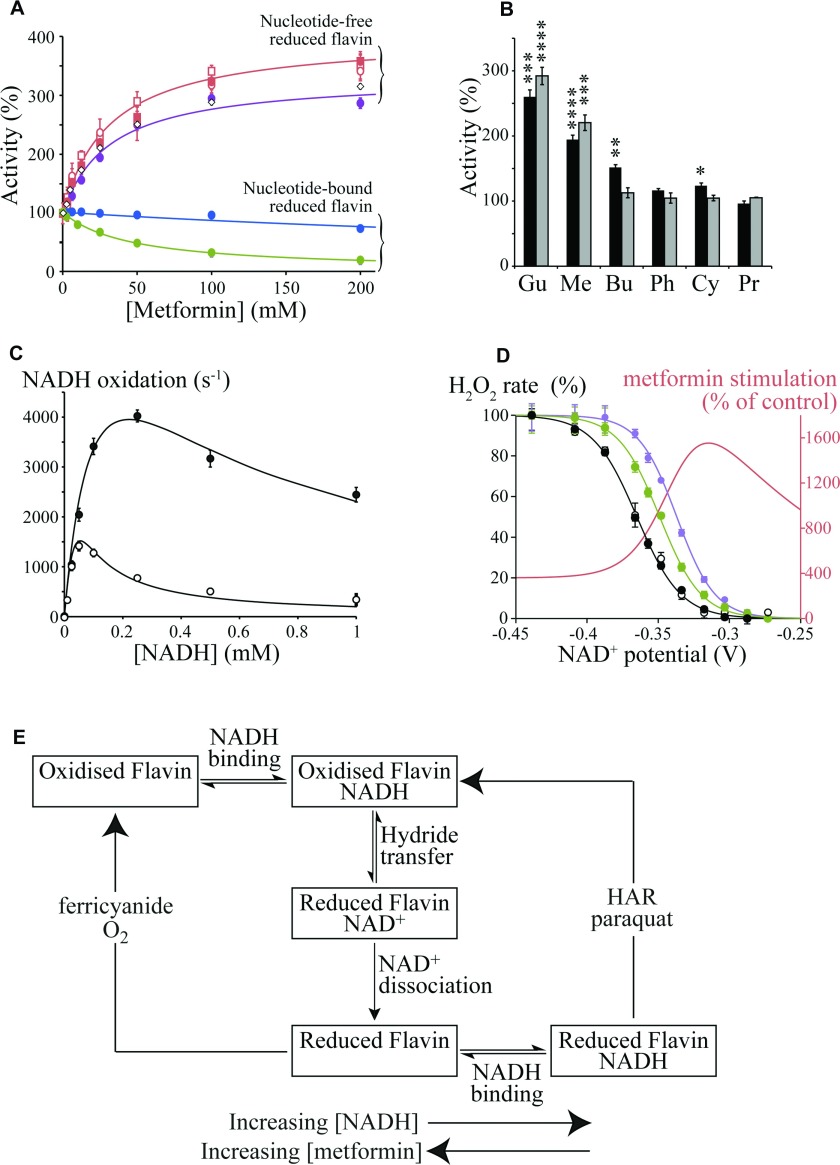
Figure 2 Biguanide effects on the flavin site of complex I (A) Effects of metformin on flavin site reactions that require nucleotide binding to the reduced flavin [NADH:HAR (blue) and NADH:paraquat (green) oxidoreduction] [27], and nucleotide-free reduced flavin [NADH:FeCN (purple) and NADH:O2 (red) oxidoreduction] [26]. NADH:O2 oxidoreduction (H2O2 production) was detected directly (by NADH oxidation, circles) or as H2O2 by the Amplex Red assay (squares) [15], with/without (open/closed symbols respectively) 1 μM rotenone. Data for H2O2 production by subcomplex Iλ are shown as open diamonds (◇). Data points are means±S.E.M., n=3–5. (B) Stimulation of flavin-site reactions, measured at the inhibitory IC50 concentrations (black, H2O2 production measured using Amplex Red; grey, NADH:FeCN oxidoreduction). Gu, 25 mM guanidinium; Me, 25 mM metformin; Bu, 5 mM buformin; Ph, 0.5 mM phenformin; Cy, 0.7 mM cycloguanil; Pr, 0.05 mM proguanil. Data are presented as means±S.E.M., n=3–5. (C) NADH-dependence of NADH:FeCN catalysis by isolated bovine complex I. ●, 100 mM metformin; ○, control. Data points are means±S.E.M., n=3–5 and data were fit as described previously [26] with KMNADH=220 μM, kcatNADH=1500 s−1, kcatFeCN=3.7×10⁷ M−1·s−1 and KNADHRed (the dissociation constant for NADH binding to the reduced flavin)=KNADHSemi=11 μM (control) or 181 μM (metformin). (D) Dependence of H2O2 production on the NAD+ potential, set by the Nernst equation with 30 μM NADH and variable NAD+ [15]. Control [with (●) and without (○) 200 mM NaCl], 20 mM phenformin (green) and 200 mM metformin (purple). Data points are means±S.E.M., n=3–5. The stimulation of H2O2 production by metformin (red) is also represented as the non-normalized percentage activity relative to the control (see A for the effect of metformin in NADH only). (E) Simplified scheme illustrating how metformin affects the different flavin site reactions differently. The boxes represent different states of the complex I flavin site, with the flavin oxidized or reduced, and with or without NADH or NAD+ bound.
Figure 2 Biguanide effects on the flavin site of complex I (A) Effects of metformin on flavin site reactions that require nucleotide binding to the reduced flavin [NADH:HAR (blue) and NADH:paraquat (green) oxidoreduction] [27], and nucleotide-free reduced flavin [NADH:FeCN (purple) and NADH:O2 (red) oxidoreduction] [26]. NADH:O2 oxidoreduction (H2O2 production) was detected directly (by NADH oxidation, circles) or as H2O2 by the Amplex Red assay (squares) [15], with/without (open/closed symbols respectively) 1 μM rotenone. Data for H2O2 production by subcomplex Iλ are shown as open diamonds (◇). Data points are means±S.E.M., n=3–5. (B) Stimulation of flavin-site reactions, measured at the inhibitory IC50 concentrations (black, H2O2 production measured using Amplex Red; grey, NADH:FeCN oxidoreduction). Gu, 25 mM guanidinium; Me, 25 mM metformin; Bu, 5 mM buformin; Ph, 0.5 mM phenformin; Cy, 0.7 mM cycloguanil; Pr, 0.05 mM proguanil. Data are presented as means±S.E.M., n=3–5. (C) NADH-dependence of NADH:FeCN catalysis by isolated bovine complex I. ●, 100 mM metformin; ○, control. Data points are means±S.E.M., n=3–5 and data were fit as described previously [26] with KMNADH=220 μM, kcatNADH=1500 s−1, kcatFeCN=3.7×10⁷ M−1·s−1 and KNADHRed (the dissociation constant for NADH binding to the reduced flavin)=KNADHSemi=11 μM (control) or 181 μM (metformin). (D) Dependence of H2O2 production on the NAD+ potential, set by the Nernst equation with 30 μM NADH and variable NAD+ [15]. Control [with (●) and without (○) 200 mM NaCl], 20 mM phenformin (green) and 200 mM metformin (purple). Data points are means±S.E.M., n=3–5. The stimulation of H2O2 production by metformin (red) is also represented as the non-normalized percentage activity relative to the control (see A for the effect of metformin in NADH only). (E) Simplified scheme illustrating how metformin affects the different flavin site reactions differently. The boxes represent different states of the complex I flavin site, with the flavin oxidized or reduced, and with or without NADH or NAD+ bound.CC BY · PubMed Central
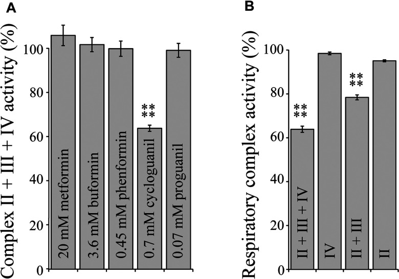
Figure 3 Biguanide interactions with respiratory complexes II, III and IV (A) Rates of complex II + III + IV activity in bovine SMPs measured by a coupled enzyme assay [28] in the presence of biguanide concentrations equivalent to IC50 for NADH:decylubiquinone catalysis. Results are means±S.E.M. as a percentage of the biguanide-free control, n=3–4. (B) Rates of respiratory complex activity in the presence of 0.7 mM cycloguanil as a percentage of their respective cycloguanil-free controls. ****P<0.0001.
Figure 3 Biguanide interactions with respiratory complexes II, III and IV (A) Rates of complex II + III + IV activity in bovine SMPs measured by a coupled enzyme assay [28] in the presence of biguanide concentrations equivalent to IC50 for NADH:decylubiquinone catalysis. Results are means±S.E.M. as a percentage of the biguanide-free control, n=3–4. (B) Rates of respiratory complex activity in the presence of 0.7 mM cycloguanil as a percentage of their respective cycloguanil-free controls. ****P<0.0001.CC BY · PubMed Central
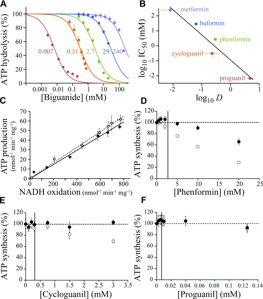
Figure 4 Biguanide inhibition of F1F0-ATP synthase in bovine SMPs (A) Inhibition of ATP hydrolysis by SMPs (colours as in B). Data points are means±S.E.M., n=3–5, and IC50 values are indicated in mM. (B) Relationship between the IC50 values for ATP hydrolysis and the log D values. IC50 values are in mM with 95% confidence intervals; log D values are means±S.E.M., n=3. Linear fit with R2=0.948. (C) Relative inhibition of ATP production in SMPs in the presence of piericidin A (○) or metformin (●). ATP synthesis was driven using 200 μM NADH; NADH oxidation was measured spectroscopically, and the concentration of ATP determined after 3.5 min (during this time a linear rate is observed). NADH oxidation rates were adjusted using 0–50 nM piericidin A or 0–250 mM metformin, with the ionic strength kept constant at 250 mM using NaCl. (D–F) Dose-dependent effects of biguanides on succinate-driven ATP production. Data points are means±S.E.M., n=3, as a percentage of ATP production in the absence of biguanide. ATP concentrations were determined after 3.5 min (○) then corrected for the rate of succinate oxidation, detected spectroscopically using a coupled assay system [28] (●). The IC50 values for hydrolysis are marked with vertical lines.
Figure 4 Biguanide inhibition of F1F0-ATP synthase in bovine SMPs (A) Inhibition of ATP hydrolysis by SMPs (colours as in B). Data points are means±S.E.M., n=3–5, and IC50 values are indicated in mM. (B) Relationship between the IC50 values for ATP hydrolysis and the log D values. IC50 values are in mM with 95% confidence intervals; log D values are means±S.E.M., n=3. Linear fit with R2=0.948. (C) Relative inhibition of ATP production in SMPs in the presence of piericidin A (○) or metformin (●). ATP synthesis was driven using 200 μM NADH; NADH oxidation was measured spectroscopically, and the concentration of ATP determined after 3.5 min (during this time a linear rate is observed). NADH oxidation rates were adjusted using 0–50 nM piericidin A or 0–250 mM metformin, with the ionic strength kept constant at 250 mM using NaCl. (D–F) Dose-dependent effects of biguanides on succinate-driven ATP production. Data points are means±S.E.M., n=3, as a percentage of ATP production in the absence of biguanide. ATP concentrations were determined after 3.5 min (○) then corrected for the rate of succinate oxidation, detected spectroscopically using a coupled assay system [28] (●). The IC50 values for hydrolysis are marked with vertical lines.CC BY · PubMed Central
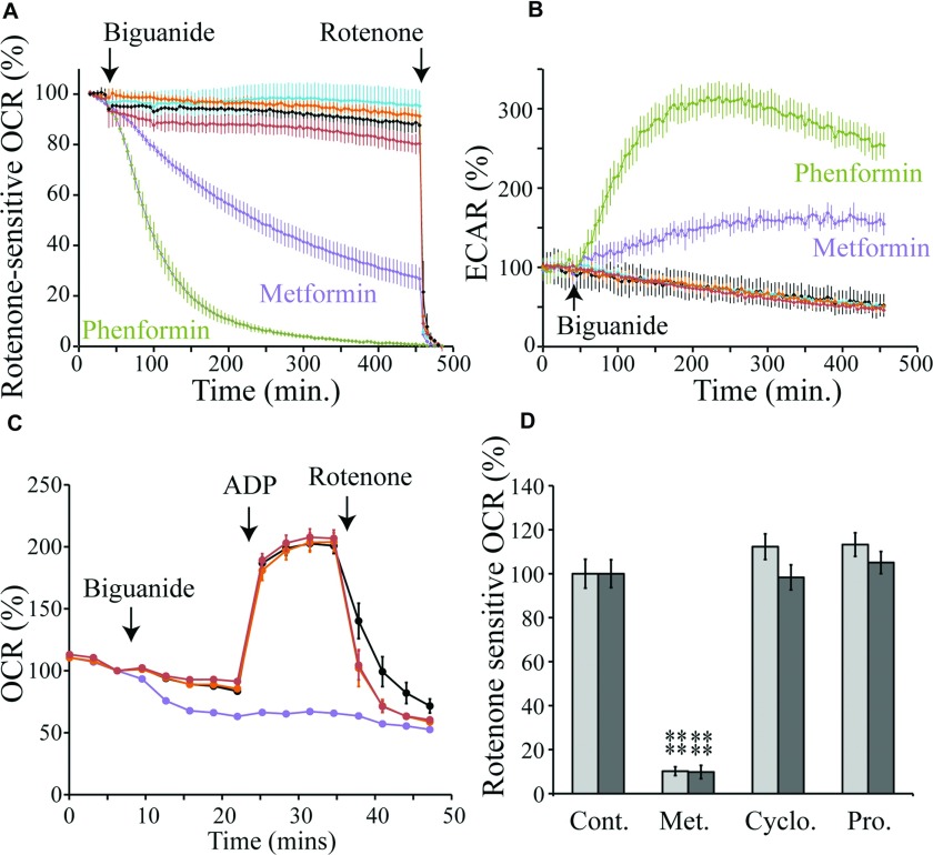
Figure 5 Selective uptake of biguanides into mitochondria (A) Effects of biguanides on the rotenone-sensitive OCR of Hep G2 cells. The traces are the means±S.D. of multiple traces. The biguanide concentrations are equal to the complex I IC50/10 values. Purple, 1.9 mM metformin; green, 0.04 mM phenformin; orange, 0.07 mM cycloguanil added in DMSO; red, 0.007 mM proguanil in DMSO; black, control (no biguanide); cyan, control (DMSO). (B) The effects of biguanides on the ECAR of Hep G2 cells. Conditions and colours as in (A). (C) Mitochondrial respiratory coupling test on 143B cells permeabilized with 2 nM PMP, respiring on pyruvate and malate and using biguanides at concentrations equal to their complex I IC50/10 values. Black, control; purple, 2 mM metformin; orange, 0.07 mM cycloguanil; red, 0.007 mM proguanil. Data points are means±S.E.M., n=5–6. (D) Rates of rotenone-sensitive ADP-stimulated NADH-linked respiration in permeabilized 143B cells (light grey) and rat liver mitochondria (dark grey) after 15 min of incubation with 2 mM metformin (Met.), 0.07 mM cycloguanil (Cyclo.) or 0.007 mM proguanil (Pro.). Values are mean percentage of the control±S.E.M., n=5–6. ****P< 0.01. Cont. control.
Figure 5 Selective uptake of biguanides into mitochondria (A) Effects of biguanides on the rotenone-sensitive OCR of Hep G2 cells. The traces are the means±S.D. of multiple traces. The biguanide concentrations are equal to the complex I IC50/10 values. Purple, 1.9 mM metformin; green, 0.04 mM phenformin; orange, 0.07 mM cycloguanil added in DMSO; red, 0.007 mM proguanil in DMSO; black, control (no biguanide); cyan, control (DMSO). (B) The effects of biguanides on the ECAR of Hep G2 cells. Conditions and colours as in (A). (C) Mitochondrial respiratory coupling test on 143B cells permeabilized with 2 nM PMP, respiring on pyruvate and malate and using biguanides at concentrations equal to their complex I IC50/10 values. Black, control; purple, 2 mM metformin; orange, 0.07 mM cycloguanil; red, 0.007 mM proguanil. Data points are means±S.E.M., n=5–6. (D) Rates of rotenone-sensitive ADP-stimulated NADH-linked respiration in permeabilized 143B cells (light grey) and rat liver mitochondria (dark grey) after 15 min of incubation with 2 mM metformin (Met.), 0.07 mM cycloguanil (Cyclo.) or 0.007 mM proguanil (Pro.). Values are mean percentage of the control±S.E.M., n=5–6. ****P< 0.01. Cont. control.CC BY · PubMed Central
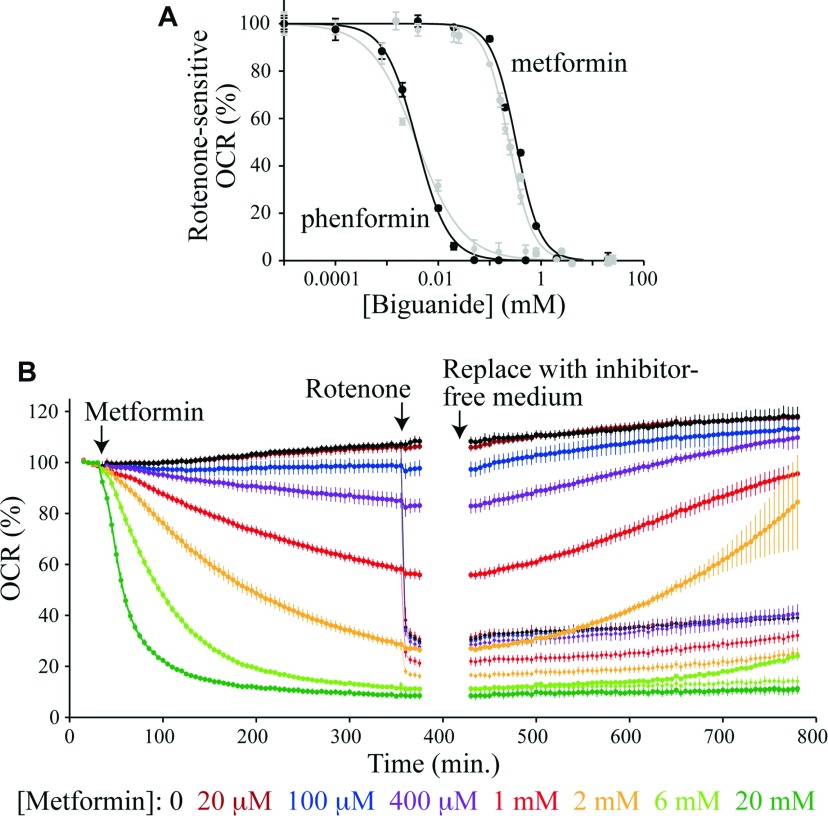
Figure 6 Biguanide effects on cells in culture: accumulation and reversibility (A) The normalized rotenone-sensitive OCR 6 h after the addition of metformin or phenformin to Hep G2 (black) or 143B (grey) cells. Results are means±S.E.M., n=3–4. The IC50 values (with 95% confidence intervals) are metformin, 240±10 μM (143B) and 330±20 μM (Hep G2); phenformin, 3.9±1.0 μM (143B) and 3.8±0.4 μM (Hep G2). (B) Metformin inhibition of the OCR by Hep G2 cells. Results are means of multiple traces±S.E.M., n=3–4. Rotenone (200 nM) was added to half of the samples part way through the experiment; pairs of traces with and without rotenone are coloured the same. Then, the assay medium in each experiment was exchanged for a metformin and rotenone-free medium. All data were measured using a Seahorse Extracellular Flux Analyzer at 37°C.
Figure 6 Biguanide effects on cells in culture: accumulation and reversibility (A) The normalized rotenone-sensitive OCR 6 h after the addition of metformin or phenformin to Hep G2 (black) or 143B (grey) cells. Results are means±S.E.M., n=3–4. The IC50 values (with 95% confidence intervals) are metformin, 240±10 μM (143B) and 330±20 μM (Hep G2); phenformin, 3.9±1.0 μM (143B) and 3.8±0.4 μM (Hep G2). (B) Metformin inhibition of the OCR by Hep G2 cells. Results are means of multiple traces±S.E.M., n=3–4. Rotenone (200 nM) was added to half of the samples part way through the experiment; pairs of traces with and without rotenone are coloured the same. Then, the assay medium in each experiment was exchanged for a metformin and rotenone-free medium. All data were measured using a Seahorse Extracellular Flux Analyzer at 37°C.CC BY · PubMed Central

Verification

Claims the paper makes

The claims and statistics of this paper haven’t been checked yet.

Run the check — free, no account needed. It reads the open-access text and checks the sources it cites.

A few free checks a day · papers already verified open instantly · results are public

Claims other papers make about this one

Each is checked against this paper’s own text: does it say what the citing paper says it does?

Self-check

Percentages, sums and reported differences re-computed from the paper’s own numbers.

The self-check needs the paper’s full text.

Citations

Cites

1 retracted

Show all 79 references

Cited by

Sorted by citations

See all 661

Metadata

Authors
Bridges, H. R., Jones, A. J. Y., Pollak, M. N., & Hirst, J.
Affiliations
  • MRC Mitochondrial Biology Unit (GB)
  • Department of Oncology, McGill University and Lady Davis Research Institute of the Jewish General Hospital, Montreal, Quebec, Canada, H3T 1E2
Corresponding author
Hirst, Judy
Venue
Biochemical Journal
Published
2014-08-22
Volume
462
Issue
3
Pages
475-487
ISSN
  • 0264-6021 (ISSN-L)
  • 1470-8728
OpenAlex
W2012442095
Cited by
661
References
79
Citations by year
2026: 30 · 2025: 53 · 2024: 45 · 2023: 41 · 2022: 61 · 2021: 95 · 2020: 72 · 2019: 66 · 2018: 64 · 2017: 60 · 2016: 43 · 2015: 22 · 2014: 9
FWCI
18.33
Type
journal article
Language
en
Open access
Locations
  • Biochemical Journal (publishedVersion, cc-by)
  • Europe PMC (PubMed Central) (publishedVersion, cc-by)
  • PubMed Central (publishedVersion, cc-by)
  • PubMed (publishedVersion)
Funders
  • Canadian Institutes of Health Research
  • Medical Research Council (U105663141)
Topics
  • Life Sciences › Biochemistry, Genetics and Molecular Biology › Molecular Biology › Metabolism, Diabetes, and Cancer
  • Life Sciences › Biochemistry, Genetics and Molecular Biology › Molecular Biology › Mitochondrial Function and Pathology
  • Life Sciences › Biochemistry, Genetics and Molecular Biology › Molecular Biology › ATP Synthase and ATPases Research
Keywords
metformin, biguanides, oxidative phosphorylation, mitochondrial complex I, ubiquinone reduction, reactive oxygen species, ATP synthase inhibition, buformin, phenformin, cycloguanil, proguanil, mitochondrial accumulation, type 2 diabetes, anticancer activity, malaria
MeSH terms
Animals, Antimalarials, Antineoplastic Agents, Biguanides, Cattle, Proguanil, Diabetes Mellitus, Type 2, Humans, Metformin, Mitochondria, Heart, Mitochondria, Liver, Oxidative Phosphorylation, Plasmodium, Triazines, Reperfusion Injury, Mitochondrial Proton-Translocating ATPases, Electron Transport Complex I, Rats, Hep G2 Cells

About these checks

Registry, retraction, Cites and statistics results are computed, not judged, with one exception: a printed reference that no index lists is read once by a model before it counts as a citation error. Claims are extracted by a model and kept only when their quote appears verbatim in the text; each cited source is then checked, with a model’s help, for whether it says what the claim relies on. Those verdicts can be wrong — read the claim’s quote and the cited source before relying on one.

Statistics and arithmetic are recomputed, not judged.

Confidence bands are shown only for fields calibrated on our annotated test set. None is calibrated yet, so no band is shown.

What counts as a claim ›How we measure confidence ›How we check this ›

Are you an author, or do you see a problem with this page? Request a review