

The DOI matches its Crossref record; 479 of the 480 references it cites were found in a registry or an index.
Seltene Erkrankungen der Orbita
Open accessAbstract
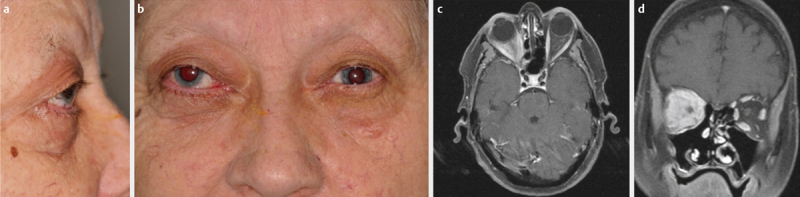
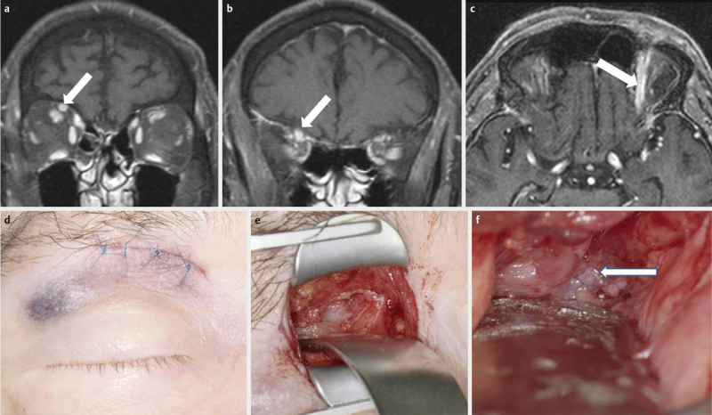
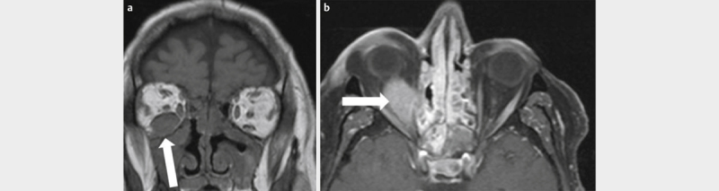
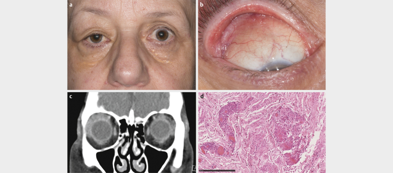
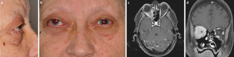
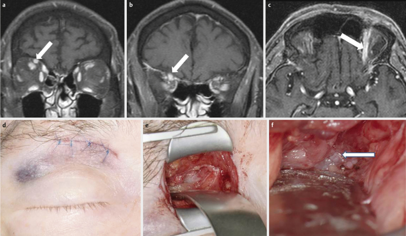
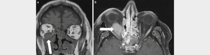
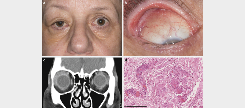
Zusammenfassung: Dieser Übersichtsartikel stellt eine Zusammenstellung seltener Erkrankungen der Orbita dar. Es werden angeborene Fehlbildungen und strukturelle Läsionen, entzündliche Erkrankungen, gutartige und bösartige epitheliale sowie nicht epitheliale Tumoren erörtert. Da in der Orbita zahlreiche Gewebetypen auf engstem Raum beieinanderliegen, kommen dort besonders viele Erkrankungen vor, sodass der Vollständigkeit und dem Tiefgang bei den einzelnen Abhandlungen natürliche Grenzen gesetzt sind. Die Kapitel beinhalten eine Übersicht über das jeweilige klinische Erscheinungsbild, wichtige Stichpunkte zur Diagnostik, Therapie und gegebenenfalls zur Prognose der Erkrankung. Dabei wurde versucht, die besonders charakteristischen Merkmale der einzelnen Erkrankungen herauszuarbeiten, sodass trotz der kurzen Darstellung die relevanten Aspekte enthalten sind.
Overview
The claims and statistics of this paper haven’t been checked yet.
Run the check — free, no account needed. It reads the open-access text and checks the sources it cites.
Integrity signals
Registry
Cites
Statistics
Figures
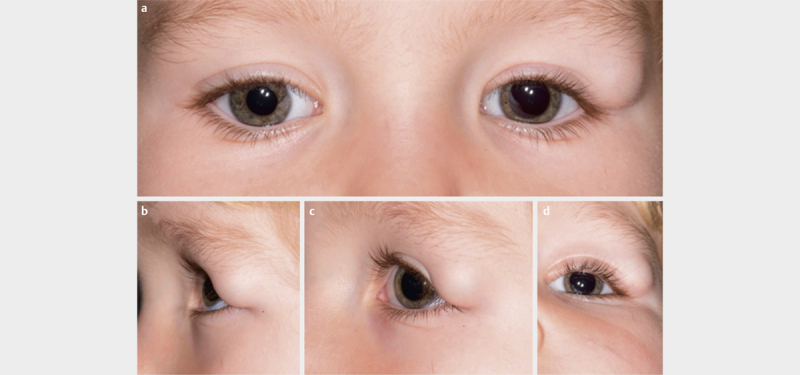
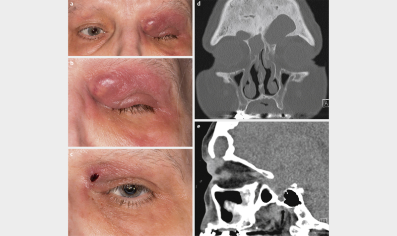
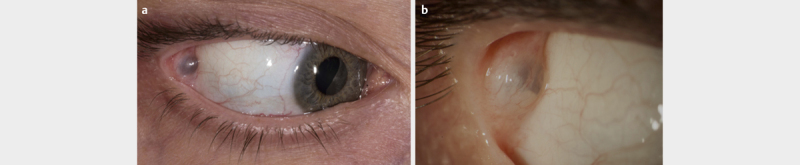
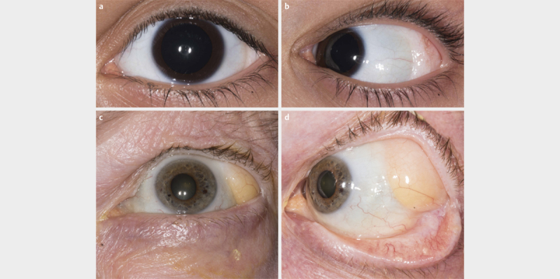
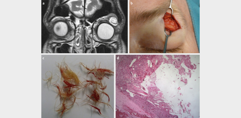


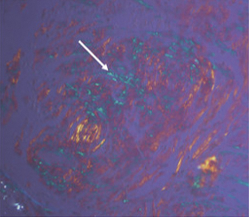
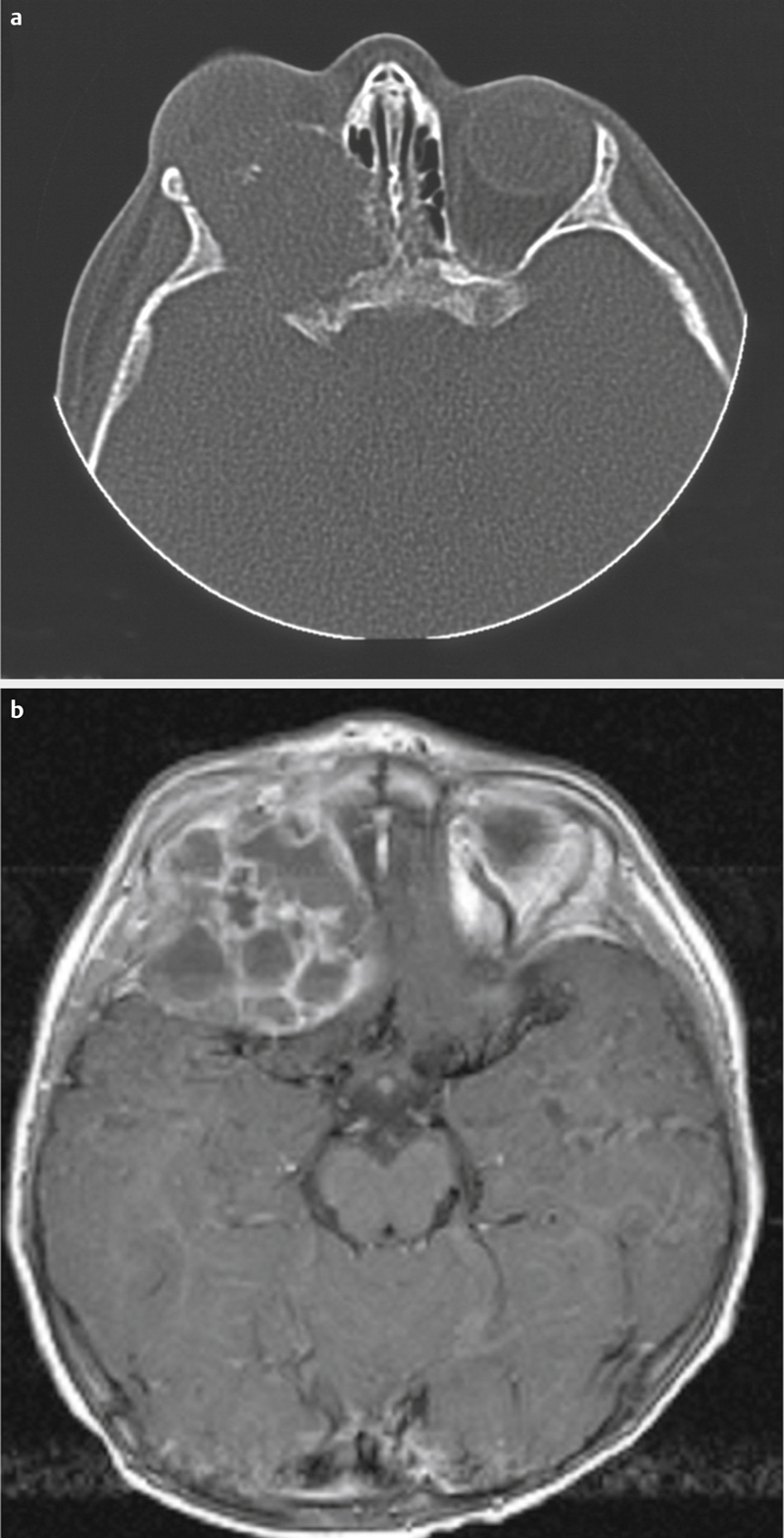
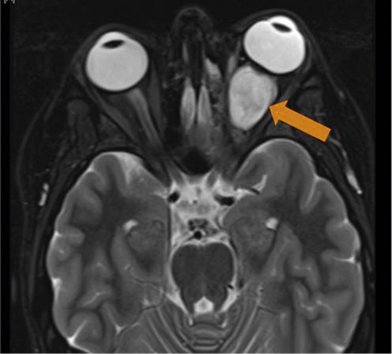
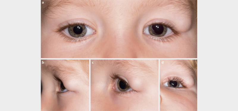
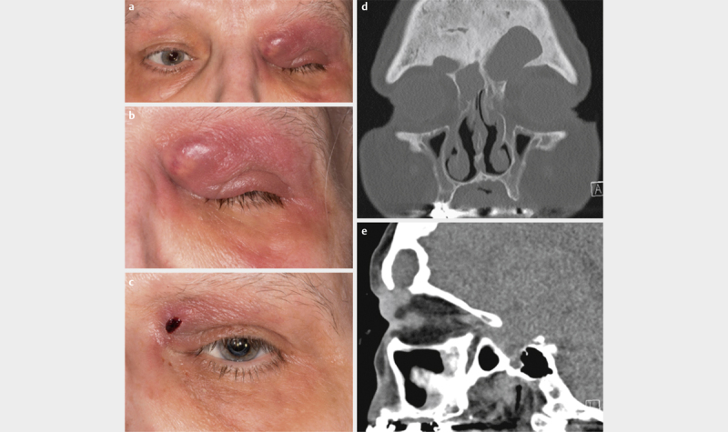
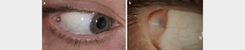
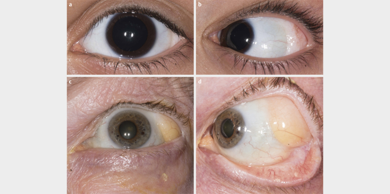
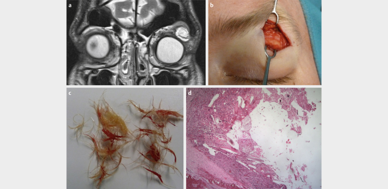


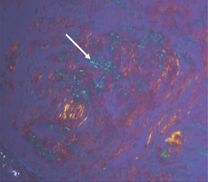
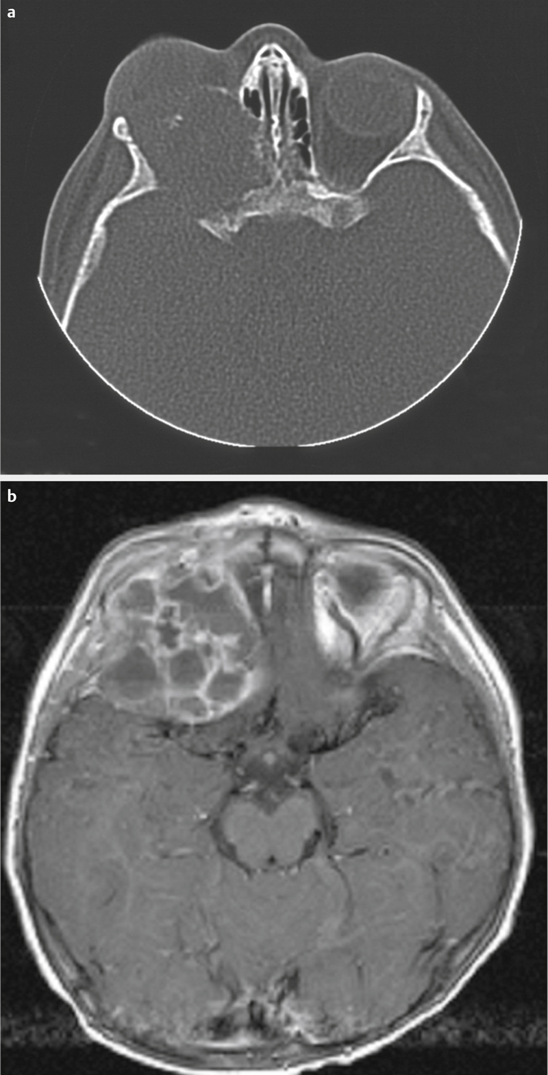
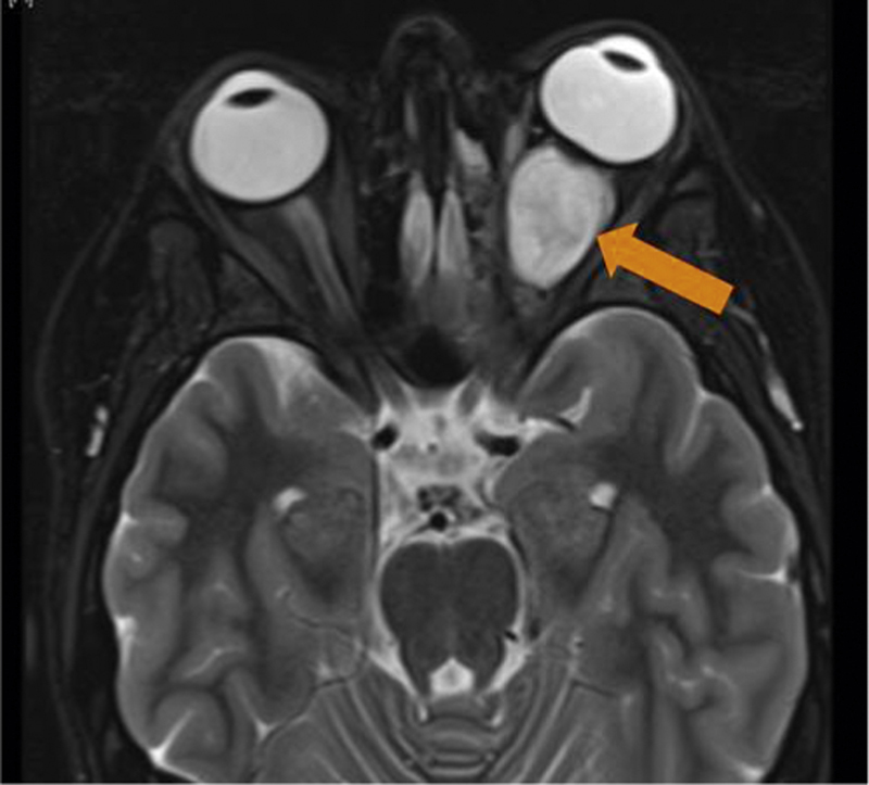
Verification
Claims the paper makes
The claims and statistics of this paper haven’t been checked yet.
Run the check — free, no account needed. It reads the open-access text and checks the sources it cites.
Claims other papers make about this one
Each is checked against this paper’s own text: does it say what the citing paper says it does?
The source backs part of the claim, not all of it — or backs it for a different population, condition or comparison.
In the citing paper
“The sample size in our study was small because the pathology treated is considered a rare disease, with a very low incidence and prevalence.”
From Serious Game for Fine Motor Control Rehabilitation for Children With Epileptic Encephalopathy: Development and Usability Study (2023) · cites this paper as [7]
Experimental — accuracy not yet measured
Self-check
Percentages, sums and reported differences re-computed from the paper’s own numbers.
The self-check needs the paper’s full text.
Citations
Cites
Masi, A. T., Hunder, G. G., Lie, J. T., Michel, B. A., Bloch, D. A., Arend, W. P., Calabrese, L. H., Edworthy, S. M., Fauci, A. S., Leavitt, R. Y., et al. (1990) · Arthritis & Rheumatism · doi:10.1002/art.1780330806 · Cited by 2,212
Cornely, O. A., Alastruey‐Izquierdo, A., Arenz, D., Chen, S. C. ‐., Dannaoui, É., Hochhegger, B., Hoenigl, M., Jensen, H. E., Lagrou, K., Lewis, R. E., et al. (2019) · The Lancet Infectious Diseases · doi:10.1016/s1473-3099(19)30312-3 · Cited by 1,915
Wassef, M., Blei, F., Adams, D. M., Alomari, A. I., Baselga, E., Berenstein, A., Burrows, P. E., Frieden, I. J., Garzón, M. C., López‐Gutiérrez, J. C., et al. (2015) · PEDIATRICS · doi:10.1542/peds.2014-3673 · Cited by 1,411
Trial of tocilizumab in giant-cell arteritis
Stone, J. H., Tuckwell, K., Dimonaco, S., Klearman, M., Aringer, M., Blockmans, D., Brouwer, E., Cid, M. C., Dasgupta, B., Rech, J., et al. (2017) · New England Journal of Medicine · doi:10.1056/nejmoa1613849 · Cited by 1,386
Sinus histiocytosis with massive lymphadenopathy (Rosai-Dorfman disease): review of the entity.
Foucar, E., Rosaí, J., & Dorfman, R. F. (1990) · PubMed · Cited by 1,232
Rosaí, J., & Dorfman, R. F. (1969) · PubMed · Cited by 1,227
Survey of 1264 patients with orbital tumors and simulating lesions
Shields, J. A., Shields, C. L., & Scartozzi, R. (2004) · Ophthalmology · doi:10.1016/j.ophtha.2003.01.002 · Cited by 1,055
Guillevin, L., Lhote, F., Gayraud, M., Cohen, P., Jarrousse, B., Lortholary, O., Thibult, N., & Casassus, P. (1996) · Medicine · doi:10.1097/00005792-199601000-00003 · Cited by 992
Necrotizing fasciitis: clinical presentation, microbiology, and determinants of mortality.
Wong, C., Chang, H.-C., Pasupathy, S., Khin, L., Tan, J.-L., & Low, C.-O. (2003) · PubMed · Cited by 975
NECROTIZING FASCIITIS: CLINICAL PRESENTATION, MICROBIOLOGY, AND DETERMINANTS OF MORTALITY
WONG, C.-H., CHANG, H.-C., PASUPATHY, S., KHIN, L.-W., TAN, J.-L., & LOW, C.-O. (2003) · The Journal of Bone and Joint Surgery-American Volume · doi:10.2106/00004623-200308000-00005 · Cited by 909
Anatomical classification of facial, cranio-facial and latero-facial clefts
Tessier, P. L. (1976) · Journal of Maxillofacial Surgery · doi:10.1016/s0301-0503(76)80013-6 · Cited by 882
Leigh syndrome: Clinical features and biochemical and DNA abnormalities
Rahman, S., Blok, R. B., Dahl, H. ‐. M., Danks, D. M., Kirby, D. M., Chow, C. W., Christodoulou, J., & Thorburn, D. R. (1996) · Annals of Neurology · doi:10.1002/ana.410390311 · Cited by 792
Erdheim-Chester Disease Clinical and Radiologic Characteristics of 59 Cases
Veyssier-Belot, C., Cacoub, P., Caparros-Lefebvre, D., Wechsler, J., Brun, B., Remy, M., Wallaert, B., Petit, H., Grimaldi, A., Wechsler, B., et al. (1996) · Medicine · doi:10.1097/00005792-199605000-00005 · Cited by 702
Therapeutic Outcome in Invasive Aspergillosis
Denning, D. W. (1996) · Clinical Infectious Diseases · doi:10.1093/clinids/23.3.608 · Cited by 685
Atypical and Malignant Solitary Fibrous Tumors in Extrathoracic Locations
Decouvelaere, A., Dry, S., & Fletcher, C. D. �. (1998) · The American Journal of Surgical Pathology · doi:10.1097/00000478-199812000-00007 · Cited by 641
Posaconazole Is Effective as Salvage Therapy in Zygomycosis: A Retrospective Summary of 91 Cases
van Burik, J. H., Hare, R. S., Solomon, H. F., Corrado, M. L., & Kontoyiannis, D. P. (2006) · Clinical Infectious Diseases · doi:10.1086/500212 · Cited by 614
Aneurysmal bone cysts.A clinicopathologic study of 66 cases
Biesecker, J. L., Marcove, R. C., Huvos, A. G., & Miké, V. (1970) · Cancer · doi:10.1002/1097-0142(197009)26:3 · Cited by 555
Tumors of the conjunctiva and cornea
Shields, C. L., & Shields, J. A. (2004) · Survey of Ophthalmology · doi:10.1016/j.survophthal.2003.10.008 · Cited by 535
Leigh syndrome: One disorder, more than 75 monogenic causes
Lake, N. J., Compton, A. G., Rahman, S., & Thorburn, D. R. (2015) · Annals of Neurology · doi:10.1002/ana.24551 · Cited by 499
Stark, Z., & Savarirayan, R. (2009) · Orphanet Journal of Rare Diseases · doi:10.1186/1750-1172-4-5 · Cited by 463
van Rhee, F., Wong, R. S., Munshi, N., Rossi, J.-F., Ke, X.-Y., Fosså, A., Simpson, D., Capra, M., Liu, T., Hsieh, R. K., et al. (2014) · The Lancet Oncology · doi:10.1016/s1470-2045(14)70319-5 · Cited by 454
Mukhtyar, C. B., Floßmann, O., Hellmich, B., Bacon, P., Cid, M. C., Tervaert, J.-W. C., Gross, W. L., Guillevin, L., Jayne, D. R. W., Mahr, A., et al. (2007) · Annals of the Rheumatic Diseases · doi:10.1136/ard.2007.071936 · Cited by 418
Cleidocranial dysplasia: clinical and molecular genetics
Mundlos, S. (1999) · Journal of Medical Genetics · doi:10.1136/jmg.36.3.177 · Cited by 407
Lymphoma of the Ocular Adnexa: A Study of 353 Cases
Ferry, J. A., Fung, C. Y., Zukerberg, L. R., Lucarelli, M. J., Hasserjian, R. P., Preffer, F. I., & Harris, N. L. (2007) · The American Journal of Surgical Pathology · doi:10.1097/01.pas.0000213350.49767.46 · Cited by 400
Sharif, S., Ferner, R. E., Birch, J. M., Gillespie, J. E., Gattamaneni, H. R., Baser, M. E., & Evans, D. G. (2006) · Journal of Clinical Oncology · doi:10.1200/jco.2005.03.8349 · Cited by 384
Classification and Incidence of Space-Occupying Lesions of the Orbit
Shields, J. A. (1984) · Archives of Ophthalmology · doi:10.1001/archopht.1984.01040031296011 · Cited by 369
Surgery in Castleman's Disease
Talat, N., Belgaumkar, A. P., & Schulte, K. (2012) · Annals of Surgery · doi:10.1097/sla.0b013e318249dcdc · Cited by 358
Dubourg, C., Bendavid, C., Pasquier, L., Henry, C., Odent, S., & David, V. (2007) · Orphanet Journal of Rare Diseases · doi:10.1186/1750-1172-2-8 · Cited by 355
Localized mesothelioma of the pleura
Okike, N., Bernatz, P. E., & Woolner, L. B. (1978) · Journal of Thoracic and Cardiovascular Surgery · doi:10.1016/s0022-5223(19)41262-2 · Cited by 342
Fajgenbaum, D. C., van Rhee, F., & Nabel, C. S. (2014) · Blood · doi:10.1182/blood-2013-12-545087 · Cited by 336
Huber, A. M., Lam, P., Duffy, C. M., Yeung, R. S. M., Ditchfield, M. R., Laxer, D. A., Cole, W. G., Graham, H. K., Allen, R. C., & Laxer, R. M. (2002) · The Journal of Pediatrics · doi:10.1067/mpd.2002.126457 · Cited by 321
Localized mesothelioma of the pleura: benign and malignant variants.
Okike, N., Bernatz, P. E., & Woolner, L. B. (1978) · PubMed · Cited by 319
Central giant cell granuloma of the jaw: a review of the literature with emphasis on therapy options
de Lange, J., van den Akker, H. P., & van den Berg, H. (2007) · Oral Surgery Oral Medicine Oral Pathology Oral Radiology and Endodontology · doi:10.1016/j.tripleo.2007.04.003 · Cited by 310
Unusual granulation combined with hyperplastic change of lymphatic tissue
Kimura, T. (1948) · Cited by 293
Pasadhika, S., & Rosenbaum, J. T. (2015) · Clinics in Chest Medicine · doi:10.1016/j.ccm.2015.08.009 · Cited by 289
Mombaerts, I., Goldschmeding, R., Schlingemann, R. O., & Koornneef, L. (1996) · Survey of Ophthalmology · doi:10.1016/s0039-6257(97)81996-0 · Cited by 278
Extrapleural solitary fibrous tumor: a clinicopathologic study of 24 cases.
Brunnemann, R. B., Ro, J. Y., Ordóǹez, N. G., Mooney, J., El‐Naggar, A. K., & Ayala, A. G. (1999) · PubMed · Cited by 272
Therapy of Parenchymal Brain Cysticercosis with Praziquantel
Sotelo, J., Escobedo, F., Rodríguez‐Carbajal, J., Torres‐Álvarez, B., & Rubio‐Donnadieu, F. (1984) · New England Journal of Medicine · doi:10.1056/nejm198404193101601 · Cited by 268
Optic nerve sheath meningiomas
Saeed, P., Rootman, J., Nugent, R. A., White, V. A., Mackenzie, I. R., & Koornneef, L. (2003) · Ophthalmology · doi:10.1016/s0161-6420(03)00787-5 · Cited by 264
Cogan Syndrome: A Retrospective Review of 60 Patients Throughout a Half Century
Gluth, M. B., Baratz, K. H., Matteson, E. L., & Driscoll, C. L. W. (2006) · Mayo Clinic Proceedings · doi:10.4065/81.4.483 · Cited by 256
The clinical spectrum of Erdheim-Chester disease: an observational cohort study
Estrada‐Veras, J. I., O’Brien, K., Boyd, L. C., Dave, R. H., Durham, B. H., Xi, L., Malayeri, A. A., Chen, M. Y., Gardner, P. J., Enriquez, J. R. A., et al. (2017) · Blood Advances · doi:10.1182/bloodadvances.2016001784 · Cited by 255
Typical and atypical Cogan's syndrome: 32 cases and review of the literature
Grasland, A. (2004) · British journal of rheumatology · doi:10.1093/rheumatology/keh228 · Cited by 249
Oral glutamine reduces the duration and severity of stomatitis after cytotoxic cancer chemotherapy
Anderson, P. M., Schroeder, G. S., & Skubitz, K. M. (1998) · Cancer · doi:10.1002/(sici)1097-0142(19981001)83:7 · Cited by 243
Fibrous Dysplasia Involving the Skull Base and Temporal Bone
Lustig, L. R., Holliday, M., McCarthy, E. F., & Nager, G. T. (2001) · Archives of Otolaryngology - Head and Neck Surgery · doi:10.1001/archotol.127.10.1239 · Cited by 239
Primary malignant neoplasms of the lacrimal gland.
Wright, J. E., Rose, G. E., & Garner, A. (1992) · British Journal of Ophthalmology · doi:10.1136/bjo.76.7.401 · Cited by 236
USP6-induced neoplasms: the biologic spectrum of aneurysmal bone cyst and nodular fasciitis
Oliveira, A. M., & Chou, M. M. (2013) · Human Pathology · doi:10.1016/j.humpath.2013.03.005 · Cited by 236
Turbin, R. E., Thompson, C. R., Kennerdell, J. S., Cockerham, K. P., & Kupersmith, M. J. (2002) · Ophthalmology · doi:10.1016/s0161-6420(02)01017-5 · Cited by 233
Ophthalmic Pathology: An Atlas and Textbook
Perlman, J. I. (1996) · JAMA · doi:10.1001/jama.1996.03540220071036 · Cited by 229
Hindsø, T. G., & Heegaard, S. (2018) · Survey of Ophthalmology · doi:10.1016/j.survophthal.2018.08.002 · Cited by 221
Tumors and Tumorlike Lesions of Bone
Schajowicz, F. (1994) · Springer eBooks · doi:10.1007/978-3-642-49954-8 · Cited by 216
Idiopathic orbital inflammatory syndrome: Clinical features and treatment outcomes
Swamy, B. N., McCluskey, P., Nemet, A., Crouch, R., Martin, P. A., Benger, R. S., Ghabriel, R., & Wakefield, D. (2007) · British Journal of Ophthalmology · doi:10.1136/bjo.2007.124156 · Cited by 214
Governale, L. S. (2015) · Pediatric Neurology · doi:10.1016/j.pediatrneurol.2015.07.006 · Cited by 212
Clinical genetics of craniosynostosis
Wilkie, A. O. M., Johnson, D., & Wall, S. A. (2017) · Current Opinion in Pediatrics · doi:10.1097/mop.0000000000000542 · Cited by 211
Mold Infections of the Central Nervous System
McCarthy, M. W., Rosengart, A. J., Schuetz, A. N., Kontoyiannis, D. P., & Walsh, T. J. (2014) · New England Journal of Medicine · doi:10.1056/nejmra1216008 · Cited by 207
Clinicopathologic Review of 142 Cases of Lacrimal Gland Lesions
Shields, C. L., Shields, J. A., Eagle, R. C., & Rathmell, J. P. (1989) · Ophthalmology · doi:10.1016/s0161-6420(89)32873-9 · Cited by 207
Erdheim–Chester disease: A systematic review
Cives, M., Simone, V. D., Rizzo, F. M., Dicuonzo, F., Lacalamita, M. C., Ingravallo, G., Silvestris, F., & Dammacco, F. (2015) · Critical Reviews in Oncology/Hematology · doi:10.1016/j.critrevonc.2015.02.004 · Cited by 203
Vascular Anomalies (Part I): Classification and Diagnostics of Vascular Anomalies
Sadick, M., Müller‐Wille, R., Wildgruber, M., & Wohlgemuth, W. A. (2018) · RöFo - Fortschritte auf dem Gebiet der Röntgenstrahlen und der bildgebenden Verfahren · doi:10.1055/a-0620-8925 · Cited by 199
Pleomorphic adenoma of the lacrimal gland.
Rose, G. E., & Wright, J. E. (1992) · British Journal of Ophthalmology · doi:10.1136/bjo.76.7.395 · Cited by 197
Auclair, P. L., Cuenin, P., Kratochvil, F. J., Slater, L. J., & Ellis, G. L. (1988) · Oral Surgery Oral Medicine Oral Pathology · doi:10.1016/0030-4220(88)90094-1 · Cited by 196
Infliximab for Intravenous Immunoglobulin Resistance in Kawasaki Disease: A Retrospective Study
Son, M. B. F., Gauvreau, K., Burns, J. C., Corinaldesi, E., Tremoulet, A. H., Watson, V. E., Baker, A., Fulton, D. R., Sundel, R. P., & Newburger, J. W. (2010) · The Journal of Pediatrics · doi:10.1016/j.jpeds.2010.10.012 · Cited by 194
Googe, P. B., Harris, N. L., & Mihm, M. C. (1987) · Journal of Cutaneous Pathology · doi:10.1111/j.1600-0560.1987.tb00498.x · Cited by 191
Carubbi, F., Cipriani, P., Marrelli, A., Benedetto, P. D., Ruscitti, P., Berardicurti, O., Pantano, I., Liakouli, V., Alvaro, S., Alunno, A., et al. (2013) · Arthritis Research & Therapy · doi:10.1186/ar4359 · Cited by 186
Jaspers, H. C. J., Verbist, B. M., Schoffelen, R., Mattijssen, V., Slootweg, P. J., van der Graaf, W. T. A., & van Herpen, C. M. L. (2011) · Journal of Clinical Oncology · doi:10.1200/jco.2010.32.8351 · Cited by 179
Orbital Inflammation With IgG4-Positive Plasma Cells
Plaza, J. A. (2011) · Archives of Ophthalmology · doi:10.1001/archophthalmol.2011.16 · Cited by 175
Seyahi, E. (2016) · Current Opinion in Rheumatology · doi:10.1097/bor.0000000000000343 · Cited by 168
The spectrum of orbital aspergillosis: a clinicopathological review
Levin, L. A., Avery, R. K., Shore, J. W., Woog, J. J., & Baker, A. S. (1996) · Survey of Ophthalmology · doi:10.1016/s0039-6257(96)80004-x · Cited by 165
Siddiqui, A. A., Shah, A. A., & Bashir, S. H. (2004) · Neurosurgery · doi:10.1227/01.neu.0000134597.94269.48 · Cited by 163
Granulocytic sarcoma: a systematic review.
Yılmaz, A. F., Saydam, G., Şahin, F., & Baran, Y. (2013) · Europe PMC (PubMed Central) · Cited by 161
The kaleidoscopic world of rare craniofacial clefts: order out of chaos (Tessier classification).
Hk, K. (1976) · PubMed · Cited by 161
Fungus ball of the paranasal sinuses: Experience in 160 patients treated with endoscopic surgery
Nicolai, P., Lombardi, D., Tomenzoli, D., Villaret, A. B., Piccioni, M., Mensi, M., & Maroldi, R. (2009) · The Laryngoscope · doi:10.1002/lary.20578 · Cited by 157
de Leval, L., & Jaffe, E. S. (2020) · The Cancer Journal · doi:10.1097/ppo.0000000000000451 · Cited by 155
Green, W. R. (1969) · Archives of Ophthalmology · doi:10.1001/archopht.1969.00990020304002 · Cited by 155
Bahrami, A., & Folpe, A. L. (2010) · The American Journal of Surgical Pathology · doi:10.1097/pas.0b013e3181ef70b6 · Cited by 153
Gnekow, A. K., Kortmann, R.-D., Pietsch, T., & Emser, A. (2004) · Klinische Pädiatrie · doi:10.1055/s-2004-832355 · Cited by 152
Gamel, J. W., & Font, R. L. (1982) · Human Pathology · doi:10.1016/s0046-8177(82)80180-9 · Cited by 151
Localised invasive sino-orbital aspergillosis: characteristic features
Sivak-Callcott, J. A. (2004) · British Journal of Ophthalmology · doi:10.1136/bjo.2003.021725 · Cited by 150
Idiopathic Sclerosing Inflammation of the Orbit
Rootman, J., McCarthy, M., White, V. A., Harris, G. F., & Kennerdell, J. S. (1994) · Ophthalmology · doi:10.1016/s0161-6420(94)31298-x · Cited by 149
The Classification and Management of Acute Orbital Pseudotumors
Rootman, J., & Nugent, R. A. (1982) · Ophthalmology · doi:10.1016/s0161-6420(82)34683-7 · Cited by 149
Chang, S. Y., Keogh, K. A., Lewis, J. E., Ryu, J. H., Cornell, L. D., Garrity, J. A., & Yi, E. S. (2013) · Human Pathology · doi:10.1016/j.humpath.2013.05.023 · Cited by 149
Periorbital necrotising fasciitis
Lazzeri, D., Lazzeri, S., Figus, M., Tascini, C., Bocci, G., Colizzi, L., Giannotti, G., Lorenzetti, F., Gandini, D., Danesi, R., et al. (2009) · British Journal of Ophthalmology · doi:10.1136/bjo.2009.167486 · Cited by 143
Optic nerve sheath meningioma: current diagnosis and treatment
Eddleman, C. S., & Liu, J. K. (2007) · Neurosurgical FOCUS · doi:10.3171/foc-07/11/e4 · Cited by 143
Orbital dermoids: clinical presentation and management.
Sherman, R. P., Rootman, J., & Lapointe, J. S. (1984) · British Journal of Ophthalmology · doi:10.1136/bjo.68.9.642 · Cited by 142
Epithelial Lacrimal Gland Tumors
Weis, E. (2009) · Archives of Ophthalmology · doi:10.1001/archophthalmol.2009.209 · Cited by 141
Diverse Clinical Presentations of Orbital Sarcoid
Mavrikakis, I., & Rootman, J. (2007) · American Journal of Ophthalmology · doi:10.1016/j.ajo.2007.07.019 · Cited by 139
Primary bone tumors of the orbit
Selva, D. N., White, V. A., OʼConnell, J. X., & Rootman, J. (2004) · Survey of Ophthalmology · doi:10.1016/j.survophthal.2004.02.011 · Cited by 139
Periocular and Orbital AmyloidosisClinical Characteristics, Management, and Outcome
Leibovitch, I., Selva, D. N., Goldberg, R. A., Sullivan, T. J., Saeed, P., Davis, G. J., McCann, J. D., McNab, A. A., & Rootman, J. (2006) · Ophthalmology · doi:10.1016/j.ophtha.2006.03.052 · Cited by 131
Orbital inflammatory pseudotumor and ischemic vasculitis in Churg-Strauss syndrome
Takanashi, T., Uchida, S., Arita, M., Okada, M., & Kashii, S. (2001) · Ophthalmology · doi:10.1016/s0161-6420(01)00557-7 · Cited by 129
Early diagnosis of invasive mould infections and disease
Lamoth, F., & Calandra, T. (2017) · Journal of Antimicrobial Chemotherapy · doi:10.1093/jac/dkx030 · Cited by 128
Ahmad, S. Z., Esmaeli, B., Williams, M. D., Nguyen, J., Fay, A. M., Woog, J. J., Selvadurai, D., Rootman, J., Weis, E., Selva, D. N., et al. (2009) · Ophthalmology · doi:10.1016/j.ophtha.2008.12.049 · Cited by 128
Management of the orbit in malignant sinonasal tumors
Suarez, C. M., Ferlito, A., Lund, V. J., Silver, C. E., Fagan, J. J., Rodrigo, J. P., Llorente, J. L., Cantù, G., Politi, M., Wei, W., et al. (2007) · Head & Neck · doi:10.1002/hed.20736 · Cited by 128
Fromm, M., Littman, P., Raney, R. B., Nelson, L. P., Handler, S. D., Diamond, G. R., & Stanley, C. A. (1986) · Cancer · doi:10.1002/1097-0142(19860515)57:10 · Cited by 125
The Clinical and Pathologic Constellation of Wegener Granulomatosis of the Orbit
Perry, S. R., Rootman, J., & White, V. A. (1997) · Ophthalmology · doi:10.1016/s0161-6420(97)30251-6 · Cited by 124
Periorbital necrotizing fasciitis - a review
Amrith, S., Pai, V. H., & Ling, W. W. (2012) · Acta Ophthalmologica · doi:10.1111/j.1755-3768.2012.02420.x · Cited by 124
Shields, J. A., Kaden, I. H., Eagle, R. C., & Shields, C. L. (1997) · Ophthalmic Plastic and Reconstructive Surgery · doi:10.1097/00002341-199712000-00007 · Cited by 122
Orbital lymphoma: diagnostic approach and treatment outcome
Eckardt, A. M., Lemound, J., Rana, M., & Gellrich, N. (2013) · World Journal of Surgical Oncology · doi:10.1186/1477-7819-11-73 · Cited by 121
Immunoglobulin G4–Related Disease of the Orbit: Imaging Features in 27 Patients
Tiegs‐Heiden, C. A., Eckel, L. J., Hunt, C., Diehn, F. E., Schwartz, K. M., Kallmes, D. F., Salomão, D. R., Witzig, T. E., & Garrity, J. A. (2014) · American Journal of Neuroradiology · doi:10.3174/ajnr.a3865 · Cited by 118
Demirci, H., & Christianson, M. D. (2011) · American Journal of Ophthalmology · doi:10.1016/j.ajo.2010.12.011 · Cited by 117
Epithelial tumors of the lacrimal gland: an update
Bernardini, F. P., Devoto, M. H., & Croxatto, J. O. (2008) · Current Opinion in Ophthalmology · doi:10.1097/icu.0b013e32830b13e1 · Cited by 116
Nagel, M. A., White, T., Khmeleva, N., Rempel, A., Boyer, P. J., Bennett, J. L., Haller, A., Lear-Kaul, K. C., Kandasmy, B., Amato, M. M., et al. (2015) · JAMA Neurology · doi:10.1001/jamaneurol.2015.2101 · Cited by 115
Vascular tumors in infants and adolescents
Wildgruber, M., Sadick, M., Müller‐Wille, R., & Wohlgemuth, W. A. (2019) · Insights into Imaging · doi:10.1186/s13244-019-0718-6 · Cited by 115
Ocular Manifestations of Granulomatosis with Polyangiitis: A Review of the Literature
Sfiniadaki, E., Tsiara, I., THEODOSSIADIS, P. G., & Chatziralli, I. (2019) · Ophthalmology and Therapy · doi:10.1007/s40123-019-0176-8 · Cited by 114
Salivary gland tumours: diagnostic challenges and an update on the latest WHO classification
Speight, P. M., & Barrett, A. W. (2020) · Diagnostic histopathology · doi:10.1016/j.mpdhp.2020.01.001 · Cited by 113
Orbital inflammation: Biopsy first
Mombaerts, I., Rose, G. E., & Garrity, J. A. (2016) · Survey of Ophthalmology · doi:10.1016/j.survophthal.2016.03.002 · Cited by 110
Mucoceles of the Paranasal Sinuses with Intracranial and Intraorbital Extension
Delfini, R., Missori, P., Iannetti, G., Ciappetta, P., & Cantore, G. (1993) · Neurosurgery · doi:10.1227/00006123-199306000-00003 · Cited by 109
Clinical mini-review: systemic lupus erythematosus and the eye
Read, R. W. (2004) · Ocular Immunology and Inflammation · doi:10.1080/09273940490895308 · Cited by 108
Hamper, K., Lazar, F., Dietel, M., Caselitz, J., Berger, J., Arps, H., Falkmer, S., Auer, G., & Seifert, G. (1990) · Journal of Oral Pathology and Medicine · doi:10.1111/j.1600-0714.1990.tb00806.x · Cited by 105
Pero, M. M. D., Chaudhry, A., Jones, R. B., Sivasothy, P. R., Jani, P., & Jayne, D. R. W. (2009) · Clinical Otolaryngology · doi:10.1111/j.1749-4486.2009.01968.x · Cited by 104
Diffuse Normolipemic Plane Xanthoma
Altman, J. (1962) · Archives of Dermatology · doi:10.1001/archderm.1962.01590050063012 · Cited by 104
Orbital Tuberculosis: A Review of the Literature
Madge, S. N., Prabhakaran, V. C., Shome, D., Kim, U. R., Honavar, S. G., & Selva, D. N. (2008) · Orbit · doi:10.1080/01676830802225152 · Cited by 103
A Clinicopathologic Study of Primary Adenoid Cystic Carcinoma of the Lacrimal Gland
Lee, D. A., Bell, C. M., Waller, R. R., & Ilstrup, D. M. (1985) · Ophthalmology · doi:10.1016/s0161-6420(85)34081-2 · Cited by 103
von Holstein, S. L., Coupland, S. E., Briscoe, D., Tourneau, C. L., & Heegaard, S. (2012) · Acta Ophthalmologica · doi:10.1111/j.1755-3768.2012.02402.x · Cited by 102
Morales, A. G., Croxatto, J. O., Crovetto, L., & Ebner, R. (1988) · Ophthalmology · doi:10.1016/s0161-6420(88)33067-8 · Cited by 99
Necrobiotic Xanthogranuloma with Paraproteinemia
Bullock, J. D., Bartley, G. B., Bell, C. M., Yanes, B., Connelly, P. J., & Funkhouser, J. W. (1986) · Ophthalmology · doi:10.1016/s0161-6420(86)33605-4 · Cited by 98
Tse, D. T., Benedetto, P. W., Dubovy, S. R., Schiffman, J., & Feuer, W. J. (2005) · American Journal of Ophthalmology · doi:10.1016/j.ajo.2005.08.068 · Cited by 98
Lannon, D. A., & Earley, M. J. (2001) · British Journal of Plastic Surgery · doi:10.1054/bjps.2001.3701 · Cited by 97
Vaglio, A., Moosig, F., & Zwerina, J. (2011) · Current Opinion in Rheumatology · doi:10.1097/bor.0b013e32834d85ce · Cited by 95
Immunoglobulin G4-related diseases in the head and neck: A systematic review
Mulholland, G. B., Jeffery, C. C., Satija, P., & Côté, D. W. J. (2015) · Journal of Otolaryngology - Head and Neck Surgery · doi:10.1186/s40463-015-0071-9 · Cited by 93
Ocular-Adnexal Lymphoid Tumors
Johnson, T. E., Tse, D. T., Byrne, G. E., Restrepo, A., Whitcomb, C. C., Voigt, W., Benedetto, P. W., & Curtin, V. T. (1999) · Ophthalmic Plastic and Reconstructive Surgery · doi:10.1097/00002341-199905000-00006 · Cited by 93
Borruat, F.-X., Bogousslavsky, J., Uffer, S., Klainguti, G., & Schatz, N. J. (1993) · Ophthalmology · doi:10.1016/s0161-6420(93)31606-4 · Cited by 93
The role of postoperative radiation therapy in carcinoma ex pleomorphic adenoma of the parotid gland
Chen, A. M., García, J. J., Bucci, M. K., Quivey, J. M., & Eisele, D. W. (2006) · International Journal of Radiation Oncology*Biology*Physics · doi:10.1016/j.ijrobp.2006.07.1380 · Cited by 86
Complications Following Ex-Press Glaucoma Shunt Implantation
Stewart, R. M., Diamond, J. G., Ashmore, E. D., & Ayyala, R. S. (2005) · American Journal of Ophthalmology · doi:10.1016/j.ajo.2005.02.033 · Cited by 86
Holle, J. U., Voigt, C., Both, M., Holl‐Ulrich, K., Nölle, B., Laudien, M., Moosig, F., & Gross, W. L. (2013) · Rheumatology · doi:10.1093/rheumatology/kes382 · Cited by 85
An Update on Tumors of the Lacrimal Gland
Andreasen, S., Esmaeli, B., von Holstein, S. L., Mikkelsen, L. H., Rasmussen, P. K., & Heegaard, S. (2017) · Asia-Pacific Journal of Ophthalmology · doi:10.22608/apo.201707 · Cited by 85
Periocular and Orbital Amyloidosis: Clinical Characteristics, Management, and Outcome
Eagle, R. C. (2007) · Yearbook of Ophthalmology · doi:10.1016/s0084-392x(08)70195-3 · Cited by 84
Clinical and radiological presentation of 95 orbital lymphoid tumors
Polito, E., Galieni, P., & Leccisotti, A. (1996) · Graefe s Archive for Clinical and Experimental Ophthalmology · doi:10.1007/bf00184859 · Cited by 83
Giant Cell Reparative Granuloma
Yamaguchi, T., & Dorfman, H. D. (2001) · International Journal of Surgical Pathology · doi:10.1177/106689690100900304 · Cited by 83
Vascular Anomalies (Part II): Interventional Therapy of Peripheral Vascular Malformations
Müller‐Wille, R., Wildgruber, M., Sadick, M., & Wohlgemuth, W. A. (2018) · RöFo - Fortschritte auf dem Gebiet der Röntgenstrahlen und der bildgebenden Verfahren · doi:10.1055/s-0044-101266 · Cited by 83
Hedrich, C. M., Schnabel, A., & Hospach, T. (2018) · Frontiers in Pediatrics · doi:10.3389/fped.2018.00198 · Cited by 83
Primary liposarcoma of the orbit: A clinicopathologic study of seven cases
Cai, Y. (2001) · Annals of Diagnostic Pathology · doi:10.1053/adpa.2001.27911 · Cited by 82
Plaque Radiotherapy for Selected Orbital Malignancies: Preliminary Observations
Shields, J. A., Shields, C. L., Freire, J. E., Brady, L. W., & Komarnicky, L. T. (2003) · Ophthalmic Plastic and Reconstructive Surgery · doi:10.1097/01.iop.0000056020.66654.33 · Cited by 82
New Treatment Approaches to Arteriovenous Malformations
Gilbert, P., Dubois, J., Giroux, M.-F., & Soulez, G. (2017) · Seminars in Interventional Radiology · doi:10.1055/s-0037-1604299 · Cited by 81
One year in review 2019: Sjögren's syndrome.
Cafaro, G., Croia, C., Argyropoulou, O. D., Leone, M. C., Orlandi, M., Finamore, F., Cecchettini, A., Ferro, F., Baldini, C., & Bartoloni, E. (2019) · PubMed · Cited by 80
Dagan, O., Barak, Y., & Metzker, A. (1995) · Pediatric Dermatology · doi:10.1111/j.1525-1470.1995.tb00122.x · Cited by 79
Orbital Pseudotumor: Histopathological Classification and Treatment
Fujii, H., Fujisada, H., Kondo, T., Takahashi, T., & Okada, S. (2010) · Ophthalmologica · doi:10.1159/000309523 · Cited by 79
Rosai dorfman disease of the orbit
Vemuganti, G. K., Naik, M. N., & Honavar, S. G. (2008) · Journal of Hematology & Oncology · doi:10.1186/1756-8722-1-7 · Cited by 78
Extrapleural Solitary Fibrous Tumor: A Clinicopathologic Study of 19 Cases
Insabato, L., Siano, M., Somma, A., Gentile, R., Santangelo, M. L., & Pettinato, G. (2009) · International Journal of Surgical Pathology · doi:10.1177/1066896909333779 · Cited by 77
Recent advances in the management of Takayasu arteritis
Misra, D. P., Wakhlu, A., Agarwal, V., & Danda, D. (2019) · International Journal of Rheumatic Diseases · doi:10.1111/1756-185x.13285 · Cited by 77
Green, W. R. (1967) · Archives of Ophthalmology · doi:10.1001/archopht.1967.00980030320010 · Cited by 76
Orbital Hemangiopericytoma and Solitary Fibrous Tumor: A Morphologic Continuum
Goldsmith, J. D., van de Rijn, M., & Syed, N. A. (2001) · International Journal of Surgical Pathology · doi:10.1177/106689690100900406 · Cited by 75
Sino-orbital Aspergillosis in Acquired Immunodeficiency Syndrome
Johnson, T. E. (1999) · Archives of Ophthalmology · doi:10.1001/archopht.117.1.57 · Cited by 73
Primary Ductal Adenocarcinoma of the Lacrimal Gland
Katz, S. E., Rootman, J., Dolman, P. J., White, V. A., & Berean, K. W. (1996) · Ophthalmology · doi:10.1016/s0161-6420(96)30746-x · Cited by 72
Tolosa-Hunt Syndrome Extending in the Cerebellopontine Angle
İaconetta, G., Stella, L., Esposito, M., & Cappabianca, P. (2005) · Cephalalgia · doi:10.1111/j.1468-2982.2005.00924.x · Cited by 72
Bonavolontà, G., Tranfa, F., de Conciliis, C., & Strianese, D. (1995) · Ophthalmic Plastic and Reconstructive Surgery · doi:10.1097/00002341-199509000-00005 · Cited by 72
Clinical and Pathological Manifestations with Differential Diagnosis in Behçet's Disease
Köktürk, A. (2011) · Pathology Research International · doi:10.1155/2012/690390 · Cited by 71
Clinical and Imaging Findings of Lymphoma in Patients with Sjögren Syndrome
Tonami, H., Matoba, M., Kuginuki, Y., Yokota, H., Higashi, K., Yamamoto, I., & Sugai, S. (2003) · Journal of Computer Assisted Tomography · doi:10.1097/00004728-200307000-00011 · Cited by 69
Myoepithelial carcinoma of the salivary glands: behavior and management.
俞光岩, 马大权, 孙开华, 李铁军, & Ye, Z. (2003) · PubMed · Cited by 67
Myoepithelial carcinoma of the salivary glands: behavior and management.
Yu, G., Ma, D.-Q., Sun, K., Li, T., & Zhang, Y. (2003) · PubMed · Cited by 67
Clinical and Imaging Characteristics of Orbital Leukemic Tumors
Bidar, M., Wilson, M. W., Laquis, S. J., Wilson, T. D., Fleming, J. C., Wesley, R. E., Ribeiro, R. C., & Haik, B. G. (2007) · Ophthalmic Plastic and Reconstructive Surgery · doi:10.1097/iop.0b013e3180333a85 · Cited by 66
Hepatic veno-occlusive disease due to DTIC
Asbury, R. F., Rosenthal, S. N., Descalzi, M. E., Ratcliffe, R. L., & Arseneau, J. C. (1980) · Cancer · doi:10.1002/1097-0142(19800515)45:10 · Cited by 66
Solitary Fibrous Tumor of the Lacrimal Gland Fossa
Scott, I. U., Tanenbaum, M., Rubin, D., & Lores, E. (1996) · Ophthalmology · doi:10.1016/s0161-6420(96)30455-7 · Cited by 66
Schittkowski, M. (2006) · British Journal of Ophthalmology · doi:10.1136/bjo.2006.092478 · Cited by 65
Cottin, V., & Cordier, J.-F. (1999) · Allergy · doi:10.1034/j.1398-9995.1999.t01-1-00091.x · Cited by 64
Mucoepidermoid tumors of the major salivary glands
Thorvaldsson, S. E., Beahrs, O. H., Woolner, L. B., & Simons, J. N. (1970) · The American Journal of Surgery · doi:10.1016/s0002-9610(70)80002-2 · Cited by 64
The treatment outcomes in IgG4‐related orbital disease: a systematic review of the literature
Detiger, S. E., Karim, F., Verdijk, R. M., van Hagen, P. M., van Laar, J. A. M., & Paridaens, D. A. (2019) · Acta Ophthalmologica · doi:10.1111/aos.14048 · Cited by 63
Acute invasive fungal rhinosinusitis: our experience with 18 cases
Bakhshaee, M., Bojdi, A., Allahyari, A., Majidi, M. R., Tavakol, S., Najafzadeh, M. J., & Asghari, M. (2016) · European Archives of Oto-Rhino-Laryngology · doi:10.1007/s00405-016-4109-z · Cited by 63
Histopathological classification of 272 primary epithelial tumors of the lacrimal gland.
Ni, C., Kuo, P. K., & Dryja, T. P. (1992) · PubMed · Cited by 62
Congenital Microphthalmos With Orbital Cysts
Chaudhry, I. A., Arat, Y. Ö., Shamsi, F. A., & Boniuk, M. (2004) · Ophthalmic Plastic and Reconstructive Surgery · doi:10.1097/01.iop.0000143716.12643.98 · Cited by 62
Heike, C. L., Hing, A. V., Aspinall, C., Bartlett, S. P., Birgfeld, C. B., Drake, A. F., Pimenta, L. A. F., Sie, K. C. Y., Urata, M. M., Vivaldi, D., et al. (2013) · American Journal of Medical Genetics Part C Seminars in Medical Genetics · doi:10.1002/ajmg.c.31373 · Cited by 60
de Lange, J., van Rijn, R. R., van den Berg, H., & van den Akker, H. P. (2008) · British Journal of Oral and Maxillofacial Surgery · doi:10.1016/j.bjoms.2008.05.013 · Cited by 59
Cogan's Syndrome—Clinical Guidelines and Novel Therapeutic Approaches
Tayer-Shifman, O. E., Ilan, O., Tovi, H., & Tal, Y. (2014) · Clinical Reviews in Allergy & Immunology · doi:10.1007/s12016-013-8406-7 · Cited by 58
Late-adult onset Leigh syndrome
McKelvie, P. A., Infeld, B., Marotta, R., Chin, J., Thorburn, D. R., & Collins, S. J. (2012) · Journal of Clinical Neuroscience · doi:10.1016/j.jocn.2011.09.009 · Cited by 58
Wolkow, N., Jakobiec, F. A., Lee, H., & Sutula, F. C. (2018) · American Journal of Ophthalmology · doi:10.1016/j.ajo.2018.07.024 · Cited by 57
Melanotic Neuroectodermal Tumor of Infancy
Soles, B. S., Wilson, A. M., Lucas, D. R., & Heider, A. (2018) · Archives of Pathology & Laboratory Medicine · doi:10.5858/arpa.2018-0241-ra · Cited by 57
Friedrich, R. E., & Bleckmann, V. (2003) · PubMed · Cited by 56
Progressive facial hemiatrophy and epilepsy
Dupont, S., Catala, M., Hasboun, D., Semah, F., & Baulac, M. (1997) · Neurology · doi:10.1212/wnl.48.4.1013 · Cited by 56
Malignant Lymphoma of the Lacrimal Gland
Rasmussen, P. K. (2011) · Archives of Ophthalmology · doi:10.1001/archophthalmol.2011.270 · Cited by 56
Lymphoid hyperplasia of the orbit and ocular adnexa: A clinical pathologic review
Andrew, N. H., Coupland, S. E., Pirbhai, A., & Selva, D. N. (2016) · Survey of Ophthalmology · doi:10.1016/j.survophthal.2016.04.004 · Cited by 56
Albendazole Therapy in the Treatment of Hydatid Liver Disease
Şenyüz, O. F., Yeşildağ, E., & Celayir, S. (2001) · Surgery Today · doi:10.1007/s005950170106 · Cited by 55
Bettiol, A., Silvestri, E., Scala, G. D., Amedei, A., Becatti, M., Fiorillo, C., Lopalco, G., Salvarani, C., Cantarini, L., Soriano, A., et al. (2019) · Rheumatology International · doi:10.1007/s00296-019-04259-y · Cited by 54
Wilhelm, H. M. (2009) · Current Opinion in Neurology · doi:10.1097/wco.0b013e32831fd9f5 · Cited by 53
The role of elective neck dissection in patients with adenoid cystic carcinoma of the head and neck
Xiao, R., Sethi, R. K. V., Feng, A. L., Fontanarosa, J., & Deschler, D. G. (2019) · The Laryngoscope · doi:10.1002/lary.27814 · Cited by 53
Primary Malignant Mixed Tumors of the Lacrimal Gland
Henderson, J. W., & Farrow, G. M. (1980) · Ophthalmology · doi:10.1016/s0161-6420(80)35206-8 · Cited by 52
Propranolol for Isolated Orbital Infantile Hemangioma
Fay, A. M. (2010) · Archives of Ophthalmology · doi:10.1001/archophthalmol.2009.375 · Cited by 52
Ocular adnexal and orbital amyloidosis: a case series and literature review
Mora-Horna, E. R., Rojas-Padilla, R., López, V. G., Guzmán, M. J., Ceriotto, A., & Salcedo, G. (2015) · International Ophthalmology · doi:10.1007/s10792-015-0138-7 · Cited by 52
Malignant Rhabdoid Tumor of the Orbit
Rootman, J., Damji, K. F., & Dimmick, J. E. (1989) · Ophthalmology · doi:10.1016/s0161-6420(89)32666-2 · Cited by 52
Primary Adenocarcinoma of the Lacrimal Gland
Heaps, R. S., Miller, N. R., Albert, D. M., Green, W. R., & Vitale, S. (1993) · Ophthalmology · doi:10.1016/s0161-6420(93)31385-0 · Cited by 50
Local Corticosteroid Treatment of Eyelid and Orbital Xanthogranuloma
Elner, V. M., Shtein, R. M., Demirci, H., & Hassan, A. S. (2006) · Ophthalmic Plastic and Reconstructive Surgery · doi:10.1097/01.iop.0000192645.24814.e8 · Cited by 50
Pediatric paranasal sinus mucoceles: Etiologic factors, management and outcome
Nicollas, R., Facon, F., Sudre-Levillain, I., Forman, C., Roman, S., & Triglia, J. (2005) · International Journal of Pediatric Otorhinolaryngology · doi:10.1016/j.ijporl.2005.10.002 · Cited by 49
Solitary Fibrous Tumor of the Lacrimal Sac
Woo, K. I., Suh, Y., & Kim, Y. (1999) · Ophthalmic Plastic and Reconstructive Surgery · doi:10.1097/00002341-199911000-00018 · Cited by 48
Melkersson-Rosenthal Syndrome in the Periocular Area: A Review of the Literature and Case Report
Shapiro, M. J., Peters, S. B., & Spinelli, H. M. (2003) · Annals of Plastic Surgery · doi:10.1097/01.sap.0000069068.03742.48 · Cited by 48
Rosenbaum, J. T., Choi, D., Wilson, D. J., Grossniklaus, H. E., Harrington, C. A., Sibley, C. H., Dailey, R. A., Ng, J. D., Steele, E. A., Czyz, C. N., et al. (2015) · JAMA Ophthalmology · doi:10.1001/jamaophthalmol.2015.0726 · Cited by 47
Retinoblastoma: Update on Current Management
Cassoux, N., Rouïc, L. L.-L., Lévy‐Gabriel, C., Aerts, I., Doz, F., & Desjardins, L. (2017) · Asia-Pacific Journal of Ophthalmology · doi:10.22608/apo.201778 · Cited by 47
Epithelioid hemangioma of the orbit
McEachren, T., Brownstein, S., Jordan, D. R., Montpetit, V. A. J., & Font, R. L. (2000) · Ophthalmology · doi:10.1016/s0161-6420(00)00011-7 · Cited by 46
Orbital complications in children: differential diagnosis of a challenging disease
Welkoborsky, H.-J., Graß, S. K., Deichmüller, C., Bertram, O., & Hinni, M. L. (2014) · European Archives of Oto-Rhino-Laryngology · doi:10.1007/s00405-014-3195-z · Cited by 46
Overview of benign and malignant lacrimal gland tumors
Gündüz, K., Yeşiltaş, Y. S., & Shields, C. L. (2018) · Current Opinion in Ophthalmology · doi:10.1097/icu.0000000000000515 · Cited by 46
Orbital Rosai-Dorfman Disease: A Literature Review
McClellan, S. F., & Ainbinder, D. J. (2013) · Orbit · doi:10.3109/01676830.2013.814689 · Cited by 45
Factors that influence Tolosa–Hunt syndrome and the short-term response to steroid pulse treatment
Zhang, X., Zhang, W., Liu, R., Dong, Z., & Yu, S. (2014) · Journal of the Neurological Sciences · doi:10.1016/j.jns.2014.03.031 · Cited by 44
Orbital Exenteration: A Dilemma in Mucormycosis Presented with Orbital Apex Syndrome
Songu, M., Ünlü, H., Günhan, K., İlker, S. S., & Neşe, N. (2008) · American Journal of Rhinology · doi:10.2500/ajr.2008.22.3121 · Cited by 44
Ophthalmological manifestations in patients with Leigh syndrome
Han, J., Lee, Y., Kim, S. M., Han, S. Y., Lee, J. B., & Han, S. (2014) · British Journal of Ophthalmology · doi:10.1136/bjophthalmol-2014-305704 · Cited by 44
Ocular Complications of Primary Sjögren Syndrome in Men
Mathews, P. M., Hsu, S. H., Hessen, M., Kim, J., Grader‐Beck, T., Birnbaum, J., Baer, A. N., & Akpek, E. K. (2015) · American Journal of Ophthalmology · doi:10.1016/j.ajo.2015.06.004 · Cited by 43
The Use of Radiotherapy for the Treatment of Localized Orbital Amyloidosis
Khaira, M., Mutamba, A., Meligonis, G., Rose, G. E., Plowman, P. N., & O'Donnell, H. (2008) · Orbit · doi:10.1080/01676830802350216 · Cited by 43
Malignant Rhabdoid Tumor of the Orbit
Gündüz, K., Shields, J. A., Eagle, R. C., Shields, C. L., Potter, P. D., & Klombers, L. (1998) · Archives of Ophthalmology · doi:10.1001/archopht.116.2.243 · Cited by 42
Mucoepidermoid Carcinoma of the Lacrimal Gland
Eviatar, J. A., & Hornblass, A. (1993) · Ophthalmic Plastic and Reconstructive Surgery · doi:10.1097/00002341-199309000-00002 · Cited by 42
INTERNATIONAL HEAD AND NECK SCIENTIFIC GROUP (2016) · The Journal of Laryngology & Otology · doi:10.1017/s0022215116009373 · Cited by 41
Parry Romberg syndrome with a wide range of ocular manifestations: a case report
Fea, A. M., Aragno, V., Briamonte, C., Franzone, M., Putignano, D., & Grignolo, F. M. (2015) · BMC Ophthalmology · doi:10.1186/s12886-015-0093-0 · Cited by 40
Fearon, J. A. (2008) · Journal of Craniofacial Surgery · doi:10.1097/scs.0b013e31815ca1ba · Cited by 40
Khezri, F., Gibson, L. E., & Tefferi, A. (2010) · Archives of Dermatology · doi:10.1001/archdermatol.2010.378 · Cited by 40
The role of biopsy in lacrimal gland inflammation: A clinicopathologic study
Luemsamran, P., Rootman, J., White, V. A., Nassiri, N., & Heran, M. K. S. (2017) · Orbit · doi:10.1080/01676830.2017.1352608 · Cited by 40
Disseminated cysticercosis involving orbit, brain and subcutaneous tissue
Pushker, N., Bajaj, M., & Balasubramanya, R. (2005) · Journal of Infection · doi:10.1016/j.jinf.2005.04.011 · Cited by 39
Adult Extrarenal Rhabdoid Tumor of the Lacrimal Gland
Niffenegger, J. H., Jakobiec, F. A., Shore, J. W., & Albert, D. M. (1992) · Ophthalmology · doi:10.1016/s0161-6420(92)31948-7 · Cited by 39
Research works printed but not linked: 29
- C M Poloschek, 2011, [Clinical and neuroradiological diagnostics of orbital tumors], Ophthalmologe, 108, 510
- S K Plontke, 2020, [Diseases and Surgery of the Orbit], Laryngorhinootologie, 99, 896
- J Heichel, 2019, [Pediatric acute dacryocystitis due to frontoethmoidal mucocele], HNO, 67, 458
- C Holtmann, 2016, [Dacryolith in a lacrimal duct], Ophthalmologe, 113, 244
- A Junemann, 1998, [Epithelial giant inclusion cyst 50 years after enucleation without orbital implant], Klin Monbl Augenheilkd, 212, 127
- H J Kuchle, 1986, [Congenital cystic eye], Klin Monbl Augenheilkd, 188, 239
- B Kotrikova, 2004, [Surgery of craniofacial deformities], Klin Monbl Augenheilkd, 221, 970
- S Grass, 2018, [Orbital complications], HNO, 66, 800
- G Lehnerdt, 2011, [Orbital and intracranial complications of acute sinusitis. Diagnostics and therapy in children and adolescents], HNO, 59, 75
- J Heichel, 2020, [Bilateral periocular necrotizing fasciitis with unilateral orbital involvement], Ophthalmologe, 117, 1121
- L Huber, 2020, [Necrotizing fasciitis in the head and neck region-three case reports and a review of the literature], HNO, 68, 935
- P Federspil, 2009, German Society of Oto-Rhino-Laryngology H, Neck S. [Guidelines: Antibiotic treatment of infections of the head and neck: consensus report on behalf of the presidency of the German Society of Oto-Rhino-Laryngology, Head and Neck Surgery edited by P. Federspil, Homburg/Saar], . HNO, 57, 377
- W R Green, 1969, Asperillosis of the orbit, Report of ten cases and review of the literature. Arch Ophthalmol, 82, 302
- M M Nentwich, 2016, [International ophthalmology and travel medicine], Ophthalmologe, 113, 83
- M M Speth, Tolosa-Hunt-Syndrom, das sich als migrierende, auf Kortikosteroid reagierende CN3- und CN6-Lähmung bei verschatteten Keilbeinhöhlen darstellt
- T Kimura, 1948, On the unusual granulations combined with hyperplastic changes of lymphatic tissue, Trans Soc Pathol Jpn, 37, 179
- K Manousaridis, 2010, [Erdheim-Chester disease of the orbit with compressive optic neuropathy], Ophthalmologe, 107, 266
- A Schrock, 2007, [Castleman's disease in ear, nose, and throat practice], HNO, 55, E29
- C Kesper, 2020, [Orbital Amyloidosis - Comparison of Two Different Clinical Courses], Klin Monbl Augenheilkd, 237, 35
- S Naxer, 2011, [Amyloidosis--a rare differential diagnosis of an orbital tumour], Klin Monbl Augenheilkd, 228, 555
- S Ladner-Merz, 2008, [Amyloidoses], Z Rheumatol, 67, 677
- C Wittekind, 2017, TNM-Klassifikation maligner Tumoren. Achte Auflage. Aufl
- 2016, 130, 1093
- I Bördlein, 2004, Mit schweren Ionen gegen hartnäckige Tumoren, Deutsches Ärzteblatt, 101, 1718
- T Xu, 2014, Myoepithelial carcinoma of the headand neck: A report of 23 casesand literature review. Cancer Treatment, Communications, 2, 24
- A Glien, 2007, [Orbital aneurysmal bone cyst in a 2-year-old child], HNO, 55, 281
- A Smolarz-Wojnowska, 2010, [Orbital tumours in children and adolescents. Differential diagnostics and clinical symptoms], Ophthalmologe, 107, 543
- W Daus, 1988, [Cholesterol granuloma of the orbits--clinicopathologic study], Klin Monbl Augenheilkd, 193, 195
- S Waheed, 2018, Glioblastoma involving the optic nerve, Am J Neuroradiol, 41, e
Cited by
Chandrabhatla, A. S., Horgan, T. M., Cotton, C. C., Ambati, N. R., & Shildkrot, Y. (2023) · Investigative Ophthalmology & Visual Science · doi:10.1167/iovs.64.10.29 · Cited by 22
Endoscopic surgery for intraconal orbital tumors
Mueller, S. K., & Bleier, B. S. (2022) · HNO · doi:10.1007/s00106-022-01156-y · Cited by 20
Nurses’ Knowledge of Rare Diseases: A Systematic Review
Sánchez, I. M., Martínez‐Linares, J. M., Rodríguez-Blanque, R., Cortés‐Martín, J., Reinoso‐Cobo, A., Carrasco, B. L., & Sánchez-García, J. C. (2025) · Nursing Reports · doi:10.3390/nursrep15090321 · Cited by 6
Vidal, E., Castro-Gutierrez, E., Arisaca, R., Paz-Valderrama, A., & Albiol-Pérez, S. (2023) · JMIR Formative Research · doi:10.2196/50492 · Cited by 4
“Rare diseases are a type of pathology with a low incidence and prevalence worldwide [7].”
BackgroundDifferential diagnosis of IgG4-related disease
Timakova, A., & Radenska-Lopovok, S. G. (2023) · Russian Journal of Archive of Pathology · doi:10.17116/patol20238502160 · Cited by 4
Metadata
- Authors
- Kisser, U., Heichel, J., & Glien, A.
- Affiliations
- University Hospital in Halle (DE)
- Universitätsklinik und Poliklinik für Augenheilkunde, Halle/S. (Klinikdirektor: Prof. Dr. med. A. Viestenz)
- Corresponding author
- Glien, Alexander
- Venue
- Laryngo-Rhino-Otologie
- Publisher
- Georg Thieme Verlag KG
- Published
- 2021-04
- Volume
- 100
- Issue
- S 01
- Pages
- S1-S79
- ISSN
- 0935-8943 (ISSN-L)
- 1438-8685
- PMC
- PMC8365778
- OpenAlex
- W3182185781
- Cited by
- 20
- References
- 480
- Citations by year
- 2026: 3 · 2025: 2 · 2024: 5 · 2023: 7 · 2022: 2 · 2021: 1
- FWCI
- 0.55
- Type
- journal article
- Language
- de
- Open access
- hybrid
- doi.org
- Licence
- Locations
- Laryngo-Rhino-Otologie (publishedVersion, cc-by-nc-nd)
- PubMed Central (publishedVersion, cc-by-nc-nd)
- PubMed (publishedVersion)
- Topics
- Health Sciences › Medicine › Ophthalmology › Ocular Disorders and Treatments
- Health Sciences › Medicine › Ophthalmology › Ophthalmology and Eye Disorders
- Health Sciences › Medicine › Ophthalmology › Ocular Oncology and Treatments
- Keywords
- orbital disease, rare diseases, orbital inflammation, orbital tumors, benign orbital tumors, malignant orbital tumors, diagnosis, treatment, prognosis
- MeSH terms
- Humans, Orbit, Orbital Diseases, Orbital Neoplasms, Rare Diseases
About these checks
Registry, retraction, Cites and statistics results are computed, not judged, with one exception: a printed reference that no index lists is read once by a model before it counts as a citation error. Claims are extracted by a model and kept only when their quote appears verbatim in the text; each cited source is then checked, with a model’s help, for whether it says what the claim relies on. Those verdicts can be wrong — read the claim’s quote and the cited source before relying on one.
Statistics and arithmetic are recomputed, not judged.
Confidence bands are shown only for fields calibrated on our annotated test set. None is calibrated yet, so no band is shown.
What counts as a claim ›How we measure confidence ›How we check this ›
Are you an author, or do you see a problem with this page? Request a review
Data sources
Bibliographic data: Semantic Scholar, OpenAlex, Crossref.
Contains information from the Semantic Scholar Academic Graph Datasets, made available under the ODC Attribution License (ODC-BY 1.0).
Semantic Scholar’s data is described in: Kinney, R., et al. (2023). The Semantic Scholar Open Data Platform. arXiv:2301.10140.
