Cited by 238

The DOI matches its Crossref record.

West Nile virus nonstructural protein NS1 inhibits complement activation by binding the regulatory protein factor H

Chung, Kyung Min, Liszewski, M. Kathryn, Nybakken, Grant, Davis, Alan E., Townsend, R. Reid, Fremont, Daved H., Atkinson, John P., Diamond, Michael S.·2006·Proceedings of the National Academy of Sciences

Abstract

The complement system, by virtue of its dual effector and priming functions, is a major host defense against pathogens. Flavivirus nonstructural protein (NS)-1 has been speculated to have immune evasion activity, because it is a secreted glycoprotein, binds back to cell surfaces, and accumulates to high levels in the serum of infected patients. Herein, we demonstrate an immunomodulatory function of West Nile virus NS1. Soluble and cell-surface-associated NS1 binds to and recruits the complement regulatory protein factor H, resulting in decreased complement activation in solution and attenuated deposition of C3 fragments and C5b–9 membrane attack complexes on cell surfaces. Accordingly, extracellular NS1 may function to minimize immune system targeting of West Nile virus by decreasing complement recognition of infected cells.

Overview

The claims and statistics of this paper haven’t been checked yet.

Run the check — free, no account needed. It reads the open-access text and checks the sources it cites.

A few free checks a day · papers already verified open instantly · results are public

Integrity signals

Registry

Crossref · 10.1073/pnas.0605668103

Cites

Statistics

Figures

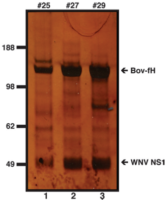
Fig. 1 Copurification of WNV NS1 and bovine fH. Supernatants were harvested from baculovirus-infected SF9 insect cells grown in 10% FBS, and NS1waspurified by nickel-affinity and size-exclusion chromatography. Silverstained SDS/PAGE of column fractions is shown. WNV NS1 and bovine fH are indicated by arrows. The numbers across the top correspond to the different column fractions. Protein size markers are indicated to the left in kilodaltons.
Fig. 1 Copurification of WNV NS1 and bovine fH. Supernatants were harvested from baculovirus-infected SF9 insect cells grown in 10% FBS, and NS1waspurified by nickel-affinity and size-exclusion chromatography. Silverstained SDS/PAGE of column fractions is shown. WNV NS1 and bovine fH are indicated by arrows. The numbers across the top correspond to the different column fractions. Protein size markers are indicated to the left in kilodaltons.From the PDF
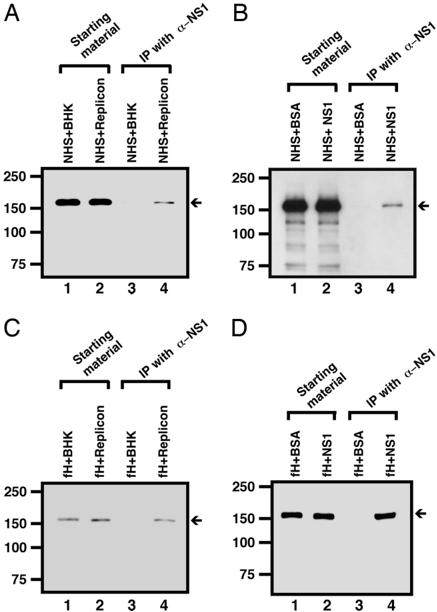
Fig. 2 Coimmunoprecipitation of WNV NS1 and human fH. Western blotting with a polyclonal anti-human fH after immunoprecipitation with antiNS1-mAb-Seph4B, NHS ( A and B ), and supernatant from WNV repliconexpressing cells ( A ) or purified NS1 ( B ). Western blotting was also performed after immunoprecipitation with anti-NS1-mAb-Seph4B ( C and D ), purified human fH, and supernatant from replicon-expressing cells ( C ) or purified NS1 ( D ). BHK cell supernatant or BSA was used as negative control, respectively. fH was identified with a sheep polyclonal anti-human fH antibody. Lanes 1 and 2ofeachimagerepresentthestartingmaterialwithoutimmunoprecipitation. The arrow points to the band that corresponds to the 150-kDa human fH protein.
Fig. 2 Coimmunoprecipitation of WNV NS1 and human fH. Western blotting with a polyclonal anti-human fH after immunoprecipitation with antiNS1-mAb-Seph4B, NHS ( A and B ), and supernatant from WNV repliconexpressing cells ( A ) or purified NS1 ( B ). Western blotting was also performed after immunoprecipitation with anti-NS1-mAb-Seph4B ( C and D ), purified human fH, and supernatant from replicon-expressing cells ( C ) or purified NS1 ( D ). BHK cell supernatant or BSA was used as negative control, respectively. fH was identified with a sheep polyclonal anti-human fH antibody. Lanes 1 and 2ofeachimagerepresentthestartingmaterialwithoutimmunoprecipitation. The arrow points to the band that corresponds to the 150-kDa human fH protein.From the PDF
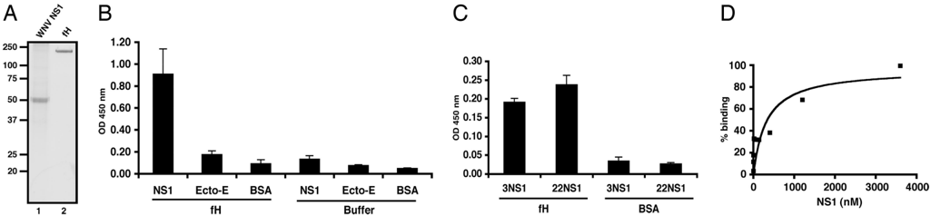
Fig. 3 WNVNS1andhumanfHbindingbyELISA.( A ) SDS/PAGE of purified WNV NS1 and human fH. WNV NS1 was purified from baculovirus-infected SF9 cells grown under serum-free conditions (lane 1). Human fH (lane 2) was commercially purchased. The positions of marker proteins are indicated to the left of the gel. ( B ) fH binds to solid-phase WNV NS1. Microtiter plates were coated overnight with purified NS1, WNV envelope (Ecto-E) protein, or BSA. After blocking, purified fH was added and detected with a polyclonal antibody against human fH. Results are representative of at least three independent experiments performed in duplicate. The error bars indicate standard deviations. ( C ) NS1 binds to solid-phase human fH. The microtiter plate was coated with human fH or BSA as control, blocked, and then incubated with purified WNV NS1. Subsequently, after washing, NS1 binding was detected with mAb 3NS1 or 22NS1. Experiments were performed in duplicate, and the results are representative of at least three independent experiments. ( D ) Saturation binding of fH and NS1. Increasing concentrations of fH were added to NS1 on microtiter plates and evaluated for binding by ELISA. The curve is representative of three independent experiments performed in duplicate.
Fig. 3 WNVNS1andhumanfHbindingbyELISA.( A ) SDS/PAGE of purified WNV NS1 and human fH. WNV NS1 was purified from baculovirus-infected SF9 cells grown under serum-free conditions (lane 1). Human fH (lane 2) was commercially purchased. The positions of marker proteins are indicated to the left of the gel. ( B ) fH binds to solid-phase WNV NS1. Microtiter plates were coated overnight with purified NS1, WNV envelope (Ecto-E) protein, or BSA. After blocking, purified fH was added and detected with a polyclonal antibody against human fH. Results are representative of at least three independent experiments performed in duplicate. The error bars indicate standard deviations. ( C ) NS1 binds to solid-phase human fH. The microtiter plate was coated with human fH or BSA as control, blocked, and then incubated with purified WNV NS1. Subsequently, after washing, NS1 binding was detected with mAb 3NS1 or 22NS1. Experiments were performed in duplicate, and the results are representative of at least three independent experiments. ( D ) Saturation binding of fH and NS1. Increasing concentrations of fH were added to NS1 on microtiter plates and evaluated for binding by ELISA. The curve is representative of three independent experiments performed in duplicate.From the PDF
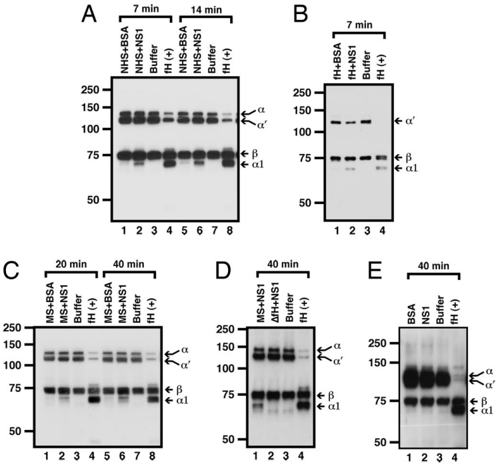
Fig. 4 WNV NS1 recruits fH in solution to degrade C3b. NS1 or BSA was incubated with 9NS1 or 22NS1 mAb Sepharose overnight at 4°C. The charged or uncharged beads were mixed with 10% NHS ( A ), purified human fH ( B ), 10% C57BL/6mouseserum(MS, C ), or 10% congenicfH -/ -MS(designatedbytriangle, D ), washed extensively, and then incubated with biotinylated C3b and fI for 7 or 14min( A and B ) or 20 or 40 min ( C and D ). In each series of experiments, a buffer negative control (lanes 3 and 7) was included that contained only biotinylated C3b, fI, and anti-NS1-mAb-Sepharose. In E , the NS1-charged beads were added directly to biotinylated C3b and fI. Reactions were stopped with SDS-reducing sample buffer, and C3b fragments were visualized after Western blot. C3b fragments are labeled at the right of the gel, and cofactor activity is established by the appearance of the 𝛂 1 fragment ( ≈ 67 kDa) of the C3b 𝛂 ′ -chain. The band marked 𝛂 represents the small amount of C3 in the preparation that was not cleaved to C3b. Its thioester bond, however, is cleaved, because it is also susceptible to degradation. Under longer exposure, the 𝛂 2degradationfragment( ≈ 41 kDa) was also observed (data not shown). Data are representative of at least two to three independent experiments.
Fig. 4 WNV NS1 recruits fH in solution to degrade C3b. NS1 or BSA was incubated with 9NS1 or 22NS1 mAb Sepharose overnight at 4°C. The charged or uncharged beads were mixed with 10% NHS ( A ), purified human fH ( B ), 10% C57BL/6mouseserum(MS, C ), or 10% congenicfH -/ -MS(designatedbytriangle, D ), washed extensively, and then incubated with biotinylated C3b and fI for 7 or 14min( A and B ) or 20 or 40 min ( C and D ). In each series of experiments, a buffer negative control (lanes 3 and 7) was included that contained only biotinylated C3b, fI, and anti-NS1-mAb-Sepharose. In E , the NS1-charged beads were added directly to biotinylated C3b and fI. Reactions were stopped with SDS-reducing sample buffer, and C3b fragments were visualized after Western blot. C3b fragments are labeled at the right of the gel, and cofactor activity is established by the appearance of the 𝛂 1 fragment ( ≈ 67 kDa) of the C3b 𝛂 ′ -chain. The band marked 𝛂 represents the small amount of C3 in the preparation that was not cleaved to C3b. Its thioester bond, however, is cleaved, because it is also susceptible to degradation. Under longer exposure, the 𝛂 2degradationfragment( ≈ 41 kDa) was also observed (data not shown). Data are representative of at least two to three independent experiments.From the PDF
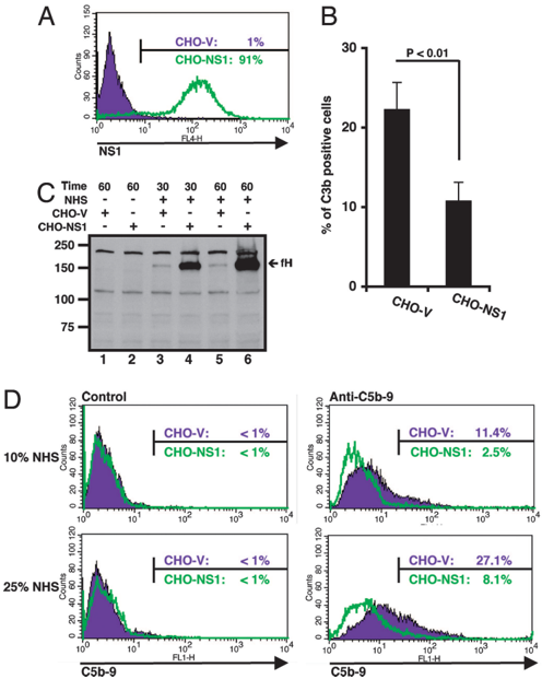
Fig. 5 Inhibition of complement deposition by cell surface-associated NS1. ( A ) Flow cytometry histograms showing cell surface-associated expression of WNV NS1 on CHO-NS1 stable cell transfectants (green line). As a negative control, CHO-V cells transfected with the parent retroviral vector were stained (purple line). ( B ) WNV NS1 inhibits C3b deposition on CHO cells. CHO-V and CHO-NS1 cells were preincubated with C7d-HS, sensitized with an anti-CHO antibody, and incubated with C7d-HS in GVB-Mg 2 + -EGTA buffer. After washing, cells were stained with mAb against human C3d and analyzed by flow cytometry. The results are the average of three independent experiments performed in duplicate, and the difference in C3b deposition between CHO-V and CHO-NS1 cells was statistically significant ( P < 0.01). ( C ) fH binding to CHO-V and CHO-NS1 cells. Cells were incubated with medium or 10% NHS for theindicatedtimes(inminutes),washedextensively,andanalyzedbyWestern blot with a sheep anti-human fH antibody. The ≈ 150 kDa fH is denoted by the arrow. ( D ) WNV NS1 inhibits C5b-9 deposition on CHO cells expressing WNV NS1. CHO-V and CHO-NS1 cells were preincubated with NHS, sensitized with an anti-CHO antibody, incubated with 10% ( Upper ) or 25% ( Lower ) NHS, and then immunostained with murine mAb against human C5b-9 ( Right ). The results are the average of three independent experiments performed in duplicate, and the difference in C5b-9 deposition between CHO-V and CHONS1 cells was statistically significant ( P < 0.01). For negative control staining ( Left ), complement-activated cell lines were stained with only anti-mouse IgG conjugated with FITC. Purple and green lines refer to CHO-V and CHO-NS1 cells, respectively.
Fig. 5 Inhibition of complement deposition by cell surface-associated NS1. ( A ) Flow cytometry histograms showing cell surface-associated expression of WNV NS1 on CHO-NS1 stable cell transfectants (green line). As a negative control, CHO-V cells transfected with the parent retroviral vector were stained (purple line). ( B ) WNV NS1 inhibits C3b deposition on CHO cells. CHO-V and CHO-NS1 cells were preincubated with C7d-HS, sensitized with an anti-CHO antibody, and incubated with C7d-HS in GVB-Mg 2 + -EGTA buffer. After washing, cells were stained with mAb against human C3d and analyzed by flow cytometry. The results are the average of three independent experiments performed in duplicate, and the difference in C3b deposition between CHO-V and CHO-NS1 cells was statistically significant ( P < 0.01). ( C ) fH binding to CHO-V and CHO-NS1 cells. Cells were incubated with medium or 10% NHS for theindicatedtimes(inminutes),washedextensively,andanalyzedbyWestern blot with a sheep anti-human fH antibody. The ≈ 150 kDa fH is denoted by the arrow. ( D ) WNV NS1 inhibits C5b-9 deposition on CHO cells expressing WNV NS1. CHO-V and CHO-NS1 cells were preincubated with NHS, sensitized with an anti-CHO antibody, incubated with 10% ( Upper ) or 25% ( Lower ) NHS, and then immunostained with murine mAb against human C5b-9 ( Right ). The results are the average of three independent experiments performed in duplicate, and the difference in C5b-9 deposition between CHO-V and CHONS1 cells was statistically significant ( P < 0.01). For negative control staining ( Left ), complement-activated cell lines were stained with only anti-mouse IgG conjugated with FITC. Purple and green lines refer to CHO-V and CHO-NS1 cells, respectively.From the PDF

Verification

Claims the paper makes

The claims and statistics of this paper haven’t been checked yet.

Run the check — free, no account needed. It reads the open-access text and checks the sources it cites.

A few free checks a day · papers already verified open instantly · results are public

Claims other papers make about this one

Each is checked against this paper’s own text: does it say what the citing paper says it does?

Self-check

Percentages, sums and reported differences re-computed from the paper’s own numbers.

The self-check needs the paper’s full text.

Citations

Cites

No references could be resolved yet.

Research works printed but not linked: 55

  1. 10.1006/viro.1996.0307 · DOI
  2. 10.1006/viro.1996.0406 · DOI
  3. 10.1128/jvi.71.12.9608-9617.1997 · DOI
  4. 10.1128/JVI.73.12.10272-10280.1999 · DOI
  5. 10.1016/0042-6822(89)90161-X · DOI
  6. 10.1016/0042-6822(89)90544-8 · DOI
  7. 10.1016/0042-6822(88)90408-4 · DOI
  8. 10.1128/JCM.40.02.376-381.2002 · DOI
  9. 10.1128/JCM.38.3.1053-1057.2000 · DOI
  10. 10.1086/343813 · DOI
  11. 10.1128/JVI.79.22.13924-13933.2005 · DOI
  12. 10.1086/500949 · DOI
  13. 10.1086/342600 · DOI
  14. 10.1007/s007050050127 · DOI
  15. 10.4049/jimmunol.174.1.395 · DOI
  16. 10.1002/jmv.10261 · DOI
  17. 10.4049/jimmunol.169.2.657 · DOI
  18. 10.1128/JVI.79.17.11403-11411.2005 · DOI
  19. JE Volanakis Curr Top Microbiol Immunol 266, 41–56 (2002).
  20. 10.1016/S0167-5699(00)01754-0 · DOI
  21. 10.1084/jem.190.8.1165 · DOI
  22. 10.1038/nm0402-373 · DOI
  23. 10.1073/pnas.96.22.12708 · DOI
  24. 10.4049/jimmunol.167.5.2446 · DOI
  25. 10.1128/iai.30.3.899-901.1980 · DOI
  26. JT Hicks, FA Ennis, E Kim, M Verbonitz J Immunol 121, 1437–1445 (1978). · DOI
  27. 10.1084/jem.20052388 · DOI
  28. 10.1128/JVI.79.12.7466-7477.2005 · DOI
  29. 10.1016/S1074-7613(02)00369-2 · DOI
  30. 10.1073/pnas.82.5.1490 · DOI
  31. 10.1084/jem.182.2.501 · DOI
  32. 10.1006/viro.1998.9205 · DOI
  33. 10.1016/S0161-5890(99)00038-3 · DOI
  34. 10.1084/jem.146.1.257 · DOI
  35. 10.1056/NEJM200104123441506 · DOI
  36. 10.1056/NEJM200104053441406 · DOI
  37. 10.1016/S0161-5890(03)00108-1 · DOI
  38. 10.1016/j.molimm.2005.06.016 · DOI
  39. 10.1128/JVI.80.3.1340-1351.2006 · DOI
  40. BJ Geiss, TC Pierson, MS Diamond Virology J 2 53, 1–13 (2005).
  41. 10.1042/bst0300971 · DOI
  42. 10.1038/nm1240 · DOI
  43. MK Liszewski, JP Atkinson J Immunol 156, 4415–4421 (1996). · DOI
  44. 10.1074/jbc.M004650200 · DOI
  45. 10.4049/jimmunol.168.12.6298 · DOI
  46. WE Brandt, D Chiewslip, DL Harris, PK Russell J Immunol 105, 1565–1568 (1970). · DOI
  47. J Rai, SK Basu, SN Ghosh Indian J Med Res 65, 167–170 (1977).
  48. 10.1056/NEJM197311082891902 · DOI
  49. 10.1073/pnas.85.5.1657 · DOI
  50. 10.4049/jimmunol.172.10.6195 · DOI
  51. 10.1128/iai.61.8.3129-3136.1993 · DOI
  52. 10.1084/jem.188.4.671 · DOI
  53. 10.1099/0022-1317-72-4-961 · DOI
  54. 10.1021/ac026136s · DOI
  55. 10.1073/pnas.0402353101 · DOI

Cited by

Sorted by citations

Metadata

Authors
Chung, K. M., Liszewski, M. K., Nybakken, G., Davis, A. E., Townsend, R. R., Fremont, D. H., Atkinson, J. P., & Diamond, M. S.
Affiliations
  • Departments of *Medicine,
  • Pathology and Immunology,
Venue
Proceedings of the National Academy of Sciences
Published
2006-12-12
Volume
103
Issue
50
Pages
19111-19116
ISSN
  • 0027-8424
  • 1091-6490
Cited by
238
References
0
Language
en

About these checks

Registry, retraction, Cites and statistics results are computed, not judged, with one exception: a printed reference that no index lists is read once by a model before it counts as a citation error. Claims are extracted by a model and kept only when their quote appears verbatim in the text; each cited source is then checked, with a model’s help, for whether it says what the claim relies on. Those verdicts can be wrong — read the claim’s quote and the cited source before relying on one.

Statistics and arithmetic are recomputed, not judged.

Confidence bands are shown only for fields calibrated on our annotated test set. None is calibrated yet, so no band is shown.

What counts as a claim ›How we measure confidence ›How we check this ›

Are you an author, or do you see a problem with this page? Request a review