

Fin ray patterns at the fin-to-limb transition
Open accessThe DOI matches its Crossref record; 64 of the 64 references it cites were found in a registry or an index; 1 of the 49 sources its claims cite were checked for support.
Abstract
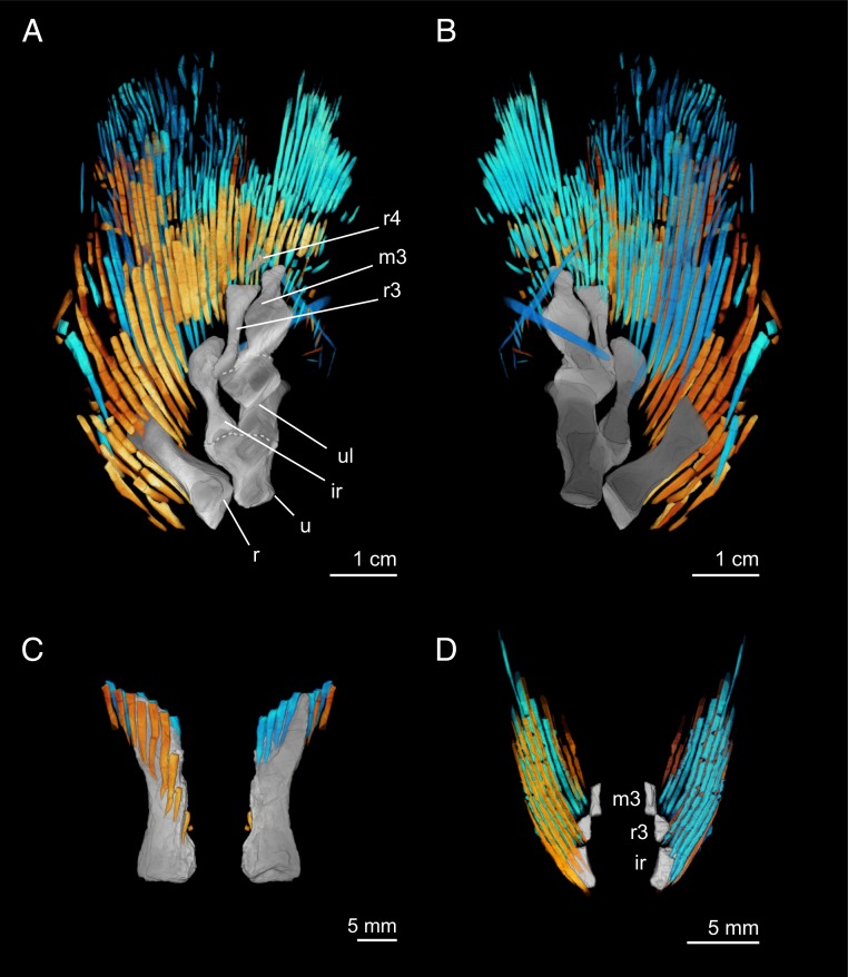
The fin-to-limb transition was marked by the origin of digits and the loss of dermal fin rays. Paleontological research into this transformation has focused on the evolution of the endoskeleton, with little attention paid to fin ray structure and function. To address this knowledge gap, we study the dermal rays of the pectoral fins of 3 key tetrapodomorph taxa— Sauripterus taylori (Rhizodontida), Eusthenopteron foordi (Tristichopteridae), and Tiktaalik roseae (Elpistostegalia)—using computed tomography. These data show several trends in the lineage leading to digited forms, including the consolidation of fin rays (e.g., reduced segmentation and branching), reduction of the fin web, and unexpectedly, the evolution of asymmetry between dorsal and ventral hemitrichia. In Eusthenopteron , dorsal rays cover the preaxial endoskeleton slightly more than ventral rays. In Tiktaalik , dorsal rays fully cover the third and fourth mesomeres, while ventral rays are restricted distal to these elements, suggesting the presence of ventralized musculature at the fin tip analogous to a fleshy “palm.” Asymmetry is also observed in cross-sectional areas of dorsal and ventral rays. Eusthenopteron dorsal rays are slightly larger than ventral rays; by contrast, Tiktaalik dorsal rays can be several times larger than ventral rays, and degree of asymmetry appears to be greater at larger sizes. Analysis of extant osteichthyans suggests that cross-sectional asymmetry in the dermal rays of paired fins is plesiomorphic to crown group osteichthyans. The evolution of dermal rays in crownward stem tetrapods reflects adaptation for a fin-supported elevated posture and resistance to substrate-based loading prior to the origin of digits.
Overview
CoChat Summary
## TL;DR
This paper uses μCT-based anatomical comparisons of dermal fin rays in tetrapodomorph fishes to show that, before digits evolved, fin rays became more consolidated, the fin web shrank, and dorsal–ventral asymmetry increased. The authors argue these changes helped fins function in substrate-based loading and likely prefigured limb-like mechanics.
## Key findings
- In Sauripterus, the pectoral fin had at least 60 dorsal hemitrichia, with 20 associated with the radius, 12 distal to the intermedium, and at least 28 distal to the ulnare; rays were unsegmented and likely represented a large fin web.
- In Eusthenopteron, the pectoral fin had at least 30 rays in CMNH 8153; dorsal hemitrichia extended more proximally than ventral hemitrichia, and dorsal–ventral size asymmetry was modest, with dorsal rays 1.10 and 1.09 times greater CSA than ventral rays in the two specimens.
- In Tiktaalik, at least 33 lepidotrichia are preserved in NUFV110, and rays are mostly unbranching and unsegmented; dorsal hemitrichia covered more of the endoskeleton than ventral hemitrichia, and dorsal rays had CSA 1.93× ventral rays in the smaller specimen and 2.67× in the larger specimen.
- Statistical comparisons found all pairwise dorsal-vs-ventral CSA comparisons within a lepidotrichium were significant (Mann–Whitney U test, P < 0.05); similar patterns held for SMA, with asymmetry greater in Tiktaalik than Eusthenopteron and greater in the larger Tiktaalik individual.
## Methods
- Comparative anatomical study of pectoral fin dermal skeletons in three tetrapodomorph taxa: Sauripterus taylori, Eusthenopteron foordi, and Tiktaalik roseae; plus comparison taxa Neoceratodus, Polypterus, and Acipenser for broader context.
- Specimens were μCT scanned, segmented, and visualized in Amira; quantitative analysis focused on cross-sectional area (CSA) and second moment of area (SMA) of dorsal and ventral hemitrichia.
- Statistical workflow: Shapiro tests for normality, then Mann–Whitney U tests for dorsal-vs-ventral comparisons; analyses were done in R.
## Limitations
- The paper states that phylogenetic patterns are difficult to determine rigorously because this type of data is currently available for only 2 tetrapodomorph species; the broader evolutionary inference is therefore limited by sparse sampling.
- The study notes that ventral hemitrichia of Tiktaalik do not show clear anatomical markers and may have been displaced or rotated during preservation; this is a stated taphonomic limitation.
- Evident from the text: some specimens are incomplete or disturbed (for example, ventral hemitrichia removed in Sauripterus, and parts of Eusthenopteron fins are damaged/missing), which constrains reconstruction and comparison.
Model-written summary. Check it against the paper before relying on it.
Integrity signals
Registry
Cites
Statistics
Figures



Verification
Claims the paper makes
0 Valid · 1 Not valid · 29 Not checked · 138 Own results and interpretations
The cited sources that could be checked couldn’t confirm or dispute this claim.
In the paper
“Detailed anatomical description of dermal rays is lacking, in part, due to taphonomic bias.”
§ Fin ray patterns at the fin-to-limb transition > Abstract
What the sources say
- Not matchedcites [19]
Couldn’t be matched to a paper in Crossref, so it can’t be checked.
- Not checked — no full text of the sourcecites [20] source
- Could not verifycites [21] sourceExperimental — accuracy not yet measured
The Abstract says pelvic material is rare and few examples have been described, but it does not say that detailed descriptions of dermal rays are lacking. Results > Pelvic Fins reports that numerous lepidotrichia were preserved and most were removed during preparation; it does not attribute a lack of descriptions to taphonomic bias. Searches for dermal rays, lepidotrichia, and taphonomic bias found no passage making the claimed connection. Page numbers are unavailable in our copy. (LLM, conf=0.72)
plainstates a possible cause- Asserted: passes — The authors put it forward as true.
- Checkable: passes — It can be checked against evidence.
- Load-bearing: passes — The paper’s conclusions depend on it.
- Based: passes — The paper gives a reason why it holds.
- Not matched
A result the paper reports from its own data — it cites no other source for it, so there is none to check. The quote shows it is in the paper.
In the paper
“On the right radius of CMNH 8153, dorsal hemitrichia extend further proximally than ventral hemitrichia (Fig. 2C), and on the intermedium and third radial of the right fin of CMNH 10526, dorsal hemitrichia extend further proximally than ventral hemitrichia (Fig. 2D).”
§ Fin ray patterns at the fin-to-limb transition > Results > Pectoral Fin Rays of Eusthenopteron.
plainSingle case- Asserted: passes — The authors put it forward as true.
- Checkable: passes — It can be checked against evidence.
- Load-bearing: unclear — It is unclear whether the paper’s conclusions depend on it.
- Based: passes — The paper gives a reason why it holds.
A result the paper reports from its own data — it cites no other source for it, so there is none to check. The quote shows it is in the paper.
In the paper
“Dorsal rays extend proximally to fully cover the radius, cover the distal half of the intermedium, and fully cover distal radials 5 through 8.”
§ Fin ray patterns at the fin-to-limb transition > Results > Pectoral Fin Rays of Sauripterus.
plainSingle case- Asserted: passes — The authors put it forward as true.
- Checkable: passes — It can be checked against evidence.
- Load-bearing: unclear — It is unclear whether the paper’s conclusions depend on it.
- Based: passes — The paper gives a reason why it holds.
A result the paper reports from its own data — it cites no other source for it, so there is none to check. The quote shows it is in the paper.
In the paper
“NUFV109 shows a similar pattern: dorsal hemitrichia cover the fifth mesomere, the anterior edge of fourth mesomere, and its anterior radial (Fig. 3C), while ventral hemitrichia are positioned distal to the fourth mesomere (Fig. 3D).”
§ Fin ray patterns at the fin-to-limb transition > Results > Pectoral Fin Rays of Tiktaalik.
plainSingle case- Asserted: passes — The authors put it forward as true.
- Checkable: passes — It can be checked against evidence.
- Load-bearing: passes — The paper’s conclusions depend on it.
- Based: passes — The paper gives a reason why it holds.
A result the paper reports from its own data — it cites no other source for it, so there is none to check. The quote shows it is in the paper.
In the paper
“These data show that, prior to the origin of digits, dermal rays were simplified, the fin web became reduced in size, and the top and bottom of the fin became more asymmetric.”
§ Fin ray patterns at the fin-to-limb transition > Abstract
plain- Asserted: passes — The authors put it forward as true.
- Checkable: passes — It can be checked against evidence.
- Load-bearing: passes — The paper’s conclusions depend on it.
- Based: passes — The paper gives a reason why it holds.
114 more claims
Create a free account to see all 168 claims and to verify the 4 references still open.
- It may state two separate claims as one. 40
- Probably not a substantial claim. 14
Citations
Cites
Fiji: an open-source platform for biological-image analysis
Schindelin, J., Arganda-Carreras, I., Frise, E., Kaynig, V., Longair, M., Pietzsch, T., Preibisch, S., Rueden, C., Saalfeld, S., Schmid, B., et al. (2012) · Nature Methods · doi:10.1038/nmeth.2019 · Cited by 66,890 · cited 1×
“0.0-cr-69/1.52n) (65)as “ Raw Sequences ” and converted to binary using the Huang method.”
InfluentialWickham, H. (2016) · Use R! · doi:10.1007/978-3-319-24277-4 · Cited by 33,115
ggplot2: Elegant Graphics for Data Analysis
Wickham, H. (2009) · doi:10.1007/978-0-387-98141-3 · Cited by 14,447
BoneJ: Free and extensible bone image analysis in ImageJ
Doube, M., Kłosowski, M. M., Arganda-Carreras, I., Cordelières, F. P., Dougherty, R. P., Jackson, J. S., Schmid, B., Hutchinson, J. R., & Shefelbine, S. J. (2010) · Bone · doi:10.1016/j.bone.2010.08.023 · Cited by 2,040 · cited 1×
“Using the package BoneJ (version 1.4.3) (66), we applied the “ Moments of Inertia ” function using default settings for all slices.”
InfluentialStudies on the structure & development of vertebrates
Goodrich, E. S. (1930) · doi:10.5962/bhl.title.82144 · Cited by 1,082
STUDIES ON THE STRUCTURE AND DEVELOPMENT OF VERTEBRATES
Goodrich, E. S. (1931) · The Journal of Nervous and Mental Disease · doi:10.1097/00005053-193111000-00026 · Cited by 1,029
Basic Structure and Evolution of Vertebrates.
Weitzman, S. H., & Jarvik, E. (1981) · Systematic Zoology · doi:10.2307/2413058 · Cited by 829
Janvier, P. (1996) · doi:10.1093/oso/9780198540472.001.0001 · Cited by 792
A Morphogenetic Approach to the Origin and Basic Organization of the Tetrapod Limb
Shubin, N. H., & Alberch, P. (1986) · Evolutionary Biology · doi:10.1007/978-1-4615-6983-1_6 · Cited by 592
Self-citationRiddle, R. D., Ensini, M., Nelson, C., Tsuchida, T., Jessell, T. M., & Tabin, C. (1995) · Cell · doi:10.1016/0092-8674(95)90103-5 · Cited by 478 · cited 1×
“, endochondral articular surfaces, distribution of musculature, and ventral vasculature grooves (SI Appendix, 3-dimensional image)] are likely patterned by Wnt signaling pathways, similar to developing limbs (60, 61).”
Research works printed but not linked: 8
- A. Romer, F. Byrne, The pes of diadectes: Notes on the primitive tetrapod limb. Paleobiologica 4, 25–48 (1931).
- W. Gregory, H. Raven, Origin of paired fins and limbs. Ann. N. Y. Acad. Sci. 42, 273–360 (1941).
- K. S. Thomson, New evidence on the evolution of the paired fins of Rhipidistia and the origin of the tetrapod limb, with description of a new genus of Osteolepididae. Postilla 157, 1–7 (1972).
- J. A. Long, T. Holland, A possible elpistostegalid from the Devonian of Gondwana. Proc. R. Soc. Vic. 120, 182–192 (2008).
- M. C. Davis, N. Shubin, E. B. Daeschler, Immature rhizodontids from the Devonian of North America. Bull. Mus. Comp. Zool. 156, 171–187 (2001).
- J. Hall, Natural History of New York. Part IV. Geology Comprising the Survey of the Fourth Geological District (D. Appleton & Company, New York, NY, 1842), p. 683.
- P. J. Millot, J. Anthony, Anatomie de Latimeria chalumnae: Tome 1: Squelette, Muscles et Formations de soutien (Éditions du Centre National de la Recherche Scientifique, Paris, France, 1958).
- E. Jarvik, On the Morphology and Taxonomy of the Middle Devonian Osteolepid Fishes of Scotland (Almqvist & Wiksells boktr., 1948).
Cited by
Evolvability in the fossil record
Love, A. C., Grabowski, M. W., Houle, D., Liow, L. H., Porto, A., Tsuboi, M., Voje, K. L., & Hunt, G. (2021) · Paleobiology · doi:10.1017/pab.2021.36 · Cited by 46
Hannard, F., Mirkhalaf, M., Ameri, A., & Barthelat, F. (2021) · Science Robotics · doi:10.1126/scirobotics.abf9710 · Cited by 40
“A prominent feature of the hemitrichs is their segmentation into ~1-mm-long bony segments connected by collagenous joints that act as flexural hinges (18).”
BackgroundMalkmus, J., Martins, L. R., Jhanwar, S., Kircher, B. K., Palacio, V., Sheth, R., Leal, F., Duchesne, A., López-Ríos, J., Peterson, K. A., et al. (2021) · Nature Communications · doi:10.1038/s41467-021-25810-1 · Cited by 36
Evolution and Development: Conceptual Issues
Love, A. C. (2024) · Cambridge University Press eBooks · doi:10.1017/9781108616751 · Cited by 39
Höch, R., Schneider, R., Kickuth, A., Meyer, A., & Woltering, J. M. (2021) · Proceedings of the National Academy of Sciences · doi:10.1073/pnas.2101783118 · Cited by 28
“Additional examples are the anterior fin spine in catfish (47) and sturgeon pectoral fins (48), robustly ossified anterior fin rays in tetrapodomorphs (49), the anterior fin spine that evolved convergently in chimaeras (50), and acanthodians (spiny sharks) and stem sharks (e.g., hybodonts) (51).”
Metadata
- Authors
- Stewart, T. A., Lemberg, J. B., Taft, N. K., Yoo, I., Daeschler, E. B., & Shubin, N. H.
- Affiliations
- University of Chicago (US)
- University of Wisconsin–Parkside (US)
- Drexel University (US)
- Corresponding author
- Stewart, Thomas A.
- Shubin, Neil H.
- Venue
- Proceedings of the National Academy of Sciences
- Publisher
- National Academy of Sciences
- Published
- 2019-12-30
- Volume
- 117
- Issue
- 3
- Pages
- 1612-1620
- ISSN
- 0027-8424 (ISSN-L)
- 1091-6490
- PMC
- PMC6983361
- OpenAlex
- W2998690626
- Cited by
- 40
- References
- 64
- Citations by year
- 2026: 6 · 2025: 8 · 2024: 9 · 2023: 5 · 2022: 2 · 2021: 8 · 2020: 1
- FWCI
- 2.09
- Type
- journal article
- Language
- en
- Open access
- hybrid
- doi.org
- Licence
- Locations
- Proceedings of the National Academy of Sciences (publishedVersion, cc-by-nc-nd)
- PubMed Central (publishedVersion, cc-by-nc-nd)
- PubMed (publishedVersion)
- Knowledge@UChicago (University of Chicago) (submittedVersion, cc-by-nc-nd)
- University of Chicago
- Funders
- NSF | GEO | Division of Earth Sciences (0208377, 0544565)
- Topics
- Physical Sciences › Earth and Planetary Sciences › Paleontology › Paleontology and Evolutionary Biology
- Physical Sciences › Earth and Planetary Sciences › Paleontology › Evolution and Paleontology Studies
- Health Sciences › Medicine › Anatomy › Comparative Animal Anatomy Studies
- Keywords
- fin-to-limb transition, pectoral fin, tetrapodomorphs, osteichthyans
- MeSH terms
- Amphibians, Animals, Biological Evolution, Extremities, Fishes, Fossils, Paleontology, Tomography, X-Ray Computed, Animal Fins
About these checks
Registry, retraction, Cites and statistics results are computed, not judged, with one exception: a printed reference that no index lists is read once by a model before it counts as a citation error. Claims are extracted by a model and kept only when their quote appears verbatim in the text; each cited source is then checked, with a model’s help, for whether it says what the claim relies on. Those verdicts can be wrong — read the claim’s quote and the cited source before relying on one.
The claim verdicts this report counts are experimental: their accuracy is not yet measured.
A language model drew the claims from the paper on 2026-10-10, under version 3 of the claim definition (model deepseek-v4.1-flash, prompt extract-v3).
A second model labelled each claim on 2026-10-10 (model jev-1.13-20260917, definition revision gold-guideline-v0.2).
Cited sources are checked with a language model’s help (gpt-6-sol).
Statistics and arithmetic are recomputed, not judged.
Confidence bands are shown only for fields calibrated on our annotated test set. None is calibrated yet, so no band is shown.
What counts as a claim ›How we measure confidence ›How we check this ›
Are you an author, or do you see a problem with this page? Request a review
How to cite
Cite CoChat for the checks on this page, and the paper for its own findings.
- This page
- CoChat. (2026). Citation check: Fin ray patterns at the fin-to-limb transition. https://app.cochat.ai/papers/10.1073/pnas.1915983117
- The paper
- Stewart, T. A., Lemberg, J. B., Taft, N. K., Yoo, I., Daeschler, E. B., & Shubin, N. H. (2020). Fin ray patterns at the fin-to-limb transition. Proceedings of the National Academy of Sciences. https://doi.org/10.1073/pnas.1915983117
Data sources
Bibliographic data: Semantic Scholar, OpenAlex, Crossref.
Contains information from the Semantic Scholar Academic Graph Datasets, made available under the ODC Attribution License (ODC-BY 1.0).
Semantic Scholar’s data is described in: Kinney, R., et al. (2023). The Semantic Scholar Open Data Platform. arXiv:2301.10140.
