

The DOI matches its Crossref record; 2 of the 2 references it cites were found in a registry or an index.
ATF6 enables pathogen infection in ticks by inducing stomatin and altering cholesterol dynamics
Open accessPublished version of a preprint
Abstract
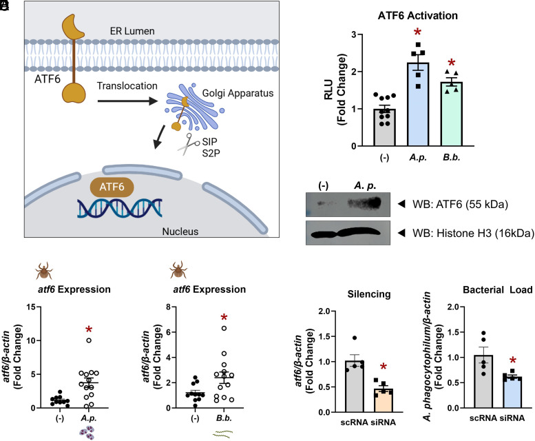
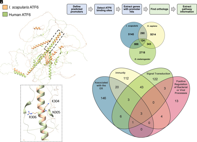
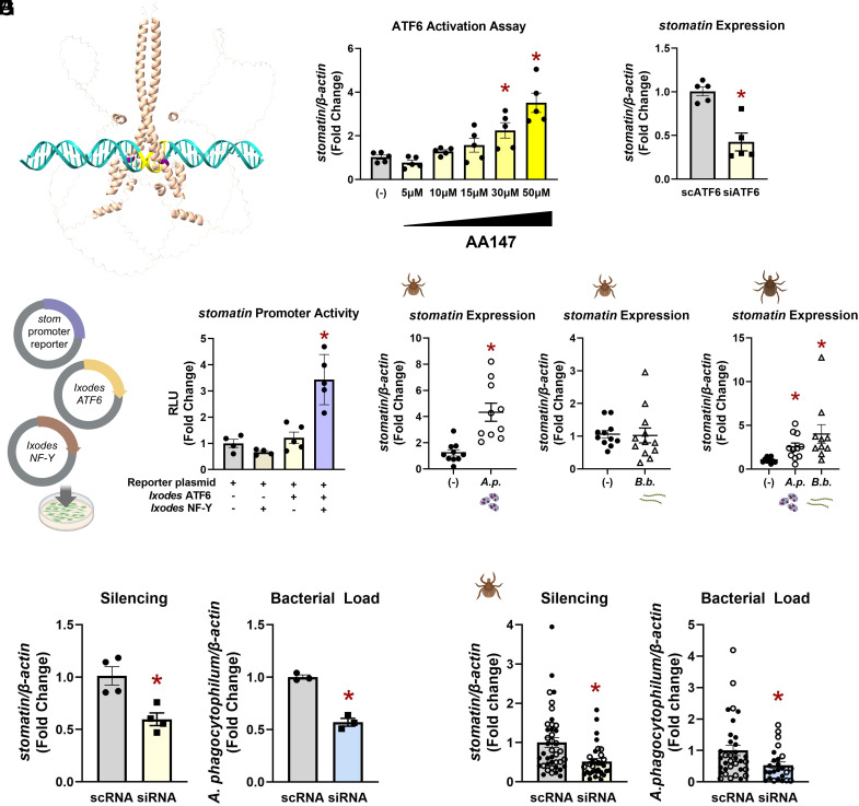
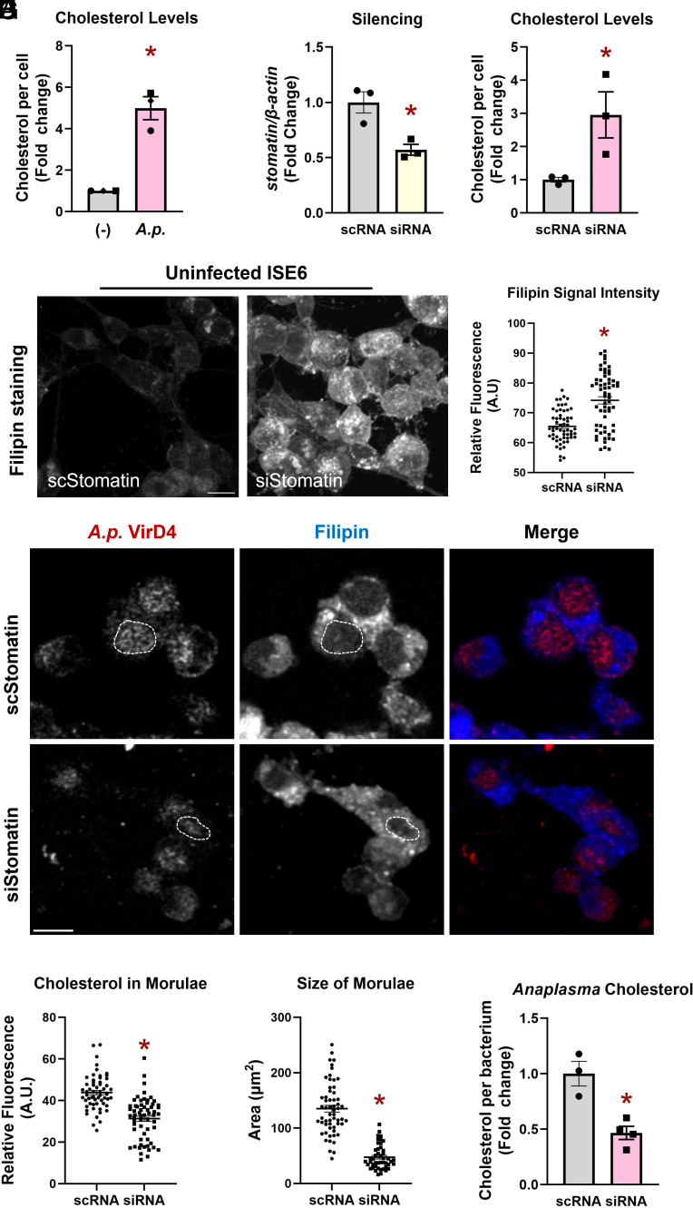
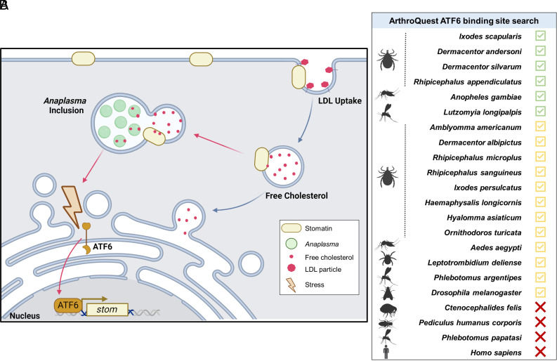
How tick-borne pathogens interact with their hosts has been primarily studied in vertebrates where disease is observed. Comparatively less is known about pathogen interactions within the tick. Here, we report that Ixodes scapularis ticks infected with either Anaplasma phagocytophilum (causative agent of anaplasmosis) or Borrelia burgdorferi (causative agent of Lyme disease) show activation of the ATF6 branch of the unfolded protein response (UPR). Disabling ATF6 functionally restricts pathogen survival in ticks. When stimulated, ATF6 functions as a transcription factor, but is the least understood out of the three UPR pathways. To interrogate the Ixodes ATF6 transcriptional network, we developed a custom R script to query tick promoter sequences. This revealed stomatin as a potential gene target, which has roles in lipid homeostasis and vesical transport. Ixodes stomatin was experimentally validated as a bona fide ATF6-regulated gene through luciferase reporter assays, pharmacological activators, RNA interference transcriptional repression, and immunofluorescence microscopy. Silencing stomatin decreased A. phagocytophilum colonization in Ixodes and disrupted cholesterol dynamics in tick cells. Furthermore, blocking stomatin restricted cholesterol availability to the bacterium, thereby inhibiting growth and survival. Taken together, we have identified the Ixodes ATF6 pathway as a contributor to vector competence through Stomatin-regulated cholesterol homeostasis. Moreover, our custom, web-based transcription factor binding site search tool “ArthroQuest” revealed that the ATF6-regulated nature of stomatin is unique to blood-feeding arthropods. Collectively, these findings highlight the importance of studying fundamental processes in nonmodel organisms.
Overview
The claims and statistics of this paper haven’t been checked yet.
Run the check — free, no account needed. It reads the open-access text and checks the sources it cites.
Integrity signals
Registry
Cites
Statistics
Figures
Verification
Claims the paper makes
The claims and statistics of this paper haven’t been checked yet.
Run the check — free, no account needed. It reads the open-access text and checks the sources it cites.
Self-check
Percentages, sums and reported differences re-computed from the paper’s own numbers.
The self-check needs the paper’s full text.
Citations
Cites
Highly accurate protein structure prediction with AlphaFold
Jumper, J., Evans, R., Pritzel, A., Green, T., Figurnov, M., Ronneberger, O., Tunyasuvunakool, K., Bates, R., Žídek, A., Potapenko, A., et al. (2021) · Nature · doi:10.1038/s41586-021-03819-2 · Cited by 43,764
Accurate structure prediction of biomolecular interactions with AlphaFold 3
Abramson, J., Adler, J., Dunger, J., Evans, R., Green, T., Pritzel, A., Ronneberger, O., Willmore, L., Ballard, A. J., Bambrick, J., et al. (2024) · Nature · doi:10.1038/s41586-024-07487-w · Cited by 15,861
Research works printed but not linked: 64
- 10.15585/mmwr.mm6717e1 · DOI
- D. E. Sonenshine, Range expansion of tick disease vectors in North America: Implications for spread of tick-borne disease. Int. J. Environ. Res. Public Health 15, 478 (2018). · DOI · Paper page
- Institute of Medicine (US) Forum on Microbial Threats, “Vector-borne disease emergence and resurgence” in Vector-Borne Diseases: Understanding the Environmental, Human Health, and Ecological Connections: Workshop Summary (National Academies Press (US), 2008).
- C. D. Paddock, R. S. Lane, J. E. Staples, M. B. Labruna, Changing Paradigms for Tick-Borne Diseases in The Americas (National Academies Press (US), 2016).
- Critical Needs and Gaps in Understanding Prevention, Amelioration, and Resolution of Lyme and Other Tick-Borne Diseases: The Short-Term and Long-Term Outcomes: Workshop Report (National Academies Press, 2011).
- 10.1017/S0031182004005967 · DOI
- J. M. Park, A. S. Oliva Chávez, D. K. Shaw, Ticks: More than just a pathogen delivery service. Front. Cell. Infect. Microbiol. 11, 739419 (2021). · DOI
- L. C. Sidak-Loftis , The unfolded-protein response triggers the arthropod immune deficiency pathway. mBio 13, e0070322 (2022). · DOI
- K. L. Rosche , PERK-mediated antioxidant response is key for pathogen persistence in ticks. mSphere 8, e0032123 (2023). · DOI · Paper page
- 10.1016/j.tips.2011.10.002 · DOI
- R. F. Hillary, U. FitzGerald, A lifetime of stress: ATF6 in development and homeostasis. J. Biomed. Sci. 25, 48 (2018). · DOI
- 10.7554/eLife.61675 · DOI
- K. L. Rosche, L. C. Sidak-Loftis, J. Hurtado, E. A. Fisk, D. K. Shaw, Arthropods under pressure: Stress responses and immunity at the pathogen-vector interface. Front. Immunol. 11 (2021). · DOI
- 10.1038/nrmicro3393 · DOI
- A. Arensdorf, D. Diedrichs, T. Rutkowski, Regulation of the transcriptome by ER stress: Non-canonical mechanisms and physiological consequences. Front. Genet. 4, 256 (2013). · DOI
- 10.1016/S0021-9258(19)61473-0 · DOI
- 10.1093/nar/gkab1061 · DOI · Paper page
- 10.1002/pro.3943 · DOI
- 10.1074/jbc.M010486200 · DOI
- 10.1128/MCB.20.18.6755-6767.2000 · DOI · Paper page
- 10.1038/75556 · DOI · Paper page
- 10.1093/genetics/iyad031 · DOI
- 10.1093/nar/gkab1028 · DOI
- P. D. Thomas , PANTHER: Making genome-scale phylogenetics accessible to all. Protein Sci. 31, 8 (2021). · DOI · Paper page
- 10.1016/j.heliyon.2024.e25937 · DOI
- 10.1016/j.yjmcc.2012.05.005 · DOI
- D. Y. Dadey , The ATF6 pathway of the ER stress response contributes to enhanced viability in glioblastoma. Oncotarget 7, 2080 (2015). · DOI
- B. Zhou , Notch signaling pathway: Architecture, disease, and therapeutics. Signal Transduct. Target. Ther. 7, 1–33 (2022).
- U. Salzer, M. Mairhofer, R. Prohaska, Stomatin: A New Paradigm of Membrane Organization Emerges (Global Science Books, 2007).
- 10.1038/s41467-018-08129-2 · DOI
- 10.4269/ajtmh.1990.42.352 · DOI
- 10.1016/j.tcb.2007.06.005 · DOI
- 10.1371/journal.pone.0178646 · DOI
- 10.1371/journal.ppat.1000329 · DOI
- 10.1074/jbc.M110.134775 · DOI
- 10.1093/nar/gkj143 · DOI
- Q. Zhang , hTFtarget: A comprehensive database for regulations of human transcription factors and their targets. Genom. Proteomics Bioinf. 18, 120–128 (2020). · DOI
- A. Cabezas-Cruz, P. Espinosa, P. Alberdi, J. de la Fuente, Tick-pathogen interactions: The metabolic perspective. Trends Parasitol. 35, 316–328 (2019). · DOI
- 10.3389/fcimb.2017.00114 · DOI
- X.-R. Wang, B. Cull, Apoptosis and autophagy: Current understanding in tick-pathogen interactions. Front. Cell. Infect. Microbiol. 12, 784430 (2022). · DOI
- J. M. Park , An Anaplasma phagocytophilum T4SS effector, AteA, is essential for tick infection. mBio 14, e0171123 (2023).
- 10.1038/ncomms14401 · DOI
- E. E. M. Carroll , P47 licenses activation of the immune deficiency pathway in the tick Ixodes scapularis. Proc. Natl. Acad. Sci. U.S.A. 116, 205–210 (2019). · DOI
- 10.1073/pnas.2208673120 · DOI
- A. Rolandelli , Tick hemocytes have a pleiotropic role in microbial infection and arthropod fitness. Nat. Commun. 15, 2117 (2024). · DOI
- 10.1016/j.pt.2020.07.010 · DOI
- 10.1002/bit.23282 · DOI
- N. Ayllón , Systems biology of tissue-specific response to anaplasma phagocytophilum reveals differentiated apoptosis in the tick vector Ixodes scapularis. PLoS Genet. 11 (2015). · DOI
- 10.1161/ATVBAHA.108.180240 · DOI
- 10.1016/j.it.2018.09.003 · DOI
- 10.1101/cshperspect.a004697 · DOI
- M. Goldmann , The Lipid raft-associated protein stomatin is required for accumulation of dectin-1 in the phagosomal membrane and for full activity of macrophages against Aspergillus fumigatus. mSphere 8, e0052322 (2023). · DOI
- R. E. Quiroz-Castañeda, H. Aguilar-Díaz, I. Amaro-Estrada, An alternative vaccine target for bovine anaplasmosis based on enolase, a moonlighting protein. Front. Vet. Sci. 10, 1225873 (2023). · DOI
- 10.4014/jmb.1409.09063 · DOI
- 10.1128/mBio.02299-21 · DOI
- Q. Xiong, M. Lin, W. Huang, Y. Rikihisa, Infection by Anaplasma phagocytophilum requires recruitment of low-density lipoprotein cholesterol by flotillins. mBio 10, e02783–02818 (2019), 10.1128/mbio.02783-18. · DOI
- 10.1073/pnas.2405209121 · DOI
- 10.1111/1348-0421.12770 · DOI
- M. Labandeira-Rey, J. T. Skare, Decreased infectivity in Borrelia burgdorferi strain B31 is associated with loss of linear plasmid 25 or 28-1. Infect Immun. 69, 446–455 (2001). · DOI
- 10.1093/bioinformatics/btp033 · DOI
- 10.1093/nar/gkab1112 · DOI · Paper page
- D. Charif, J. R. Lobry, “SeqinR 1.0-2: A contributed package to the R project for statistical computing devoted to biological sequences retrieval and analysis” in Structural Approaches to Sequence Evolution: Molecules, Networks, Populations, Biological and Medical Physics, Biomedical Engineering, U. Bastolla, M. Porto, H. E. Roman, M. Vendruscolo, Eds. (Springer Verlag, 2007), pp. 207–232. · DOI
- D. J. Winter, Rentrez: An R package for the NCBI eUtils API. R J. 9, 520–526 (2017). · DOI
- K. A. Vosbigian “ATF6 enables pathogen infection in ticks by inducing stomatin and altering cholesterol dynamics.” GitHub. https://github.com/Shaw-Lab/Vosbigian-et-al-2025. Deposited 10 January 2025. · DOI
Metadata
- Authors
- Vosbigian, K. A., Wright, S. J., Steiert, B. P., Rosche, K. L., Fisk, E. A., Ramirez-Zepp, E., Park, J. M., Shelden, E. A., & Shaw, D. K.
- Affiliations
- Washington State University (US)
- Corresponding author
- Shaw, Dana K.
- ORCID
- Venue
- Proceedings of the National Academy of Sciences
- Publisher
- National Academy of Sciences
- Published
- 2025-06-17
- Volume
- 122
- Issue
- 25
- Pages
- e2501045122-e2501045122
- ISSN
- 0027-8424 (ISSN-L)
- 1091-6490
- PMC
- PMC12207416
- OpenAlex
- W4411360127
- Cited by
- 6
- References
- 2
- Citations by year
- 2026: 4 · 2025: 2
- FWCI
- 5.34
- Type
- journal article
- Language
- en
- Open access
- hybrid
- doi.org
- Licence
- Locations
- Proceedings of the National Academy of Sciences (publishedVersion, cc-by-nc-nd)
- bioRxiv (Cold Spring Harbor Laboratory) (acceptedVersion, cc-by-nc-nd)
- PubMed Central (publishedVersion, cc-by-nc-nd)
- PubMed (publishedVersion)
- PubMed Central (submittedVersion, cc-by-nc-nd)
- Funders
- HHS | NIH | National Institute of Allergy and Infectious Diseases (R01AI162819, R21AI139772, R21AI148578, T32GM008336, T32AI007025)
- HHS | NIH | National Institute of Biomedical Imaging and Bioengineering (R25EB027606)
- Achievement Rewards for College Scientists Foundation (ARCS)
- Topics
- Life Sciences › Immunology and Microbiology › Parasitology › Vector-borne infectious diseases
- Life Sciences › Agricultural and Biological Sciences › Insect Science › Insect symbiosis and bacterial influences
- Life Sciences › Biochemistry, Genetics and Molecular Biology › Cell Biology › Endoplasmic Reticulum Stress and Disease
- Keywords
- ATF6, unfolded protein response, Ixodes scapularis, stomatin, cholesterol homeostasis, Anaplasma phagocytophilum, Borrelia burgdorferi, tick-borne pathogens, tick vector competence, tick-borne diseases, transcription factor binding site prediction, lipid homeostasis
About these checks
Registry, retraction, Cites and statistics results are computed, not judged, with one exception: a printed reference that no index lists is read once by a model before it counts as a citation error. Claims are extracted by a model and kept only when their quote appears verbatim in the text; each cited source is then checked, with a model’s help, for whether it says what the claim relies on. Those verdicts can be wrong — read the claim’s quote and the cited source before relying on one.
Statistics and arithmetic are recomputed, not judged.
Confidence bands are shown only for fields calibrated on our annotated test set. None is calibrated yet, so no band is shown.
What counts as a claim ›How we measure confidence ›How we check this ›
Are you an author, or do you see a problem with this page? Request a review
