Student or educator? Sign up with your school email and get 6 months of Pro free.
Cited by 81

The DOI matches its Crossref record.

Proteolytic Processing Patterns of Prosaposin in Insect and Mammalian Cells

Leonova, Tatyana, Qi, Xiaoyang, Bencosme, Ana, Ponce, Elvira, Sun, Ying, Grabowski, Gregory A.·1996·Journal of Biological Chemistry

Overview

The claims and statistics of this paper haven’t been checked yet.

Run the check — free, no account needed. It reads the open-access text and checks the sources it cites.

A few free checks a day · papers already verified open instantly · results are public

Integrity signals

Registry

Crossref · 10.1074/jbc.271.29.17312

Cites

Statistics

Figures

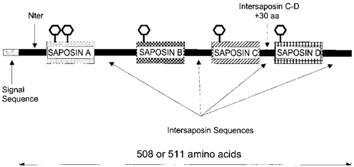
FIG. 1 Schematic diagram of the prosaposin protein organization. The mature human prosaposin is 508 (4, 46) or 511 (5) amino acids in length excluding a 16-amino acid signal sequence. The variation in amino acid sequence results from alternative splicing of a 9-base pair exon in the saposin B genomic sequence. Nter refers to the amino acid sequence that follows the signal sequence and precedes the mature saposin A NH2 terminus. The intersaposin sequences refer to amino acids that are proteolytically clipped during the processing to mature saposins and are shown in approximate proportion to their length. The individual saposins are ; 80 amino acids in length. The intersaposin C-D amino acid sequence is present in the mouse and contains 30 amino acids that are not in the human protein. The saposin A, B, C, and D proteins were derived from the human prosaposin nucleotide sequence, and the Nter and C-D proteins were from the respective mouse sequences.
FIG. 1 Schematic diagram of the prosaposin protein organization. The mature human prosaposin is 508 (4, 46) or 511 (5) amino acids in length excluding a 16-amino acid signal sequence. The variation in amino acid sequence results from alternative splicing of a 9-base pair exon in the saposin B genomic sequence. Nter refers to the amino acid sequence that follows the signal sequence and precedes the mature saposin A NH2 terminus. The intersaposin sequences refer to amino acids that are proteolytically clipped during the processing to mature saposins and are shown in approximate proportion to their length. The individual saposins are ; 80 amino acids in length. The intersaposin C-D amino acid sequence is present in the mouse and contains 30 amino acids that are not in the human protein. The saposin A, B, C, and D proteins were derived from the human prosaposin nucleotide sequence, and the Nter and C-D proteins were from the respective mouse sequences.CC-BY · From the PDF
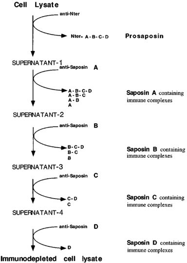
FIG. 2 Schematic diagram of the protocol for the exhaustive immunoprecipitation of cell lysates and the potential immunoprecipitable intermediates. This procedure was generalized to cellular and media sources. For example, exhaustive immunoprecipitation of a cell lysate with anti-A antisera could deplete the supernatant of a tetrasaposin (A-B-C-D), a trisaposin (A-B-C), a disaposin (A-B), and a monosaposin (A). The order of treatment with the different antisera will determine the species that are precipitated. If anti-B was used before anti-saposin A and the disaposins A-B and B-C were present, anti-B would deplete the supernatant of both these disaposins. Subsequent treatment of the supernatant with anti-A would precipitate any remaining saposin A-containing intermediates.
FIG. 2 Schematic diagram of the protocol for the exhaustive immunoprecipitation of cell lysates and the potential immunoprecipitable intermediates. This procedure was generalized to cellular and media sources. For example, exhaustive immunoprecipitation of a cell lysate with anti-A antisera could deplete the supernatant of a tetrasaposin (A-B-C-D), a trisaposin (A-B-C), a disaposin (A-B), and a monosaposin (A). The order of treatment with the different antisera will determine the species that are precipitated. If anti-B was used before anti-saposin A and the disaposins A-B and B-C were present, anti-B would deplete the supernatant of both these disaposins. Subsequent treatment of the supernatant with anti-A would precipitate any remaining saposin A-containing intermediates.CC-BY · From the PDF
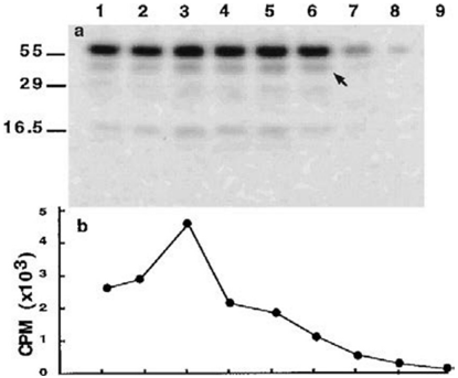
FIG. 3 Exhaustive immunoprecipitation of VL-prosap-infected Sf9 cell lysates by anti-saposin D antisera. The VL-prosapinfected insect cells were labeled for 20 h with [ 35 S]cysteine, immunoprecipitated, and analyzed by SDS-PAGE. a , image from the PhosphorImager of the immunoprecipitates after nine sequential treatments of the supernatants with the antiserum. The M r ; 60,000 dense band increases in intensity through immunoprecipitations 1-5 with diminishing signal subsequently. The pattern of lower M r species differed in intensity during the sequential antiserum treatments; i.e. the M r ; 40,000 species ( arrow ) increased in relative intensity through the fifth precipitation ( lane 6 ) and were not detectable by the eighth immunoprecipitation. b , the precipitable radioactivity of [ 35 S]cysteine associated counts in the corresponding pellets. By the ninth immunoprecipitation the counts decreased to background levels.
FIG. 3 Exhaustive immunoprecipitation of VL-prosap-infected Sf9 cell lysates by anti-saposin D antisera. The VL-prosapinfected insect cells were labeled for 20 h with [ 35 S]cysteine, immunoprecipitated, and analyzed by SDS-PAGE. a , image from the PhosphorImager of the immunoprecipitates after nine sequential treatments of the supernatants with the antiserum. The M r ; 60,000 dense band increases in intensity through immunoprecipitations 1-5 with diminishing signal subsequently. The pattern of lower M r species differed in intensity during the sequential antiserum treatments; i.e. the M r ; 40,000 species ( arrow ) increased in relative intensity through the fifth precipitation ( lane 6 ) and were not detectable by the eighth immunoprecipitation. b , the precipitable radioactivity of [ 35 S]cysteine associated counts in the corresponding pellets. By the ninth immunoprecipitation the counts decreased to background levels.CC-BY · From the PDF
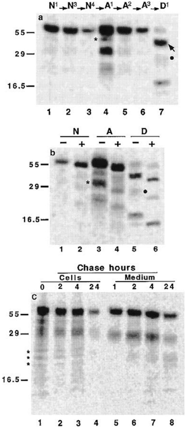
FIG. 4 Immunoprecipitation of VL-prosap-infected Sf9 cells following [ 35 S]cysteine labeling. a , insect cells were labeled for 20 h following infection with VL-prosap and then immunoprecipitated with anti-Nter ( N ), anti-A ( A ), or anti-D ( D ) antiserum. Analyses were by SDS-PAGE. The superscripts of N, A, and D refer to the number of sequential immunoprecipitations, and the arrows indicate the order of the antiserum treatments. The decreasing presence of a M r ; 60,000 species with sequential immunoprecipitations with anti-Nter ( lanes 1-3 ) and anti-A ( lanes 4-6 ) show the presence of two tetrasaposins containing saposins A, B, C, and D with ( lanes 1-3 ) or without ( lanes 4-6 ) the Nter sequence. The presence of a M r ; 43,000 and ; 40,000 species in lane 4 (anti-A) and lane 7 (anti-D), respectively, indicate two different species of trisaposin containing the saposin compositions AB-C and B-C-D, respectively. The disaposins A-B ( M r ; 31,000, lane 4 ) and C-D ( M r ; 27,000, lane 7 , ● ) differ in M r due to glycosylation differences (Fig. 1) and, potentially, intersaposin sequences. In panel b , Sf9 cells were pulse-labeled for 4 h and sequentially and exhaustively immunoprecipitated with the indicated antisera. Immunoprecipitates were untreated ( 2 ) or treated ( 1 ) with N -glycanase prior to SDS-PAGE. All of the detected labeled species increased their mobility after treatment with N -glycanase. The asterisk ( lane 3 ) and circle ( lane 5 ) indicate the disaposins A-B and C-D. c , Sf9 cells infected with the VL-prosap virus. After infection (36 h), Sf9 cells were pulse-labeled with [ 35 S]cysteine for 10 min and chased fresh, nonradioactive media for the indicated times. The immunoprecipitates were obtained with anti-A and were typical for the other antisaposin antisera. The asterisks in panel c refer to unidentified transient intermediate species.
FIG. 4 Immunoprecipitation of VL-prosap-infected Sf9 cells following [ 35 S]cysteine labeling. a , insect cells were labeled for 20 h following infection with VL-prosap and then immunoprecipitated with anti-Nter ( N ), anti-A ( A ), or anti-D ( D ) antiserum. Analyses were by SDS-PAGE. The superscripts of N, A, and D refer to the number of sequential immunoprecipitations, and the arrows indicate the order of the antiserum treatments. The decreasing presence of a M r ; 60,000 species with sequential immunoprecipitations with anti-Nter ( lanes 1-3 ) and anti-A ( lanes 4-6 ) show the presence of two tetrasaposins containing saposins A, B, C, and D with ( lanes 1-3 ) or without ( lanes 4-6 ) the Nter sequence. The presence of a M r ; 43,000 and ; 40,000 species in lane 4 (anti-A) and lane 7 (anti-D), respectively, indicate two different species of trisaposin containing the saposin compositions AB-C and B-C-D, respectively. The disaposins A-B ( M r ; 31,000, lane 4 ) and C-D ( M r ; 27,000, lane 7 , ● ) differ in M r due to glycosylation differences (Fig. 1) and, potentially, intersaposin sequences. In panel b , Sf9 cells were pulse-labeled for 4 h and sequentially and exhaustively immunoprecipitated with the indicated antisera. Immunoprecipitates were untreated ( 2 ) or treated ( 1 ) with N -glycanase prior to SDS-PAGE. All of the detected labeled species increased their mobility after treatment with N -glycanase. The asterisk ( lane 3 ) and circle ( lane 5 ) indicate the disaposins A-B and C-D. c , Sf9 cells infected with the VL-prosap virus. After infection (36 h), Sf9 cells were pulse-labeled with [ 35 S]cysteine for 10 min and chased fresh, nonradioactive media for the indicated times. The immunoprecipitates were obtained with anti-A and were typical for the other antisaposin antisera. The asterisks in panel c refer to unidentified transient intermediate species.CC-BY · From the PDF
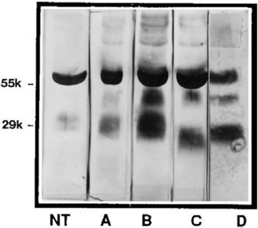
FIG. 5 Western blots of partially purified prosaposin-processing intermediates from media of High-5 cells. Media from High-5 cells were harvested and subjected to partial purification on concanavalin A-Sepharose. The fractions enriched in prosaposin and processing intermediates were collected and subjected to SDS-PAGE followed by transfer to nitrocellulose membranes and treatment with the indicated antisera. Alkaline phosphatase-conjugated second antibody was used for detection. NT , A , B , C , and D refer to the specific antisera used ( i.e. NT represents anti-Nter).
FIG. 5 Western blots of partially purified prosaposin-processing intermediates from media of High-5 cells. Media from High-5 cells were harvested and subjected to partial purification on concanavalin A-Sepharose. The fractions enriched in prosaposin and processing intermediates were collected and subjected to SDS-PAGE followed by transfer to nitrocellulose membranes and treatment with the indicated antisera. Alkaline phosphatase-conjugated second antibody was used for detection. NT , A , B , C , and D refer to the specific antisera used ( i.e. NT represents anti-Nter).CC-BY · From the PDF
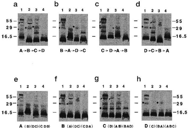
FIG. 6 Processing of prosaposin in cultured skin fibroblasts. HSF were labeled with [ 35 S]cysteine for 20 h, harvested, and washed. Aliquots of the same cell lysates were subjected to sequential, exhaustive immunoprecipitation with the indicated antisaposin antisera and analyzed by SDS-PAGE. In panels a-d are shown the individual typical experiments in which the order of antiserum addition was varied according to the indicated sequence below each panel . For example, in panel a , the cell lysates were exhaustively immunoprecipitated with anti-A ( A ) followed by anti-B ( B ), etc. So in panel a , lane 2 , the fluorographic image of the immunoprecipitates shows the saposin B-containing species that had remained in the supernatants following exhaustive treatment with anti-A. In panels e-h the individual lanes of panels a-d were rearranged to group all the immunoprecipitates obtained with one antiserum in a single panel . Pretreatment with other antisaposin antisera is shown below each lane . For example, panel e , lane 1 , was from a lysate that had been exhaustively immunoprecipitated with anti-A only. The lysate in lane 2 was immunoprecipitated with anti-A after exhaustive immunoprecipitation with anti-B. Lane 3 is after sequential treatment with anti-D and anti-C, etc. Thus, in panel e , the fluorographic images of the immunoprecipitates resolved on SDS-PAGE show the saposin A-containing intermediates that had remained in the supernatants after treatment with the indicated antisera. The arrows show a minor trisaposin consistent with an A-B-C composition. The asterisk is the Nter-saposin A component. l and ● , the disaposins consistent with compositions of B-C and C-D, respectively. f , an unidentifiable species.
FIG. 6 Processing of prosaposin in cultured skin fibroblasts. HSF were labeled with [ 35 S]cysteine for 20 h, harvested, and washed. Aliquots of the same cell lysates were subjected to sequential, exhaustive immunoprecipitation with the indicated antisaposin antisera and analyzed by SDS-PAGE. In panels a-d are shown the individual typical experiments in which the order of antiserum addition was varied according to the indicated sequence below each panel . For example, in panel a , the cell lysates were exhaustively immunoprecipitated with anti-A ( A ) followed by anti-B ( B ), etc. So in panel a , lane 2 , the fluorographic image of the immunoprecipitates shows the saposin B-containing species that had remained in the supernatants following exhaustive treatment with anti-A. In panels e-h the individual lanes of panels a-d were rearranged to group all the immunoprecipitates obtained with one antiserum in a single panel . Pretreatment with other antisaposin antisera is shown below each lane . For example, panel e , lane 1 , was from a lysate that had been exhaustively immunoprecipitated with anti-A only. The lysate in lane 2 was immunoprecipitated with anti-A after exhaustive immunoprecipitation with anti-B. Lane 3 is after sequential treatment with anti-D and anti-C, etc. Thus, in panel e , the fluorographic images of the immunoprecipitates resolved on SDS-PAGE show the saposin A-containing intermediates that had remained in the supernatants after treatment with the indicated antisera. The arrows show a minor trisaposin consistent with an A-B-C composition. The asterisk is the Nter-saposin A component. l and ● , the disaposins consistent with compositions of B-C and C-D, respectively. f , an unidentifiable species.CC-BY · From the PDF
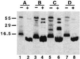
FIG. 7 Deglycosylation of immunoprecipitates from cultured skin fibroblasts following exhaustive immunoprecipitation with anti-saposin antisera. HSF were pulse-labeled for 20 h with [ 35 S]cysteine, immunoprecipitated with the indicated antisaposin antiserum. The immunoprecipitates were untreated ( 2 ) or treated ( 1 ) with N -glycanase and then subjected to SDS-PAGE and fluorography. In these experiments the cell lysates were treated directly with the specific antisera and were not subject to sequential, exhaustive immunoprecipitation.
FIG. 7 Deglycosylation of immunoprecipitates from cultured skin fibroblasts following exhaustive immunoprecipitation with anti-saposin antisera. HSF were pulse-labeled for 20 h with [ 35 S]cysteine, immunoprecipitated with the indicated antisaposin antiserum. The immunoprecipitates were untreated ( 2 ) or treated ( 1 ) with N -glycanase and then subjected to SDS-PAGE and fluorography. In these experiments the cell lysates were treated directly with the specific antisera and were not subject to sequential, exhaustive immunoprecipitation.CC-BY · From the PDF
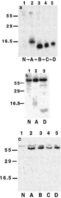
FIG. 8 Pulse labeling of NT2 neuronal precursor cells. NT2 cells were pulsed for 4 h with 20 h of chase ( a ) or no chase ( b ) prior to harvesting and sequential immunoprecipitation with the indicated antisera. N , A , B , C , and D refer to antisera to Nter, saposin A, saposin B, saposin C, and saposin D, respectively. In panel c , spent media from NT2-derived differentiated neurons following 4 h of labeling were individually, not sequentially, immunoprecipitated with the indicated antisera, and the immunoprecipitates were analyzed by fluorography.
FIG. 8 Pulse labeling of NT2 neuronal precursor cells. NT2 cells were pulsed for 4 h with 20 h of chase ( a ) or no chase ( b ) prior to harvesting and sequential immunoprecipitation with the indicated antisera. N , A , B , C , and D refer to antisera to Nter, saposin A, saposin B, saposin C, and saposin D, respectively. In panel c , spent media from NT2-derived differentiated neurons following 4 h of labeling were individually, not sequentially, immunoprecipitated with the indicated antisera, and the immunoprecipitates were analyzed by fluorography.CC-BY · From the PDF
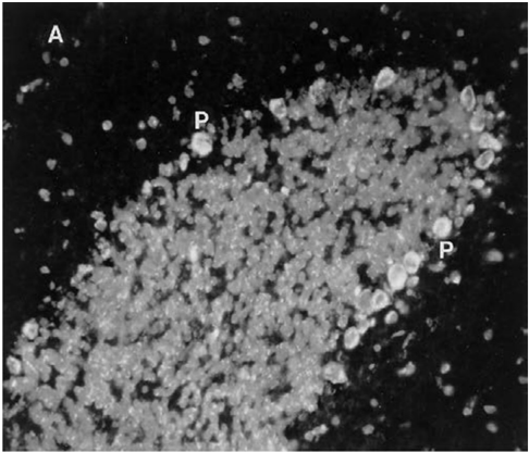
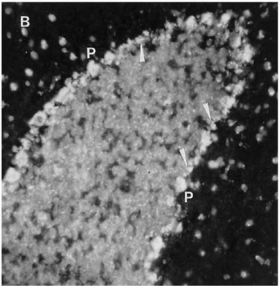
FIG. 9 Immunofluorescence of adult mouse cerebellum using prosaposin- or saposin-specific antisera. Fixed sections of adult mouse cerebellum were treated with anti-mouse intersaposin C-D ( a ) or anti-human saposin A ( b ) antisera and subjected to immunofluorescence microscopy ( 3 400; see 'Experimental Procedures.' In panel a only Purkinje cell bodies ( P ) are brightly stained with the anti-intersaposin C-D antiserum. The same results were obtained with the antiNter antiserum. On immunoblots, anti-Nter reacts only with the prosaposin precursors containing the Nter sequence (human or mouse). Similarly, anti-intersaposin C-D antiserum reacts with mouse precursors composed of Nter-A-B-C-D or A-B-C-D only (data not shown). In panel b Purkinje ( P ) and Bergmann glial ( arrow ) cells in the Purkinje cell layer are brightly stained with the anti-A. The same results were obtained with anti-B or anti-D. Due to low homology, anti-human saposin C antiserum reacts poorly with the mouse saposin C.
FIG. 9 Immunofluorescence of adult mouse cerebellum using prosaposin- or saposin-specific antisera. Fixed sections of adult mouse cerebellum were treated with anti-mouse intersaposin C-D ( a ) or anti-human saposin A ( b ) antisera and subjected to immunofluorescence microscopy ( 3 400; see 'Experimental Procedures.' In panel a only Purkinje cell bodies ( P ) are brightly stained with the anti-intersaposin C-D antiserum. The same results were obtained with the antiNter antiserum. On immunoblots, anti-Nter reacts only with the prosaposin precursors containing the Nter sequence (human or mouse). Similarly, anti-intersaposin C-D antiserum reacts with mouse precursors composed of Nter-A-B-C-D or A-B-C-D only (data not shown). In panel b Purkinje ( P ) and Bergmann glial ( arrow ) cells in the Purkinje cell layer are brightly stained with the anti-A. The same results were obtained with anti-B or anti-D. Due to low homology, anti-human saposin C antiserum reacts poorly with the mouse saposin C.CC-BY · From the PDF
Image 10
CC-BY · From the PDF
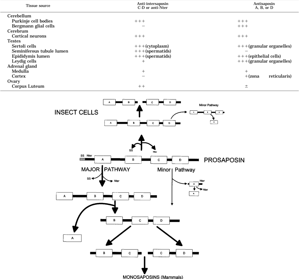
FIG. 10 The proteolytic processing pathways of prosaposin in insect and mammalian cells. The open rectangles refer to the mature saposin amino acid sequences indicated by the contained letter . The thinner black rectangles refer to the intersaposin sequences including Nter. The signal sequence ( SS ) is clipped during passage of the nascent polypeptide through the endoplasmic reticulum. For the intermediates, the amounts of intersaposin sequence shown refer only to potential, not actual, additional amino acids in the tetra-, tri-, and disaposins. The major and minor pathways in the lower part of the figure differ quantitatively by the initial step. The initial step in the minor pathway ( lower ) for both mammalian and insect cells is the cleavage of Nter-saposin A, whereas that in the major pathway is cleavage of Nter. The trisaposin containing B-C-D is a major product in mammalian or insect cells. From the trisaposin, either saposin B or D is removed to generate the disaposins B-C or C-D. These disaposins are processed to monosaposins in mammalian cells. In insect cells another significant pathway is through A-B-C-D, leading directly to the disaposins A-B and C-D that are the major end products. Only small quantities of mature saposins are produced in insect cells. Minor quantitative differences in the pathway were observed between Sf9 and High-5 cells. Another minor, but consistent pathway, in insect cells is the removal of saposin D from the tetrasaposin A-B-C-D. In mammalian cells this pathway is detectable but is very minor. The proteolytic removal of Nter from Nter-A-B-C-D occurs prior to the lysosomal compartment, since Nter-A-B-C-D is secreted into the media of insect cells ( Sf9 or High-5) and of some mammalian cells. In the latter cells the proteolytic processing of the tri- and disaposins probably occurs intralysosomally, as does the cleavage of the Nter-A fragment.
FIG. 10 The proteolytic processing pathways of prosaposin in insect and mammalian cells. The open rectangles refer to the mature saposin amino acid sequences indicated by the contained letter . The thinner black rectangles refer to the intersaposin sequences including Nter. The signal sequence ( SS ) is clipped during passage of the nascent polypeptide through the endoplasmic reticulum. For the intermediates, the amounts of intersaposin sequence shown refer only to potential, not actual, additional amino acids in the tetra-, tri-, and disaposins. The major and minor pathways in the lower part of the figure differ quantitatively by the initial step. The initial step in the minor pathway ( lower ) for both mammalian and insect cells is the cleavage of Nter-saposin A, whereas that in the major pathway is cleavage of Nter. The trisaposin containing B-C-D is a major product in mammalian or insect cells. From the trisaposin, either saposin B or D is removed to generate the disaposins B-C or C-D. These disaposins are processed to monosaposins in mammalian cells. In insect cells another significant pathway is through A-B-C-D, leading directly to the disaposins A-B and C-D that are the major end products. Only small quantities of mature saposins are produced in insect cells. Minor quantitative differences in the pathway were observed between Sf9 and High-5 cells. Another minor, but consistent pathway, in insect cells is the removal of saposin D from the tetrasaposin A-B-C-D. In mammalian cells this pathway is detectable but is very minor. The proteolytic removal of Nter from Nter-A-B-C-D occurs prior to the lysosomal compartment, since Nter-A-B-C-D is secreted into the media of insect cells ( Sf9 or High-5) and of some mammalian cells. In the latter cells the proteolytic processing of the tri- and disaposins probably occurs intralysosomally, as does the cleavage of the Nter-A fragment.CC-BY · From the PDF

Verification

Claims the paper makes

The claims and statistics of this paper haven’t been checked yet.

Run the check — free, no account needed. It reads the open-access text and checks the sources it cites.

A few free checks a day · papers already verified open instantly · results are public

Claims other papers make about this one

Each is checked against this paper’s own text: does it say what the citing paper says it does?

Self-check

Percentages, sums and reported differences re-computed from the paper’s own numbers.

The self-check needs the paper’s full text.

Citations

Cites

No references could be resolved yet.

Research works printed but not linked: 44

  1. Inui, 1985, Hum. Genet., 69, 197, 10.1007/BF00293023 · DOI
  2. Fujibayashi, 1985, Am. J. Hum. Genet., 37, 741
  3. O'Brien, 1988, Science, 241, 1098, 10.1126/science.2842863 · DOI
  4. Gavrieli-Rorman, 1989, Genomics, 5, 486, 10.1016/0888-7543(89)90014-1 · DOI
  5. Nakano, 1989, J. Biochem. (Tokyo), 105, 152, 10.1093/oxfordjournals.jbchem.a122629 · DOI
  6. Collard, 1988, Biochemistry, 27, 4557, 10.1021/bi00412a050 · DOI
  7. Reiner, 1989, J. Mol. Neuroscience, 1, 225
  8. Schnabel, 1991, FEBS Lett., 284, 57, 10.1016/0014-5793(91)80760-Z · DOI
  9. Zhang, 1990, Proc. Natl. Acad. Sci. U. S. A., 87, 1426, 10.1073/pnas.87.4.1426 · DOI
  10. Schnabel, 1992, J. Biol. Chem., 267, 3312, 10.1016/S0021-9258(19)50733-5 · DOI
  11. Zhang, 1991, Hum. Genet., 87, 211, 10.1007/BF00204185 · DOI
  12. Rafi, 1990, Biochem. Biophys. Res. Commun., 166, 1017, 10.1016/0006-291X(90)90912-7 · DOI
  13. Rafi, 1993, Somat. Cell Mol. Genet., 19, 1, 10.1007/BF01233949 · DOI
  14. Klein, 1994, Biochem. Biophys. Res. Commun., 200, 1440, 10.1006/bbrc.1994.1612 · DOI
  15. Furst, 1992, Biochim. Biophys. Acta, 1126, 1, 10.1016/0005-2760(92)90210-M · DOI
  16. Qi, 1994, J. Biol. Chem., 269, 16746, 10.1016/S0021-9258(19)89454-1 · DOI
  17. Vaccaro, 1995, J. Biol. Chem., 270, 9953, 10.1074/jbc.270.17.9953 · DOI
  18. Munford, 1996, J. Lipid Res., 36, 1653, 10.1016/S0022-2275(20)41485-3 · DOI
  19. Sprecher-Levy, 1993, Cell. Mol. Biol., 39, 287
  20. Sun, 1994, Am. J. Pathol., 145, 1390
  21. Fujibayashi, 1986, Biochim. Biophys. Acta, 875, 554, 10.1016/0005-2760(86)90077-9 · DOI
  22. Fujibayashi, 1986, J. Biol. Chem., 261, 15339, 10.1016/S0021-9258(18)66873-5 · DOI
  23. Hiraiwa, 1993, Arch. Biochem. Biophys., 303, 326, 10.1006/abbi.1993.1291 · DOI
  24. Sano, 1992, Biochim. Biophys. Acta, 1120, 75, 10.1016/0167-4838(92)90426-E · DOI
  25. Morimoto, 1989, Proc. Natl. Acad. Sci. U. S. A., 86, 3389, 10.1073/pnas.86.9.3389 · DOI
  26. Morimoto, 1988, Biochem. Biophys. Res. Commun., 156, 403, 10.1016/S0006-291X(88)80855-6 · DOI
  27. Furst, 1988, Biol. Chem. Hoppe Seyler, 369, 317, 10.1515/bchm3.1988.369.1.317 · DOI
  28. Morimoto, 1990, Proc. Natl. Acad. Sci. U. S. A., 87, 3493, 10.1073/pnas.87.9.3493 · DOI
  29. Kondoh, 1991, Biochem. Biophys. Res. Commun., 181, 286, 10.1016/S0006-291X(05)81415-9 · DOI
  30. Hineno, 1991, Biochem. Biophys. Res. Commun., 76, 668, 10.1016/S0006-291X(05)80236-0 · DOI
  31. Hiraiwa, 1992, Proc. Natl. Acad. Sci. U. S. A., 89, 11254, 10.1073/pnas.89.23.11254 · DOI
  32. Qi, 1996, J. Biol. Chem., 271, 6874, 10.1074/jbc.271.12.6874 · DOI
  33. O'Brien, 1995, FASEB J., 9, 681, 10.1096/fasebj.9.8.7768361 · DOI
  34. Kotani, 1996, J. Neurochem.
  35. Hiraiwa, 1993, Arch. Biochem. Biophys., 304, 110, 10.1006/abbi.1993.1328 · DOI
  36. Summers, 1987, Tex. Agric. Exp. Stn. Bull., 1555, 5
  37. Andrews, 1984, Dev. Biol., 103, 285, 10.1016/0012-1606(84)90316-6 · DOI · Paper page
  38. Lee, 1986, J. Neurosci., 6, 514, 10.1523/JNEUROSCI.06-02-00514.1986 · DOI
  39. Pleasure, 1992, J. Neurosci., 12, 1802, 10.1523/JNEUROSCI.12-05-01802.1992 · DOI
  40. Xu, 1996, Pediatr. Res., 39, 313, 10.1203/00006450-199602000-00021 · DOI
  41. Kondoh, 1993, J. Comp. Neurol., 334, 590, 10.1002/cne.903340407 · DOI
  42. Morreau, 1992, J. Biol. Chem., 267, 17949, 10.1016/S0021-9258(19)37135-2 · DOI
  43. Galjart, 1991, J. Biol. Chem., 266, 14754, 10.1016/S0021-9258(18)98751-X · DOI · Paper page
  44. Gavrieli-Rorman, 1992, Genomics, 13, 312, 10.1016/0888-7543(92)90247-P · DOI

Cited by

Metadata

Authors
Leonova, T., Qi, X., Bencosme, A., Ponce, E., Sun, Y., & Grabowski, G. A.
Venue
Journal of Biological Chemistry
Publisher
Elsevier BV
Published
1996-07
Volume
271
Issue
29
Pages
17312-17320
ISSN
0021-9258
Cited by
81
References
0
Language
en

About these checks

Registry, retraction, Cites and statistics results are computed, not judged, with one exception: a printed reference that no index lists is read once by a model before it counts as a citation error. Claims are extracted by a model and kept only when their quote appears verbatim in the text; each cited source is then checked, with a model’s help, for whether it says what the claim relies on. Those verdicts can be wrong — read the claim’s quote and the cited source before relying on one.

Statistics and arithmetic are recomputed, not judged.

Confidence bands are shown only for fields calibrated on our annotated test set. None is calibrated yet, so no band is shown.

What counts as a claim ›How we measure confidence ›How we check this ›

Are you an author, or do you see a problem with this page? Request a review