Cited by 46

The DOI matches its Crossref record; 2 of the 2 references it cites were found in a registry or an index.

SHIP1, an SH2 Domain Containing Polyinositol-5-phosphatase, Regulates Migration through Two Critical Tyrosine Residues and Forms a Novel Signaling Complex with DOK1 and CRKL

Sattler, Martin, Verma, Shalini, Pride, Yuri B., Salgia, Ravi, Rohrschneider, Larry R., Griffin, James D.·2001·Journal of Biological Chemistry

Overview

The claims and statistics of this paper haven’t been checked yet.

Run the check — free, no account needed. It reads the open-access text and checks the sources it cites.

A few free checks a day · papers already verified open instantly · results are public

Integrity signals

Registry

Crossref · 10.1074/jbc.m006250200

Cites

2 of 2 references verified0 citation errors · 0 not checkable

Statistics

Figures

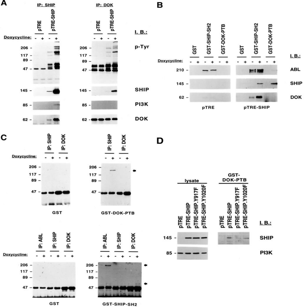
FIG. 1 SHIP1 and DOK1 form a complex in Ba/F3.p210 cells re-expressing SHIP1. A-C, SHIP1 expression was induced by doxycycline treatment of Ba/F3.p210.pTRE-SHIP cells. Lysates of untreated cells ( 2 ) or cells treated for 18 h with 1 m g/ml doxycycline ( 1 ) were used for precipitations. A, lysates of 20 3 10 6 Ba/F3.p210.pTRE and Ba/F3.p210.pTRE-SHIP cells were incubated with anti-SHIP1 and anti-DOK1 antibodies and SHIP1, DOK1, or tyrosine-phosphorylated proteins were detected by immunoblotting ( I.B .). B, lysates of 20 3 10 6 Ba/ F3.p210.pTRE-SHIP cells were incubated with 5 m g of GST and GST fusion protein of the SHIP1 SH2 or the DOK1 PTB domain immobilized on glutathione beads. Co-precipitation of ABL, SHIP1, or DOK1 was detected by immunoblotting ( I.B .). C, lysates of 20 3 10 6 Ba/F3.p210.pTRE-SHIP cells were immunoprecipitated with antibodies against SHIP1, DOK1, and BCR/ABL as indicated. Proteins were separated by SDS-PAGE and transferred to nitrocellulose membrane. Specific direct binding of GST and a GST fusion protein of the DOK1 PTB domain or the SHIP1 SH2 domain to proteins in the immunoprecipitates was detected in a protein overlay assay. The molecular mass of the proteins is indicated in kDa on the left of each figure. D, Ba/F3.p210.TetON cell were transfected with the empty vector (pTRE), a vector containing full-length SHIP1 (pTRE-SHIP), or SHIP1 containing a Y917F (pTRE-SHIP.Y917F) or a Y1020F (pTRE-SHIP.Y1020F) point mutation. SHIP1 protein was induced 1 day after transfectionly by doxycycline treatment for 24 h. SHIP1 expression was detected by immunoblotting ( I.B .) in whole cell lysate (2.5 3 10 5 cells) or in precipitations using a GST-DOK1 PTB domain fusion protein and lysates of 25 3 10 6 cells. The molecular mass of the proteins is as indicated in kDa on the left of each figure.
FIG. 1 SHIP1 and DOK1 form a complex in Ba/F3.p210 cells re-expressing SHIP1. A-C, SHIP1 expression was induced by doxycycline treatment of Ba/F3.p210.pTRE-SHIP cells. Lysates of untreated cells ( 2 ) or cells treated for 18 h with 1 m g/ml doxycycline ( 1 ) were used for precipitations. A, lysates of 20 3 10 6 Ba/F3.p210.pTRE and Ba/F3.p210.pTRE-SHIP cells were incubated with anti-SHIP1 and anti-DOK1 antibodies and SHIP1, DOK1, or tyrosine-phosphorylated proteins were detected by immunoblotting ( I.B .). B, lysates of 20 3 10 6 Ba/ F3.p210.pTRE-SHIP cells were incubated with 5 m g of GST and GST fusion protein of the SHIP1 SH2 or the DOK1 PTB domain immobilized on glutathione beads. Co-precipitation of ABL, SHIP1, or DOK1 was detected by immunoblotting ( I.B .). C, lysates of 20 3 10 6 Ba/F3.p210.pTRE-SHIP cells were immunoprecipitated with antibodies against SHIP1, DOK1, and BCR/ABL as indicated. Proteins were separated by SDS-PAGE and transferred to nitrocellulose membrane. Specific direct binding of GST and a GST fusion protein of the DOK1 PTB domain or the SHIP1 SH2 domain to proteins in the immunoprecipitates was detected in a protein overlay assay. The molecular mass of the proteins is indicated in kDa on the left of each figure. D, Ba/F3.p210.TetON cell were transfected with the empty vector (pTRE), a vector containing full-length SHIP1 (pTRE-SHIP), or SHIP1 containing a Y917F (pTRE-SHIP.Y917F) or a Y1020F (pTRE-SHIP.Y1020F) point mutation. SHIP1 protein was induced 1 day after transfectionly by doxycycline treatment for 24 h. SHIP1 expression was detected by immunoblotting ( I.B .) in whole cell lysate (2.5 3 10 5 cells) or in precipitations using a GST-DOK1 PTB domain fusion protein and lysates of 25 3 10 6 cells. The molecular mass of the proteins is as indicated in kDa on the left of each figure.CC-BY-NC-ND · From the PDF
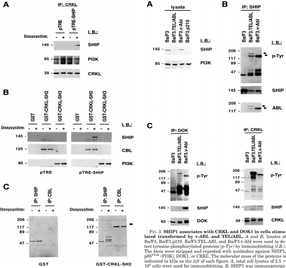
FIG. 2 SHIP1 and CRKL form a complex in Ba/F3.p210 cells re-expressing SHIP1. A-C, SHIP1 expression was induced by doxycycline treatment of Ba/F3.p210.pTRE-SHIP cells. Lysates of untreated cells ( 2 ) or cells treated for 18 h with 1 m g/ml doxycycline ( 1 ) were used for precipitations. The molecular mass of the proteins is indicated in kDa on the left of each figure. A, lysates of 20 3 10 6 Ba/F3.p210.pTRESHIP and Ba/F3.p210.pTRE-SHIP cells were incubated with antiCRKL antibodies and SHIP, CRKL, or p85 PI3K (PI3K) protein detected by immunoblotting ( I.B .). B, lysates of 20 3 10 6 Ba/F3.p210.pTRE-SHIP cells were incubated with 5 m g of GST and GST fusion protein of the CRKL SH2 or the CRKL SH3 domain immobilized on glutathione beads. Co-precipitation of SHIP, CBL, or p85 PI3K (PI3K) was detected by immunoblotting ( I.B .). C, lysates of 20 3 10 6 Ba/F3.p210.pTRE-SHIP cells were immunoprecipitated with antibodies against SHIP1 and CBL as indicated. Proteins were separated by SDS-PAGE, and transferred to polyvinylidene difluoride membrane. Specific direct binding of GST and a GST fusion protein of the CRKL SH2 domain to proteins in the immunoprecipitates was detected in a protein overlay assay.
FIG. 2 SHIP1 and CRKL form a complex in Ba/F3.p210 cells re-expressing SHIP1. A-C, SHIP1 expression was induced by doxycycline treatment of Ba/F3.p210.pTRE-SHIP cells. Lysates of untreated cells ( 2 ) or cells treated for 18 h with 1 m g/ml doxycycline ( 1 ) were used for precipitations. The molecular mass of the proteins is indicated in kDa on the left of each figure. A, lysates of 20 3 10 6 Ba/F3.p210.pTRESHIP and Ba/F3.p210.pTRE-SHIP cells were incubated with antiCRKL antibodies and SHIP, CRKL, or p85 PI3K (PI3K) protein detected by immunoblotting ( I.B .). B, lysates of 20 3 10 6 Ba/F3.p210.pTRE-SHIP cells were incubated with 5 m g of GST and GST fusion protein of the CRKL SH2 or the CRKL SH3 domain immobilized on glutathione beads. Co-precipitation of SHIP, CBL, or p85 PI3K (PI3K) was detected by immunoblotting ( I.B .). C, lysates of 20 3 10 6 Ba/F3.p210.pTRE-SHIP cells were immunoprecipitated with antibodies against SHIP1 and CBL as indicated. Proteins were separated by SDS-PAGE, and transferred to polyvinylidene difluoride membrane. Specific direct binding of GST and a GST fusion protein of the CRKL SH2 domain to proteins in the immunoprecipitates was detected in a protein overlay assay.CC-BY-NC-ND · From the PDF
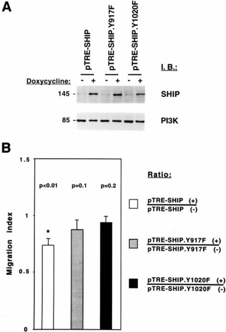
FIG. 4 Tyr 917 and Tyr 1020 in SHIP1 regulate spontaneous Transwell migration. A and B, Ba/F3.p210.TetON cells transfected with a wild type SHIP1 expression vector (pTRE-SHIP) and vectors containing the SHIP1 mutants Y917F (pTRE-SHIP.Y917F) or Y1020F (pTRE-SHIP.Y1020F) were either left untreated ( 2 ) or treated with doxycycline ( 1 ) and used for immunoblotting ( I.B .) or Transwell migration. A, expression of wild type SHIP1 and the two SHIP1 mutants was detected in total cell lysates (2.5 3 10 5 cells) by immunoblotting ( I.B .) with anti-SHIP1 antibodies. The blot was stripped and reprobed with anti-PI3K antibodies. The molecular mass of the proteins is indicated in kDa on the left of the figure. B, cells were used for Transwell migration assays and the number of viable cells in the lower chamber was determined after 2.5 h. The change in migration of four independent experiments was calculated as a migration index. The error bars indicate the standard error of the mean. The p values were calculated and a significant decrease ( p , 0.01) indicated by an asterisk (*).
FIG. 4 Tyr 917 and Tyr 1020 in SHIP1 regulate spontaneous Transwell migration. A and B, Ba/F3.p210.TetON cells transfected with a wild type SHIP1 expression vector (pTRE-SHIP) and vectors containing the SHIP1 mutants Y917F (pTRE-SHIP.Y917F) or Y1020F (pTRE-SHIP.Y1020F) were either left untreated ( 2 ) or treated with doxycycline ( 1 ) and used for immunoblotting ( I.B .) or Transwell migration. A, expression of wild type SHIP1 and the two SHIP1 mutants was detected in total cell lysates (2.5 3 10 5 cells) by immunoblotting ( I.B .) with anti-SHIP1 antibodies. The blot was stripped and reprobed with anti-PI3K antibodies. The molecular mass of the proteins is indicated in kDa on the left of the figure. B, cells were used for Transwell migration assays and the number of viable cells in the lower chamber was determined after 2.5 h. The change in migration of four independent experiments was calculated as a migration index. The error bars indicate the standard error of the mean. The p values were calculated and a significant decrease ( p , 0.01) indicated by an asterisk (*).CC-BY-NC-ND · From the PDF

Verification

Claims the paper makes

The claims and statistics of this paper haven’t been checked yet.

Run the check — free, no account needed. It reads the open-access text and checks the sources it cites.

A few free checks a day · papers already verified open instantly · results are public

Claims other papers make about this one

Each is checked against this paper’s own text: does it say what the citing paper says it does?

Self-check

Percentages, sums and reported differences re-computed from the paper’s own numbers.

The self-check needs the paper’s full text.

Citations

Cites

Research works printed but not linked: 58

  1. Damen, 1996, Proc. Natl. Acad. Sci. U. S. A., 93, 1689, 10.1073/pnas.93.4.1689 · DOI · Paper page
  2. Pesesse, 1997, Biochem. Biophys. Res. Commun., 239, 697, 10.1006/bbrc.1997.7538 · DOI
  3. Hejna, 1995, Genomics, 29, 285, 10.1006/geno.1995.1247 · DOI
  4. Lucas, 1999, Blood, 93, 1922, 10.1182/blood.V93.6.1922.406k21_1922_1933 · DOI
  5. Chacko, 1996, J. Immunol., 157, 2234, 10.4049/jimmunol.157.6.2234 · DOI · Paper page
  6. Osborne, 1996, J. Biol. Chem., 271, 29271, 10.1074/jbc.271.46.29271 · DOI
  7. Habib, 1998, J. Biol. Chem., 273, 18605, 10.1074/jbc.273.29.18605 · DOI
  8. Muraille, 2000, Immunol. Lett., 72, 7, 10.1016/S0165-2478(00)00162-0 · DOI
  9. Aman, 1998, J. Biol. Chem., 273, 33922, 10.1074/jbc.273.51.33922 · DOI · Paper page
  10. Hunter, 1998, J. Immunol., 160, 4979, 10.4049/jimmunol.160.10.4979 · DOI
  11. Rameh, 1999, J. Biol. Chem., 274, 8347, 10.1074/jbc.274.13.8347 · DOI
  12. Helgason, 1998, Genes Dev., 12, 1610, 10.1101/gad.12.11.1610 · DOI · Paper page
  13. Kim, 1999, J. Clin. Invest., 104, 1751, 10.1172/JCI7310 · DOI
  14. Liu, 1997, J. Biol. Chem., 272, 8983, 10.1074/jbc.272.14.8983 · DOI
  15. Ono, 1996, Nature, 383, 263, 10.1038/383263a0 · DOI
  16. Sattler, 1999, Mol. Cell. Biol., 19, 7473, 10.1128/MCB.19.11.7473 · DOI
  17. Phee, 2000, J. Biol. Chem., 275, 19090, 10.1074/jbc.M001093200 · DOI
  18. Kavanaugh, 1996, Curr. Biol., 6, 438, 10.1016/S0960-9822(02)00511-0 · DOI · Paper page
  19. Liu, 1994, Mol. Cell. Biol., 14, 6926
  20. Liu, 1997, J. Biol. Chem., 272, 10998, 10.1074/jbc.272.17.10998 · DOI
  21. Sattler, 1997, Oncogene, 15, 2379, 10.1038/sj.onc.1201422 · DOI
  22. Sattler, 1997, Cytokine Growth Factor Rev., 8, 63, 10.1016/S1359-6101(96)00047-0 · DOI
  23. Verfaillie, 1992, J. Clin. Invest., 90, 1232, 10.1172/JCI115985 · DOI
  24. Salgia, 1997, J. Clin. Invest., 100, 46, 10.1172/JCI119520 · DOI · Paper page
  25. Noguchi, 1999, EMBO J., 18, 1748, 10.1093/emboj/18.7.1748 · DOI
  26. Uemura, 1999, J. Biol. Chem., 274, 37525, 10.1074/jbc.274.53.37525 · DOI
  27. Bhat, 1998, J. Biol. Chem., 273, 32360, 10.1074/jbc.273.48.32360 · DOI
  28. Sattler, 1997, J. Biol. Chem., 272, 10248, 10.1074/jbc.272.15.10248 · DOI
  29. Sattler, 1996, Oncogene, 12, 839
  30. Carpino, 1997, Cell, 88, 197, 10.1016/S0092-8674(00)81840-1 · DOI
  31. Yamanashi, 1997, Cell, 88, 205, 10.1016/S0092-8674(00)81841-3 · DOI
  32. Di Cristofano, 1998, J. Biol. Chem., 273, 4827, 10.1074/jbc.273.9.4827 · DOI
  33. Lemay, 2000, Mol. Cell. Biol., 20, 2743, 10.1128/MCB.20.8.2743-2754.2000 · DOI
  34. Wisniewski, 1996, Leukemia, 10, 229
  35. Bouton, 1991, Mol. Cell. Biol., 11, 945
  36. Hosomi, 1994, J. Biol. Chem., 269, 11498, 10.1016/S0021-9258(19)78151-4 · DOI
  37. Zhou, 1995, Nature, 378, 584, 10.1038/378584a0 · DOI
  38. Eck, 1996, Cell, 85, 695, 10.1016/S0092-8674(00)81236-2 · DOI
  39. Tamir, 2000, Immunity, 12, 347, 10.1016/S1074-7613(00)80187-9 · DOI
  40. Dunant, 2000, Cell Signal, 12, 317, 10.1016/S0898-6568(00)00073-5 · DOI
  41. Kashige, 2000, Proc. Natl. Acad. Sci. U. S. A., 97, 2093, 10.1073/pnas.040547997 · DOI
  42. Siegel, 1999, Oncogene, 18, 7135, 10.1038/sj.onc.1203212 · DOI
  43. Rozakis-Adcock, 1992, Nature, 360, 689, 10.1038/360689a0 · DOI
  44. Kundra, 1995, J. Cell Biol., 130, 725, 10.1083/jcb.130.3.725 · DOI
  45. Lee, 1996, Development, 122, 409, 10.1242/dev.122.2.409 · DOI
  46. Ueoka, 2000, Br. J. Cancer, 82, 891, 10.1054/bjoc.1999.1016 · DOI
  47. Suzuki, 2000, Mol. Cell. Biol., 20, 4658, 10.1128/MCB.20.13.4658-4665.2000 · DOI
  48. Oda, 1994, J. Biol. Chem., 269, 22925, 10.1016/S0021-9258(17)31596-X · DOI
  49. Nichols, 1994, Blood, 84, 2912, 10.1182/blood.V84.9.2912.2912 · DOI
  50. ten Hoeve, 1994, Blood, 84, 1731, 10.1182/blood.V84.6.1731.1731 · DOI
  51. Sattler, 1998, Leukemia, 12, 637, 10.1038/sj.leu.2401010 · DOI
  52. Sattler, 1997, J. Biol. Chem., 272, 14320, 10.1074/jbc.272.22.14320 · DOI
  53. Park, 1999, Blood, 94, 2112, 10.1182/blood.V94.6.2112 · DOI
  54. Skorski, 1995, Blood, 86, 726, 10.1182/blood.V86.2.726.bloodjournal862726 · DOI
  55. Papadopoulos, 1995, Cancer Res., 55, 34
  56. Golub, 1996, Mol. Cell. Biol., 16, 4107, 10.1128/MCB.16.8.4107 · DOI
  57. Reynolds, 1980, J. Virol., 36, 374, 10.1128/jvi.36.2.374-386.1980 · DOI
  58. Rosenberg, 1980, J. Virol., 36, 766, 10.1128/jvi.36.3.766-774.1980 · DOI

Cited by

See all 46

Metadata

Authors
Sattler, M., Verma, S., Pride, Y. B., Salgia, R., Rohrschneider, L. R., & Griffin, J. D.
Venue
Journal of Biological Chemistry
Publisher
Elsevier BV
Published
2001-01
Volume
276
Issue
4
Pages
2451-2458
ISSN
0021-9258
Cited by
46
References
2
Language
en

About these checks

Registry, retraction, Cites and statistics results are computed, not judged, with one exception: a printed reference that no index lists is read once by a model before it counts as a citation error. Claims are extracted by a model and kept only when their quote appears verbatim in the text; each cited source is then checked, with a model’s help, for whether it says what the claim relies on. Those verdicts can be wrong — read the claim’s quote and the cited source before relying on one.

Statistics and arithmetic are recomputed, not judged.

Confidence bands are shown only for fields calibrated on our annotated test set. None is calibrated yet, so no band is shown.

What counts as a claim ›How we measure confidence ›How we check this ›

Are you an author, or do you see a problem with this page? Request a review