Cited by 97

The DOI matches its Crossref record; 2 of the 2 references it cites were found in a registry or an index.

Human Papillomavirus (HPV) E7 Induces Prolonged G2 following S Phase Reentry in Differentiated Human Keratinocytes

Banerjee, N. Sanjib, Wang, Hsu-Kun, Broker, Thomas R., Chow, Louise T.·2011·Journal of Biological Chemistry

Overview

The claims and statistics of this paper haven’t been checked yet.

Run the check — free, no account needed. It reads the open-access text and checks the sources it cites.

A few free checks a day · papers already verified open instantly · results are public

Integrity signals

Registry

Crossref · 10.1074/jbc.m110.197574

Cites

2 of 2 references verified0 citation errors · 0 not checkable

Statistics

Figures

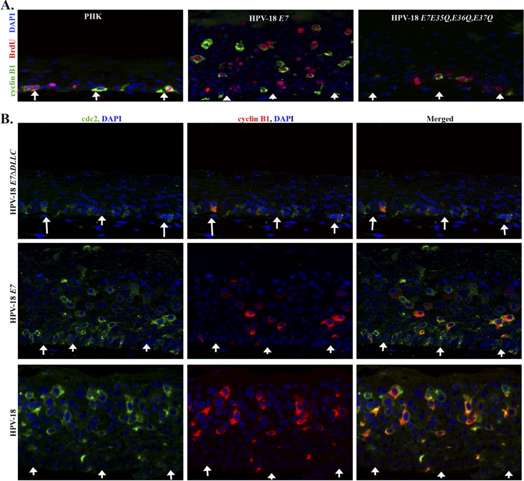
FIGURE 1 HPV-18 E7 and genomic HPV-18 plasmid induce elevated expression of cyclin B1 and cdc2 in the differentiated strata of PHK raft cultures. A, double indirect immunofluorescence microscopy was performed to detect cyclin B1 ( green ) and BrdU ( red ) in organotypic raft cultures of PHK ( left panel ), HPV-18 E7 wild type ( middle panel ), and HPV-18 E7E35Q , E36Q , E37Q ( right panel ). B , cdc2 ( green ) and cyclin B1 ( red ) in raft cultures of HPV-18 E7 𝚫 DLLC ( upper row ), the wild type HPV-18 E7 ( middle row ), and HPV-18 genomic plasmid-containing raft cultures ( lower row ). 𝚫 DLLC and E35Q , E36Q , E37Q are mutations of HPV-18 E7 lacking the pocket protein binding site or CKII motif, respectively. In this figure and Figs. 3 and 5, images were captured at × 20 magnification and arrowheads indicate the basal layer of the epithelia. All the raft cultures shown here are 10 days old.
FIGURE 1 HPV-18 E7 and genomic HPV-18 plasmid induce elevated expression of cyclin B1 and cdc2 in the differentiated strata of PHK raft cultures. A, double indirect immunofluorescence microscopy was performed to detect cyclin B1 ( green ) and BrdU ( red ) in organotypic raft cultures of PHK ( left panel ), HPV-18 E7 wild type ( middle panel ), and HPV-18 E7E35Q , E36Q , E37Q ( right panel ). B , cdc2 ( green ) and cyclin B1 ( red ) in raft cultures of HPV-18 E7 𝚫 DLLC ( upper row ), the wild type HPV-18 E7 ( middle row ), and HPV-18 genomic plasmid-containing raft cultures ( lower row ). 𝚫 DLLC and E35Q , E36Q , E37Q are mutations of HPV-18 E7 lacking the pocket protein binding site or CKII motif, respectively. In this figure and Figs. 3 and 5, images were captured at × 20 magnification and arrowheads indicate the basal layer of the epithelia. All the raft cultures shown here are 10 days old.CC-BY · From the PDF
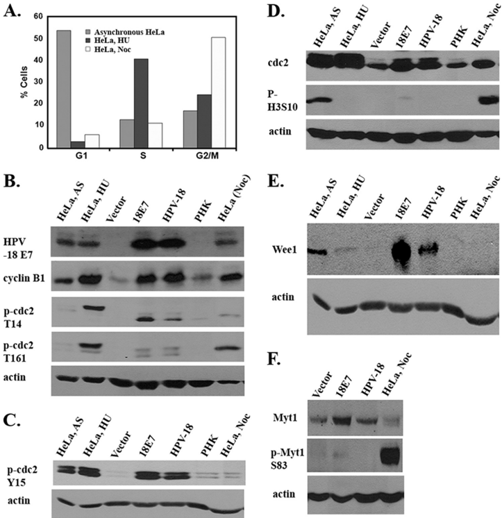
FIGURE 2 HPV-18 E7 andgenomicHPV-18plasmiddysregulate cell cycle regulatory proteins favoring a G2 phase milieu. A , FACS analysis of HeLa cells without any treatment or treated with hydroxyurea ( HU ) to enrich cells in S phase or with nocodazole ( Noc ), which enrich cells in M. B-E , Western blot analysis of lysates from asynchronous ( AS ) HeLa, hydroxyurea-treated HeLa, vector-transduced raft culture ( Vector ), HPV-18 E7 -expressing raft culture ( 18E7 ), PHK raft culture containing amplifying HPV-18 genomic plasmids ( HPV-18 ), PHK control raft culture ( PHK ), and Noc-treated HeLa. B , immunoblots showing steady state levels of HPV-18 E7, cyclin B1, p -cdc2 Thr 14 , and p -cdc2 Thr 161 . C , steady state expression levels of p -cdc2 Tyr 15 and p -cdc2 Thr 161 . D , steady state expression levels of cdc2 and phospho-histone Ser 10 (p-H3S10). E , steady state expression level of Wee1. F , steady state expression level of unphosphorylated Myt1 ( upper panel ) and phosphorylated Myt1 ( middle panel ). B-D and F represent 4 different gels. C and E are from the same gel. The blot represented in panel B wasprobed, stripped, and reprobed with different antibodies. Actin was used as a protein loading control.
FIGURE 2 HPV-18 E7 andgenomicHPV-18plasmiddysregulate cell cycle regulatory proteins favoring a G2 phase milieu. A , FACS analysis of HeLa cells without any treatment or treated with hydroxyurea ( HU ) to enrich cells in S phase or with nocodazole ( Noc ), which enrich cells in M. B-E , Western blot analysis of lysates from asynchronous ( AS ) HeLa, hydroxyurea-treated HeLa, vector-transduced raft culture ( Vector ), HPV-18 E7 -expressing raft culture ( 18E7 ), PHK raft culture containing amplifying HPV-18 genomic plasmids ( HPV-18 ), PHK control raft culture ( PHK ), and Noc-treated HeLa. B , immunoblots showing steady state levels of HPV-18 E7, cyclin B1, p -cdc2 Thr 14 , and p -cdc2 Thr 161 . C , steady state expression levels of p -cdc2 Tyr 15 and p -cdc2 Thr 161 . D , steady state expression levels of cdc2 and phospho-histone Ser 10 (p-H3S10). E , steady state expression level of Wee1. F , steady state expression level of unphosphorylated Myt1 ( upper panel ) and phosphorylated Myt1 ( middle panel ). B-D and F represent 4 different gels. C and E are from the same gel. The blot represented in panel B wasprobed, stripped, and reprobed with different antibodies. Actin was used as a protein loading control.CC-BY · From the PDF
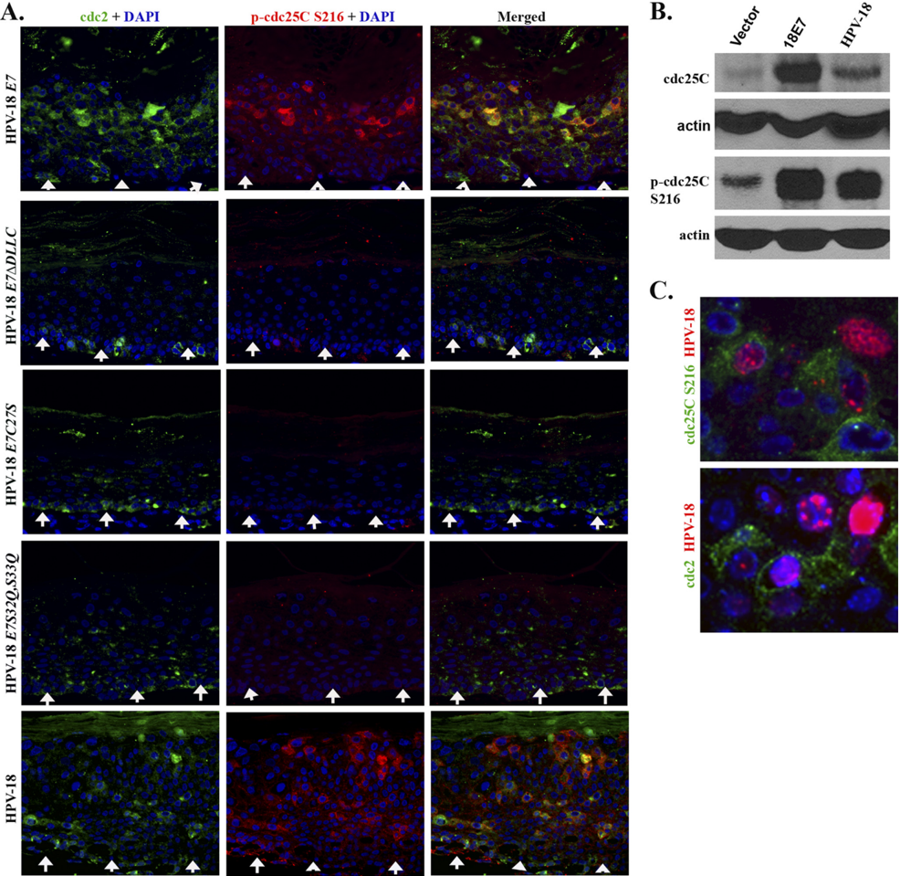
FIGURE 3 HPV-18 E7 and genomic HPV-18 plasmid induce changes in the expression and phosphorylation status of the cdc25C phosphatase. A , indirect immunofluorescence microscopy was used to detect the expression and localization of p -cdc25C Ser 216 ( red ) relative to cdc2 ( green ) in raft cultures of HPV-18 E7 , HPV-18 E7 𝚫 DLLC , HPV-18 E7C27S , HPV-18 E7S32Q , S33Q , and HPV-18 genomic plasmids. B , immunoblot analysis were performed on lysates from vector-only PHK raft cultures, HPV-18 E7-expressing raft cultures, and HPV-18 raft cultures to assess total cdc25C and the phosphorylated cdc25C Ser 216 . C , simultaneous indirect immunofluorescence and HPV-18 DNA fluorescent in situ hybridization to detect HPV-18 DNA amplification ( red ) relative to p -cdc25C Ser 216 ( green ) or cdc2 ( green ). E7C27S is mutated in the pocket protein binding motif, whereas S32Q,S33Q is mutated in the CKII substrates. Raft cultures in A and B are 10 days old and raft culture in C is 12 days old.
FIGURE 3 HPV-18 E7 and genomic HPV-18 plasmid induce changes in the expression and phosphorylation status of the cdc25C phosphatase. A , indirect immunofluorescence microscopy was used to detect the expression and localization of p -cdc25C Ser 216 ( red ) relative to cdc2 ( green ) in raft cultures of HPV-18 E7 , HPV-18 E7 𝚫 DLLC , HPV-18 E7C27S , HPV-18 E7S32Q , S33Q , and HPV-18 genomic plasmids. B , immunoblot analysis were performed on lysates from vector-only PHK raft cultures, HPV-18 E7-expressing raft cultures, and HPV-18 raft cultures to assess total cdc25C and the phosphorylated cdc25C Ser 216 . C , simultaneous indirect immunofluorescence and HPV-18 DNA fluorescent in situ hybridization to detect HPV-18 DNA amplification ( red ) relative to p -cdc25C Ser 216 ( green ) or cdc2 ( green ). E7C27S is mutated in the pocket protein binding motif, whereas S32Q,S33Q is mutated in the CKII substrates. Raft cultures in A and B are 10 days old and raft culture in C is 12 days old.CC-BY · From the PDF
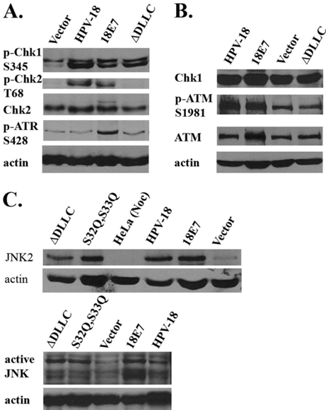
FIGURE 4 HPV-18 E7 and genomic HPV-18 plasmid activate kinases responsible for cdc25C phosphorylation on Ser 216 . Immunoblot analysis of ( A ) p -ATR Ser 428 , p -Chk1 Ser 345 , total Chk2, and p -Chk2 Thr 68 and ( B ) ATM, p -ATMSer 1981 , total Chk1 from lysates of raft cultures harboring empty vector ( Vector ), HPV-18 genomic plasmid, or expressing HPV-18 E7 ( 18E7 ) or HPV-18 E7 𝚫 DLLC ( 𝚫 DLLC ) mutation; C , JNK2 and phosphorylated JNKs in raft cultures harboring the vector-only ( Vector ), HPV-18 E7 ( 18E7 ), E7 𝚫 DLLC ( 𝚫 DLLC ), E7S32Q , E33Q ( S32Q,S33Q ), or HPV-18 genomic plasmids. HeLa ( Noc ) denotes lysates from nocodazole-treated HeLa as negative control for JNK2.
FIGURE 4 HPV-18 E7 and genomic HPV-18 plasmid activate kinases responsible for cdc25C phosphorylation on Ser 216 . Immunoblot analysis of ( A ) p -ATR Ser 428 , p -Chk1 Ser 345 , total Chk2, and p -Chk2 Thr 68 and ( B ) ATM, p -ATMSer 1981 , total Chk1 from lysates of raft cultures harboring empty vector ( Vector ), HPV-18 genomic plasmid, or expressing HPV-18 E7 ( 18E7 ) or HPV-18 E7 𝚫 DLLC ( 𝚫 DLLC ) mutation; C , JNK2 and phosphorylated JNKs in raft cultures harboring the vector-only ( Vector ), HPV-18 E7 ( 18E7 ), E7 𝚫 DLLC ( 𝚫 DLLC ), E7S32Q , E33Q ( S32Q,S33Q ), or HPV-18 genomic plasmids. HeLa ( Noc ) denotes lysates from nocodazole-treated HeLa as negative control for JNK2.CC-BY · From the PDF
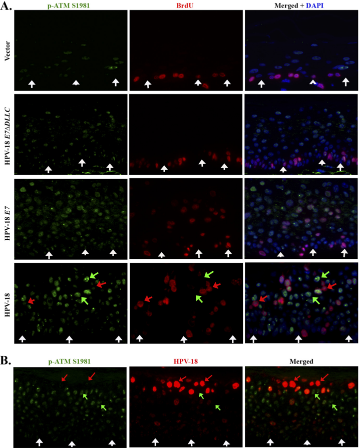
FIGURE 5 A , indirect immunofluorescent detection of p -ATMSer 1981 ( green ) and S phase reentry (BrdU incorporation, red ) in vector-only transduced PHK raft culture ( top row ), HPV-18 E7 𝚫 DLLC mutation-expressing raft culture ( second row ), HPV-18 E7-expressing raft culture ( third row ), and HPV-18 plasmid containing raft culture ( fourth row ). In the last row, the red arrow indicates the high BrdU and low p -ATMSer 1981 , whereas the green arrow points to high p -ATMSer 1981 but low or no BrdU incorporation. B , p -ATMSer 1981 expression relative to HPV-18 plasmid amplification was revealed by combined indirect immunofluorescence andHPV-18DNAfluorescent in situ hybridization. Redarrow indicate high HPV-18 DNA and low or no p -ATMSer 1981 , whereas the greenarrow points to the high p -ATM Ser 1981 but low or no HPV-18 plasmid. Raft cultures in A are 10 days old and raft culture in B is 12 days old.
FIGURE 5 A , indirect immunofluorescent detection of p -ATMSer 1981 ( green ) and S phase reentry (BrdU incorporation, red ) in vector-only transduced PHK raft culture ( top row ), HPV-18 E7 𝚫 DLLC mutation-expressing raft culture ( second row ), HPV-18 E7-expressing raft culture ( third row ), and HPV-18 plasmid containing raft culture ( fourth row ). In the last row, the red arrow indicates the high BrdU and low p -ATMSer 1981 , whereas the green arrow points to high p -ATMSer 1981 but low or no BrdU incorporation. B , p -ATMSer 1981 expression relative to HPV-18 plasmid amplification was revealed by combined indirect immunofluorescence andHPV-18DNAfluorescent in situ hybridization. Redarrow indicate high HPV-18 DNA and low or no p -ATMSer 1981 , whereas the greenarrow points to the high p -ATM Ser 1981 but low or no HPV-18 plasmid. Raft cultures in A are 10 days old and raft culture in B is 12 days old.CC-BY · From the PDF

Verification

Claims the paper makes

The claims and statistics of this paper haven’t been checked yet.

Run the check — free, no account needed. It reads the open-access text and checks the sources it cites.

A few free checks a day · papers already verified open instantly · results are public

Claims other papers make about this one

Each is checked against this paper’s own text: does it say what the citing paper says it does?

Self-check

Percentages, sums and reported differences re-computed from the paper’s own numbers.

The self-check needs the paper’s full text.

Citations

Cites

Show all 2 references

Research works printed but not linked: 58

  1. de Villiers, 2004, Virology, 324, 17, 10.1016/j.virol.2004.03.033 · DOI
  2. Chow, 2010, APMIS, 118, 422, 10.1111/j.1600-0463.2010.02625.x · DOI
  3. Cheng, 1995, Genes Dev., 9, 2335, 10.1101/gad.9.19.2335 · DOI
  4. Banerjee, 2006, J. Virol., 80, 6517, 10.1128/JVI.02499-05 · DOI
  5. Zhang, 2006, Proc. Natl. Acad. Sci. U.S.A., 103, 437, 10.1073/pnas.0510012103 · DOI · Paper page
  6. Genovese, 2008, J. Virol., 82, 4862, 10.1128/JVI.01202-07 · DOI
  7. Wang, 2009, Genes Dev., 23, 181, 10.1101/gad.1735109 · DOI
  8. Chien, 2002, J. Virol., 76, 2964, 10.1128/JVI.76.6.2964-2972.2002 · DOI
  9. Chien, 2000, Cell Growth Differ., 11, 425
  10. Hardouin, 2000, Genesis, 26, 245, 10.1002/(SICI)1526-968X(200004)26:4<245::AID-GENE50>3.0.CO;2-9 · DOI
  11. Wilson, 1992, Cell Growth Differ., 3, 471
  12. Banerjee, 2005, Methods Mol. Med., 119, 187
  13. Van Tine, 2005, Methods Mol. Biol., 292, 215
  14. Pines, 1991, J. Cell Biol., 115, 1, 10.1083/jcb.115.1.1 · DOI
  15. Noya, 2001, J. Virol., 75, 6121, 10.1128/JVI.75.13.6121-6134.2001 · DOI · Paper page
  16. Taylor, 1999, Mol. Biol. Cell, 10, 3607, 10.1091/mbc.10.11.3607 · DOI
  17. Toyoshima, 1998, EMBO J., 17, 2728, 10.1093/emboj/17.10.2728 · DOI
  18. Solomon, 1992, Mol. Biol. Cell, 3, 13, 10.1091/mbc.3.1.13 · DOI
  19. Atherton-Fessler, 1994, Mol. Biol. Cell, 5, 989, 10.1091/mbc.5.9.989 · DOI
  20. Liu, 1997, Mol. Cell. Biol., 17, 571, 10.1128/MCB.17.2.571 · DOI
  21. Liu, 1999, Mol. Cell. Biol., 19, 5113, 10.1128/MCB.19.7.5113 · DOI
  22. Parker, 1992, Science, 257, 1955, 10.1126/science.1384126 · DOI
  23. Mueller, 1995, Science, 270, 86, 10.1126/science.270.5233.86 · DOI
  24. Kornbluth, 1994, Mol. Biol. Cell, 5, 273, 10.1091/mbc.5.3.273 · DOI
  25. Booher, 1997, J. Biol. Chem., 272, 22300, 10.1074/jbc.272.35.22300 · DOI
  26. Palmer, 1998, EMBO J., 17, 5037, 10.1093/emboj/17.17.5037 · DOI
  27. Nakajima, 2003, J. Biol. Chem., 278, 25277, 10.1074/jbc.C300126200 · DOI
  28. Coleman, 1994, Curr. Opin. Cell Biol., 6, 877, 10.1016/0955-0674(94)90060-4 · DOI
  29. Dunphy, 1991, Cell, 67, 189, 10.1016/0092-8674(91)90582-J · DOI
  30. Karlsson, 1999, J. Cell Biol., 146, 573, 10.1083/jcb.146.3.573 · DOI
  31. Strausfeld, 1994, J. Biol. Chem., 269, 5989, 10.1016/S0021-9258(17)37560-9 · DOI
  32. Strausfeld, 1991, Nature, 351, 242, 10.1038/351242a0 · DOI
  33. Dalal, 1999, Mol. Cell. Biol., 19, 4465, 10.1128/MCB.19.6.4465 · DOI
  34. Peng, 1997, Science, 277, 1501, 10.1126/science.277.5331.1501 · DOI · Paper page
  35. Peng, 1998, Cell Growth Differ., 9, 197
  36. Kudoh, 2004, J. Virol., 78, 104, 10.1128/JVI.78.1.104-115.2004 · DOI
  37. Lilley, 2005, Proc. Natl. Acad. Sci. U.S.A., 102, 5844, 10.1073/pnas.0501916102 · DOI
  38. Moody, 2010, Nat. Rev. Cancer, 10, 550, 10.1038/nrc2886 · DOI
  39. Shirata, 2005, J. Biol. Chem., 280, 30336, 10.1074/jbc.M500976200 · DOI
  40. Zhao, 2008, J. Virol., 82, 5316, 10.1128/JVI.02677-07 · DOI
  41. Rogoff, 2004, Mol. Cell. Biol., 24, 2968, 10.1128/MCB.24.7.2968-2977.2004 · DOI
  42. Gutierrez, 2010, J. Biol. Chem., 285, 14217, 10.1074/jbc.M110.121848 · DOI
  43. Wang, 2009, J. Cell Physiol., 221, 213, 10.1002/jcp.21844 · DOI
  44. Garner-Hamrick, 2004, J. Virol., 78, 9041, 10.1128/JVI.78.17.9041-9050.2004 · DOI
  45. Herzinger, 1995, Oncogene, 11, 2151
  46. Gabrielli, 1997, Oncogene, 15, 749, 10.1038/sj.onc.1201254 · DOI
  47. Doorbar, 1997, Virology, 238, 40, 10.1006/viro.1997.8768 · DOI
  48. Chow, 2009, Cell Cycle, 8, 1319, 10.4161/cc.8.9.8242 · DOI
  49. Davy, 2007, Virology, 368, 219, 10.1016/j.virol.2007.05.043 · DOI
  50. Knight, 2011, Virology, 412, 196, 10.1016/j.virol.2011.01.007 · DOI
  51. Datta, 2007, Retrovirology, 4, 49, 10.1186/1742-4690-4-49 · DOI
  52. Kino, 2005, J. Virol., 79, 2780, 10.1128/JVI.79.5.2780-2787.2005 · DOI
  53. Yoshizuka, 2005, J. Virol., 79, 11366, 10.1128/JVI.79.17.11366-11381.2005 · DOI
  54. Dahl, 2005, J. Virol., 79, 13007, 10.1128/JVI.79.20.13007-13017.2005 · DOI
  55. Shi, 2005, J. Biol. Chem., 280, 40195, 10.1074/jbc.C500400200 · DOI
  56. Hein, 2009, J. Virol., 83, 117, 10.1128/JVI.01515-08 · DOI
  57. Boichuk, 2010, J. Virol., 84, 8007, 10.1128/JVI.00334-10 · DOI
  58. Luo, 2011, J. Virol., 85, 133, 10.1128/JVI.01534-10 · DOI

Metadata

Authors
Banerjee, N. S., Wang, H.-K., Broker, T. R., & Chow, L. T.
Venue
Journal of Biological Chemistry
Publisher
Elsevier BV
Published
2011-04
Volume
286
Issue
17
Pages
15473-15482
ISSN
0021-9258
Cited by
97
References
2
Language
en

About these checks

Registry, retraction, Cites and statistics results are computed, not judged, with one exception: a printed reference that no index lists is read once by a model before it counts as a citation error. Claims are extracted by a model and kept only when their quote appears verbatim in the text; each cited source is then checked, with a model’s help, for whether it says what the claim relies on. Those verdicts can be wrong — read the claim’s quote and the cited source before relying on one.

Statistics and arithmetic are recomputed, not judged.

Confidence bands are shown only for fields calibrated on our annotated test set. None is calibrated yet, so no band is shown.

What counts as a claim ›How we measure confidence ›How we check this ›

Are you an author, or do you see a problem with this page? Request a review

How to cite

Cite CoChat for the checks on this page, and the paper for its own findings.

This page
CoChat. (2026). Citation check: Human Papillomavirus (HPV) E7 Induces Prolonged G2 following S Phase Reentry in Differentiated Human Keratinocytes. https://app.cochat.ai/papers/10.1074/jbc.m110.197574
The paper
Banerjee, N. S., Wang, H.-K., Broker, T. R., & Chow, L. T. (2011). Human Papillomavirus (HPV) E7 Induces Prolonged G2 following S Phase Reentry in Differentiated Human Keratinocytes. Journal of Biological Chemistry. https://doi.org/10.1074/jbc.m110.197574