Cited by 47

The DOI matches its Crossref record.

Association of Bcr-Abl with the Proto-oncogene Vav Is Implicated in Activation of the Rac-1 Pathway

Bassermann, Florian, Jahn, Thomas, Miething, Cornelius, Seipel, Petra, Bai, Ren-Yuan, Coutinho, Sunita, Peschel, Christian, Duyster, Justus, Tybulewicz, Victor L.·2002·Journal of Biological Chemistry

Overview

The claims and statistics of this paper haven’t been checked yet.

Run the check — free, no account needed. It reads the open-access text and checks the sources it cites.

A few free checks a day · papers already verified open instantly · results are public

Integrity signals

Registry

Crossref · 10.1074/jbc.m112397200

Cites

Statistics

Figures

Image 1
CC-BY · From the PDF
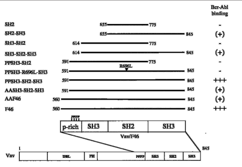
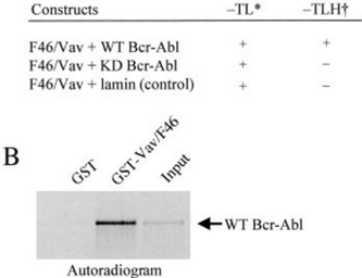
FIG. 1 A , a C-terminal Vav fragment binds to kinase-active Bcr-Abl in yeast. *, growth under tryptophan and leucine selection reflecting the transfection efficiency; † , growth under additional histidine selection reflecting the specificity of interaction. B , the Vav/F46 fragment binds directly to Bcr-Abl in vitro , Bcr-Abl was in vitro translated and S 35 -labeled. 50 𝛍 l of the translation mix was allowed to autophosphorylate and was then incubated with 5 𝛍 g of GST-Vav/F46 or GST alone as a control. Input ( right lane ) and bound fractions on GST-Vav/F46 ( middle lane ) and GST as control ( left lane ) were resolved by SDS-PAGE and visualized by autoradiography.
FIG. 1 A , a C-terminal Vav fragment binds to kinase-active Bcr-Abl in yeast. *, growth under tryptophan and leucine selection reflecting the transfection efficiency; † , growth under additional histidine selection reflecting the specificity of interaction. B , the Vav/F46 fragment binds directly to Bcr-Abl in vitro , Bcr-Abl was in vitro translated and S 35 -labeled. 50 𝛍 l of the translation mix was allowed to autophosphorylate and was then incubated with 5 𝛍 g of GST-Vav/F46 or GST alone as a control. Input ( right lane ) and bound fractions on GST-Vav/F46 ( middle lane ) and GST as control ( left lane ) were resolved by SDS-PAGE and visualized by autoradiography.CC-BY · From the PDF
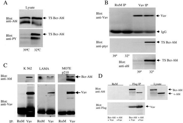
FIG. 2 Vav interacts with Bcr-Abl and v-Abl in vivo. A , a temperature-sensitive mutant of Bcr-Abl ( TS Bcr-Abl ) was transiently transfected into 293 cells. TSBcr-Abl-transfected 293 cells were kept at 39 ° C(restrictive temperature) or shifted to 32 ° C(permissive temperature) for 4 h. Cell lysates were run on SDS-PAGE and immunoblotted with an anti-phosphotyrosine antibody (PY20) ( upper panel ) or an anti-Abl antibody (8E9) ( lower panel ). B , cell lysates from 1 × 10 7 TSBcr-Abl-transfected 293 cells kept at the restrictive (39 ° C) or permissive (32 ° C) temperature were immunoprecipitated with an anti-Vav antibody or a rabbit anti-mouse ( R 𝛂 M ) antibody as control. Protein complexes were washed thoroughly and resolved by SDS-PAGE followed by immunoblotting with anti-Vav antibody ( upper panel ), anti-phosphotyrosine antibody ( anti-ptyr , middle panel ), or anti-Abl antibody ( lower panel ). C , lysates of 5 × 10 6 K562 cells ( left panel ), Lama84 cells ( middle panel ), and MO7E overexpressing Bcr-Abl ( right panel ) were immunoprecipitated with anti-Vav antibody or rabbit anti-mouse antibody as control. Bound fractions were resolved by SDS-PAGE and immunoblotted with anti-Vav antibody. D , 293 cells were transiently cotransfected with v-Abl or Bcr-Abl together with Flag-tagged full-length Vav ( Vav ). Cell lysates were prepared and immunoprecipitated with rabbit anti-mouse antibody as control or anti-Flag antibody to precipitate Flag-tagged Vav. Precipitated proteins ( upper left and lower left panels ) and lysates ( right panel ) were run on SDS-PAGE and visualized with anti-Abl and anti-Flag antibodies as indicated.
FIG. 2 Vav interacts with Bcr-Abl and v-Abl in vivo. A , a temperature-sensitive mutant of Bcr-Abl ( TS Bcr-Abl ) was transiently transfected into 293 cells. TSBcr-Abl-transfected 293 cells were kept at 39 ° C(restrictive temperature) or shifted to 32 ° C(permissive temperature) for 4 h. Cell lysates were run on SDS-PAGE and immunoblotted with an anti-phosphotyrosine antibody (PY20) ( upper panel ) or an anti-Abl antibody (8E9) ( lower panel ). B , cell lysates from 1 × 10 7 TSBcr-Abl-transfected 293 cells kept at the restrictive (39 ° C) or permissive (32 ° C) temperature were immunoprecipitated with an anti-Vav antibody or a rabbit anti-mouse ( R 𝛂 M ) antibody as control. Protein complexes were washed thoroughly and resolved by SDS-PAGE followed by immunoblotting with anti-Vav antibody ( upper panel ), anti-phosphotyrosine antibody ( anti-ptyr , middle panel ), or anti-Abl antibody ( lower panel ). C , lysates of 5 × 10 6 K562 cells ( left panel ), Lama84 cells ( middle panel ), and MO7E overexpressing Bcr-Abl ( right panel ) were immunoprecipitated with anti-Vav antibody or rabbit anti-mouse antibody as control. Bound fractions were resolved by SDS-PAGE and immunoblotted with anti-Vav antibody. D , 293 cells were transiently cotransfected with v-Abl or Bcr-Abl together with Flag-tagged full-length Vav ( Vav ). Cell lysates were prepared and immunoprecipitated with rabbit anti-mouse antibody as control or anti-Flag antibody to precipitate Flag-tagged Vav. Precipitated proteins ( upper left and lower left panels ) and lysates ( right panel ) were run on SDS-PAGE and visualized with anti-Abl and anti-Flag antibodies as indicated.CC-BY · From the PDF
Image 4
CC-BY · From the PDF
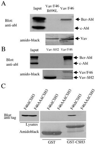
FIG. 4 The Vav SH2 domain is required but not sufficient to mediate the complex with Bcr-Abl in K562 cells. Lysates of 5 × 10 6 K562 cells were incubated with GST-Vav/F46 and GST-Vav/ F46R696L (SH2-mutant) ( A ) or GST-Vav-SH2 ( B ). Input and bound fractions were resolved by SDS-PAGE. Proteins were visualized by immunoblotting with anti-Abl antibody and by Amido Black staining as indicated. C , 293 cells were transiently transfected with Xpress ® -tagged F46 lacking the C-terminal SH3 domain ( F46 𝚫 CSH3 ) or F46 with a proline-to-alanine substitution of 4 proline residues and a deletion of the C-terminal SH3 domain (F 46AA 𝚫 CSH3 ). Cell lysates were prepared and incubated with GST-F46CSH3. Input and bound fractions were resolved by SDS-PAGE. Proteins were visualized by immunoblotting with anti-Xpress ® antibody and by Amido Black staining as indicated.
FIG. 4 The Vav SH2 domain is required but not sufficient to mediate the complex with Bcr-Abl in K562 cells. Lysates of 5 × 10 6 K562 cells were incubated with GST-Vav/F46 and GST-Vav/ F46R696L (SH2-mutant) ( A ) or GST-Vav-SH2 ( B ). Input and bound fractions were resolved by SDS-PAGE. Proteins were visualized by immunoblotting with anti-Abl antibody and by Amido Black staining as indicated. C , 293 cells were transiently transfected with Xpress ® -tagged F46 lacking the C-terminal SH3 domain ( F46 𝚫 CSH3 ) or F46 with a proline-to-alanine substitution of 4 proline residues and a deletion of the C-terminal SH3 domain (F 46AA 𝚫 CSH3 ). Cell lysates were prepared and incubated with GST-F46CSH3. Input and bound fractions were resolved by SDS-PAGE. Proteins were visualized by immunoblotting with anti-Xpress ® antibody and by Amido Black staining as indicated.CC-BY · From the PDF
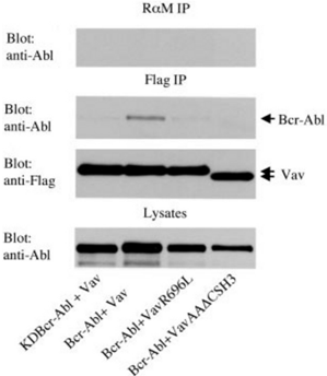
FIG. 5 The Vav SH2 and SH3 domain and a proline-rich region are required for the Vav/Bcr-Abl complex in vivo. 293 cells were transiently transfected with wild-type Bcr-Abl or kinase-defective BcrAbl ( KDBcr-Abl ) together with Flag-tagged full-length Vav ( Vav ), Flagtagged full-length Vav with a mutation in the SH2 domain ( VavR696L ), or Flag-tagged full-length Vav with a proline-to-alanine substitution of 4 proline residues and a deletion of the C-terminal SH3 domain ( VavAA 𝚫 CSH3 ). Cell lysates were prepared and immunoprecipitated ( IP ) with rabbit anti-mouse ( R 𝛂 M ) antibody as control ( upper panel ) or anti-Flag antibody to precipitate Flag-tagged Vav ( middle two panels ). Precipitated proteins and lysates ( lower panel ) were run on SDS-PAGE and visualized with anti-Abl and anti-Flag antibodies as indicated.
FIG. 5 The Vav SH2 and SH3 domain and a proline-rich region are required for the Vav/Bcr-Abl complex in vivo. 293 cells were transiently transfected with wild-type Bcr-Abl or kinase-defective BcrAbl ( KDBcr-Abl ) together with Flag-tagged full-length Vav ( Vav ), Flagtagged full-length Vav with a mutation in the SH2 domain ( VavR696L ), or Flag-tagged full-length Vav with a proline-to-alanine substitution of 4 proline residues and a deletion of the C-terminal SH3 domain ( VavAA 𝚫 CSH3 ). Cell lysates were prepared and immunoprecipitated ( IP ) with rabbit anti-mouse ( R 𝛂 M ) antibody as control ( upper panel ) or anti-Flag antibody to precipitate Flag-tagged Vav ( middle two panels ). Precipitated proteins and lysates ( lower panel ) were run on SDS-PAGE and visualized with anti-Abl and anti-Flag antibodies as indicated.CC-BY · From the PDF
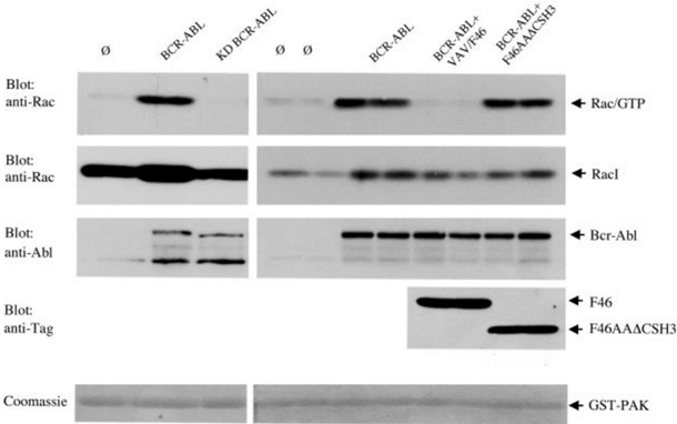
FIG. 6 Bcr-Abl utilizes Vav to activate Rac-1. 293 cells were transiently transfected with Rac-1 and empty vectors ( ø ); Rac-1, WT-Bcr-Abl, or KD-Bcr-Abl and empty vector ( Bcr-Abl, KD-Bcr-Abl ); Rac-1, Bcr-Abl, and Vav/F46 as a dominant-negative; or Rac-1, Bcr-Abl, and Vav/ F46AA 𝚫 CSH3. The amount of DNA was equal in all transfections. 72 h after transfection, lysates were prepared from 1 × 10 7 starved cells. A GST pull-down assay with the GST-p21-binding domain of PAK, which specifically precipitates Rac and Cdc42 in their GTP-bound form (43), was done in duplicates. Bound fractions were resolved by SDS-PAGE and analyzed by immunoblotting with a monoclonal anti-Rac antibody, a monoclonal anti-Abl antibody, and an anti-tag antibody to detect Flag-tagged Vav mutants as indicated. The amount of GST-PAK was analyzed by Coomassie staining of the gel. A representative experiment is shown that was done independently three times with similar results.
FIG. 6 Bcr-Abl utilizes Vav to activate Rac-1. 293 cells were transiently transfected with Rac-1 and empty vectors ( ø ); Rac-1, WT-Bcr-Abl, or KD-Bcr-Abl and empty vector ( Bcr-Abl, KD-Bcr-Abl ); Rac-1, Bcr-Abl, and Vav/F46 as a dominant-negative; or Rac-1, Bcr-Abl, and Vav/ F46AA 𝚫 CSH3. The amount of DNA was equal in all transfections. 72 h after transfection, lysates were prepared from 1 × 10 7 starved cells. A GST pull-down assay with the GST-p21-binding domain of PAK, which specifically precipitates Rac and Cdc42 in their GTP-bound form (43), was done in duplicates. Bound fractions were resolved by SDS-PAGE and analyzed by immunoblotting with a monoclonal anti-Rac antibody, a monoclonal anti-Abl antibody, and an anti-tag antibody to detect Flag-tagged Vav mutants as indicated. The amount of GST-PAK was analyzed by Coomassie staining of the gel. A representative experiment is shown that was done independently three times with similar results.CC-BY · From the PDF
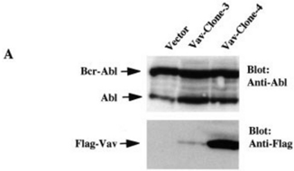
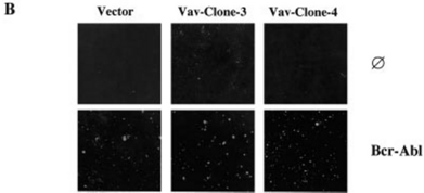
FIG. 7 Overexpression of Vav does not enhance the transformation potential of Bcr-Abl in NIH3T3 fibroblasts. A , immunoblot analysis showing the expression of Vav and Bcr-Abl proteins in stable NIH3T3 cell lines used in B . NIH3T3 cells were transfected with the pcDNA 3.1-Flag-Vav construct containing a Zeocin ® selection marker or the empty vector, respectively. Zeocin ® -resistant clones were picked, expanded, and retrovirally transduced with a Mig-Bcr-Abl construct. B , the indicated NIH3T3 cell lines were plated into soft agar, and colonies were photographed after 2 weeks.
FIG. 7 Overexpression of Vav does not enhance the transformation potential of Bcr-Abl in NIH3T3 fibroblasts. A , immunoblot analysis showing the expression of Vav and Bcr-Abl proteins in stable NIH3T3 cell lines used in B . NIH3T3 cells were transfected with the pcDNA 3.1-Flag-Vav construct containing a Zeocin ® selection marker or the empty vector, respectively. Zeocin ® -resistant clones were picked, expanded, and retrovirally transduced with a Mig-Bcr-Abl construct. B , the indicated NIH3T3 cell lines were plated into soft agar, and colonies were photographed after 2 weeks.CC-BY · From the PDF
Image 9
CC-BY · From the PDF
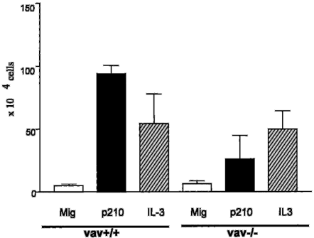
FIG. 8 Reduced transformation of Vav H11546 / H11546 compared with Vav ÷ / ÷ bone marrow by Bcr-Abl. 1 × 10 5 bone marrow cells from wild-type ( Vav + / + ) or Vav knock-out ( Vav -/ -) mice retrovirally transduced with empty vector ( Mig ) or Bcr-Abl ( p210 ) were plated in bone marrow medium as described under ' Materials and Methods. ' As a further control, growth of cells in the presence of 1 ng/ml IL-3 was measured. Bars represent the numbers of cells after 10 days of culture. Data represent means ± standard deviation of an experiment done in triplicate. Similar results were obtained in three independent experiments.
FIG. 8 Reduced transformation of Vav H11546 / H11546 compared with Vav ÷ / ÷ bone marrow by Bcr-Abl. 1 × 10 5 bone marrow cells from wild-type ( Vav + / + ) or Vav knock-out ( Vav -/ -) mice retrovirally transduced with empty vector ( Mig ) or Bcr-Abl ( p210 ) were plated in bone marrow medium as described under ' Materials and Methods. ' As a further control, growth of cells in the presence of 1 ng/ml IL-3 was measured. Bars represent the numbers of cells after 10 days of culture. Data represent means ± standard deviation of an experiment done in triplicate. Similar results were obtained in three independent experiments.CC-BY · From the PDF

Verification

Claims the paper makes

The claims and statistics of this paper haven’t been checked yet.

Run the check — free, no account needed. It reads the open-access text and checks the sources it cites.

A few free checks a day · papers already verified open instantly · results are public

Claims other papers make about this one

Each is checked against this paper’s own text: does it say what the citing paper says it does?

Self-check

Percentages, sums and reported differences re-computed from the paper’s own numbers.

The self-check needs the paper’s full text.

Citations

Cites

No references could be resolved yet.

Research works printed but not linked: 47

  1. Katzav, 1995, Oncogene, 11, 1079
  2. Adams, 1992, Oncogene, 7, 611
  3. Bustelo, 2000, Mol. Cell. Biol., 20, 1461, 10.1128/MCB.20.5.1461-1477.2000 · DOI
  4. Turner, 1997, Immunity, 7, 451, 10.1016/S1074-7613(00)80367-2 · DOI
  5. Fischer, 1998, Curr. Biol., 8, 554, 10.1016/S0960-9822(98)70224-6 · DOI
  6. Tarakhovsky, 1995, Nature, 374, 467, 10.1038/374467a0 · DOI
  7. Zhang, 1995, Nature, 374, 470, 10.1038/374470a0 · DOI
  8. Costello, 1999, Proc. Natl. Acad. Sci. U. S. A., 96, 3035, 10.1073/pnas.96.6.3035 · DOI
  9. Margolis, 1992, Nature, 356, 71, 10.1038/356071a0 · DOI
  10. Bustelo, 1992, Science, 256, 1196, 10.1126/science.256.5060.1196 · DOI
  11. Matsuguchi, 1995, EMBO J., 14, 257, 10.1002/j.1460-2075.1995.tb06999.x · DOI
  12. Alai, 1992, J. Biol. Chem., 267, 18021, 10.1016/S0021-9258(19)37146-7 · DOI
  13. Bustelo, 1992, Nature, 356, 68, 10.1038/356068a0 · DOI
  14. Crespo, 1997, Nature, 385, 169, 10.1038/385169a0 · DOI
  15. Han, 1998, Science, 279, 558, 10.1126/science.279.5350.558 · DOI
  16. Sawyers, 1999, N. Engl. J. Med., 340, 1330, 10.1056/NEJM199904293401706 · DOI
  17. Daley, 1988, Proc. Natl. Acad. Sci. U. S. A., 85, 9312, 10.1073/pnas.85.23.9312 · DOI
  18. Daley, 1990, Science, 247, 824, 10.1126/science.2406902 · DOI · Paper page
  19. McLaughlin, 1987, Proc. Natl. Acad. Sci. U. S. A., 84, 6558, 10.1073/pnas.84.18.6558 · DOI
  20. Lugo, 1989, Mol. Cell. Biol., 9, 1263
  21. Puil, 1994, EMBO J., 13, 764, 10.1002/j.1460-2075.1994.tb06319.x · DOI
  22. Pendergast, 1993, Cell, 75, 175, 10.1016/S0092-8674(05)80094-7 · DOI
  23. Bai, 1998, Oncogene, 17, 941, 10.1038/sj.onc.1202024 · DOI
  24. Coutinho, 2000, Blood, 96, 618, 10.1182/blood.V96.2.618 · DOI
  25. Cortez, 1995, Mol. Cell. Biol., 15, 5531, 10.1128/MCB.15.10.5531 · DOI
  26. Skorski, 1995, Blood, 86, 726, 10.1182/blood.V86.2.726.bloodjournal862726 · DOI
  27. Carlesso, 1996, J. Exp. Med., 183, 811, 10.1084/jem.183.3.811 · DOI
  28. Ilaria, 1996, J. Biol. Chem., 271, 31704, 10.1074/jbc.271.49.31704 · DOI
  29. Sattler, 1996, Oncogene, 12, 839
  30. Salgia, 1995, J. Biol. Chem., 270, 29145, 10.1074/jbc.270.49.29145 · DOI
  31. Oda, 1994, J. Biol. Chem., 269, 22925, 10.1016/S0021-9258(17)31596-X · DOI
  32. Shuai, 1996, Oncogene, 13, 247
  33. Jahn, 2001, J. Biol. Chem., 276, 43419, 10.1074/jbc.M107030200 · DOI
  34. Skorski, 1998, Proc. Natl. Acad. Sci. U. S. A., 95, 11858, 10.1073/pnas.95.20.11858 · DOI · Paper page
  35. Weidner, 1996, Nature, 384, 173, 10.1038/384173a0 · DOI
  36. Duyster, 1995, Proc. Natl. Acad. Sci. U. S. A., 92, 1555, 10.1073/pnas.92.5.1555 · DOI
  37. Bai, 2000, Blood, 96, 4319, 10.1182/blood.V96.13.4319 · DOI
  38. Lopez-Lago, 2000, Mol. Cell. Biol., 20, 1678, 10.1128/MCB.20.5.1678-1691.2000 · DOI
  39. Afar, 1997, Immunity, 6, 773, 10.1016/S1074-7613(00)80452-5 · DOI
  40. Songyang, 1994, Mol. Cell. Biol., 14, 2777
  41. Schindler, 2000, Science, 289, 1938, 10.1126/science.289.5486.1938 · DOI
  42. Songyang, 1995, Nature, 373, 536, 10.1038/373536a0 · DOI
  43. Benard, 1999, J. Biol. Chem., 274, 13198, 10.1074/jbc.274.19.13198 · DOI · Paper page
  44. Songyang, 1993, Cell, 72, 767, 10.1016/0092-8674(93)90404-E · DOI
  45. Kon-Kozlowski, 1996, J. Biol. Chem., 271, 3856, 10.1074/jbc.271.7.3856 · DOI
  46. Germani, 1999, Mol. Cell. Biol., 19, 3798, 10.1128/MCB.19.5.3798 · DOI
  47. Danhauser-Riedl, 1996, Cancer Res., 56, 3589

Cited by

Metadata

Authors
Bassermann, F., Jahn, T., Miething, C., Seipel, P., Bai, R.-Y., Coutinho, S., Peschel, C., Duyster, J., & Tybulewicz, V. L.
Venue
Journal of Biological Chemistry
Publisher
Elsevier BV
Published
2002-04
Volume
277
Issue
14
Pages
12437-12445
ISSN
0021-9258
Cited by
47
References
0
Language
en

About these checks

Registry, retraction, Cites and statistics results are computed, not judged, with one exception: a printed reference that no index lists is read once by a model before it counts as a citation error. Claims are extracted by a model and kept only when their quote appears verbatim in the text; each cited source is then checked, with a model’s help, for whether it says what the claim relies on. Those verdicts can be wrong — read the claim’s quote and the cited source before relying on one.

Statistics and arithmetic are recomputed, not judged.

Confidence bands are shown only for fields calibrated on our annotated test set. None is calibrated yet, so no band is shown.

What counts as a claim ›How we measure confidence ›How we check this ›

Are you an author, or do you see a problem with this page? Request a review