

The DOI matches its Crossref record; 58 of the 58 references it cites were found in a registry or an index.
Crystal Structure of the Dioxygen-bound Heme Oxygenase from Corynebacterium diphtheriae
Abstract
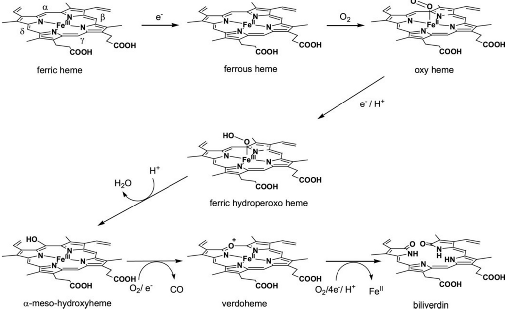
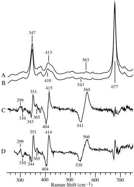
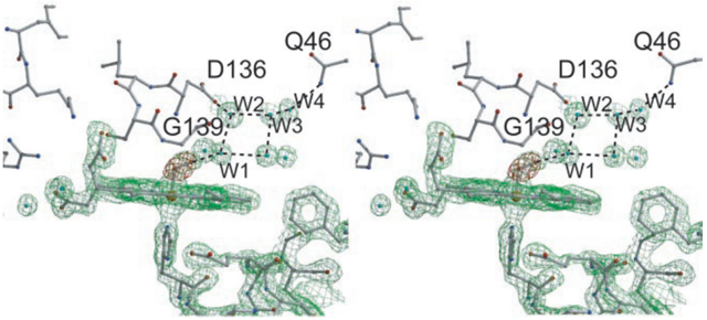
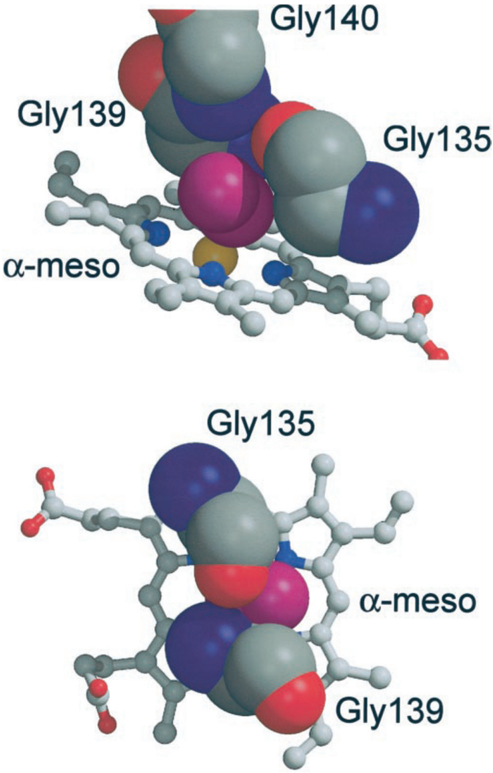
HmuO, a heme oxygenase of Corynebacterium diphtheriae, catalyzes degradation of heme using the same mechanism as the mammalian enzyme. The oxy form of HmuO, the precursor of the catalytically active ferric hydroperoxo species, has been characterized by ligand binding kinetics, resonance Raman spectroscopy, and x-ray crystallography. The oxygen association and dissociation rate constants are 5 microm(-1) s(-1) and 0.22 s(-1), respectively, yielding an O(2) affinity of 21 microm(-1), which is approximately 20 times greater than that of mammalian myoglobins. However, the affinity of HmuO for CO is only 3-4-fold greater than that for mammalian myoglobins, implying the presence of strong hydrogen bonding interactions in the distal pocket of HmuO that preferentially favor O(2) binding. Resonance Raman spectra show that the Fe-O(2) vibrations are tightly coupled to porphyrin vibrations, indicating the highly bent Fe-O-O geometry that is characteristic of the oxy forms of heme oxygenases. In the crystal structure of the oxy form the Fe-O-O angle is 110 degrees, the O-O bond is pointed toward the heme alpha-meso-carbon by direct steric interactions with Gly-135 and Gly-139, and hydrogen bonds occur between the bound O(2) and the amide nitrogen of Gly-139 and a distal pocket water molecule, which is a part of an extended hydrogen bonding network that provides the solvent protons required for oxygen activation. In addition, the O-O bond is orthogonal to the plane of the proximal imidazole side chain, which facilitates hydroxylation of the porphyrin alpha-meso-carbon by preventing premature O-O bond cleavage.
Overview
The claims and statistics of this paper haven’t been checked yet.
Run the check — free, no account needed. It reads the open-access text and checks the sources it cites.
Integrity signals
Registry
Cites
Statistics
Figures
Verification
Claims the paper makes
The claims and statistics of this paper haven’t been checked yet.
Run the check — free, no account needed. It reads the open-access text and checks the sources it cites.
Claims other papers make about this one
Each is checked against this paper’s own text: does it say what the citing paper says it does?
The cited source says what the claim attributes to it.
In the citing paper
“In the presence of an electron donor, the canonical HO proteins degrade heme to free iron, CO, and 𝛂 -biliverdin (120).”
From Overcoming the Heme Paradox: Heme Toxicity and Tolerance in Bacterial Pathogens (2010) · cites this paper as [120]
Self-check
Percentages, sums and reported differences re-computed from the paper’s own numbers.
The self-check needs the paper’s full text.
Citations
Cites
[20] Processing of X-ray diffraction data collected in oscillation mode
Otwinowski, Z., & Minor, W. (1997) · Methods in Enzymology · doi:10.1016/s0076-6879(97)76066-x · Cited by 33,475
The CCP4 suite: programs for protein crystallography
Collaborative Computational Project, N. 4. (1994) · Acta Crystallographica Section D Biological Crystallography · doi:10.1107/s0907444994003112 · Cited by 17,270
Crystallography & NMR System: A New Software Suite for Macromolecular Structure Determination
Brünger, A. T., Adams, P. D., Clore, G. M., DeLano, W. L., Gros, P., Grosse-Kunstleve, R. W., Jiang, J. S., Kuszewski, J., Nilges, M., Pannu, N. S., et al. (1998) · Acta Crystallographica Section D Biological Crystallography · doi:10.1107/s0907444998003254 · Cited by 15,715
MOLSCRIPT: a program to produce both detailed and schematic plots of protein structures
Kraulis, P. J. (1991) · Journal of Applied Crystallography · doi:10.1107/s0021889891004399 · Cited by 14,085
THE HEME OXYGENASE SYSTEM:A Regulator of Second Messenger Gases
Maines, M. D. (1997) · The Annual Review of Pharmacology and Toxicology · doi:10.1146/annurev.pharmtox.37.1.517 · Cited by 2,501
Sono, M., Roach, M. P., Coulter, E. D., & Dawson, J. H. (1996) · Chemical Reviews · doi:10.1021/cr9500500 · Cited by 2,477
The Catalytic Pathway of Cytochrome P450cam at Atomic Resolution
Schlichting, I., Berendzen, J., Chu, K., Stock, A., Maves, S. A., Benson, D. E., Sweet, R. M., Ringe, D., Petsko, G. A., & Sligar, S. G. (2000) · Science · doi:10.1126/science.287.5458.1615 · Cited by 1,349
Heme oxygenase 1 is required for mammalian iron reutilization
Poss, K. D., & Tonegawa, S. (1997) · Proceedings of the National Academy of Sciences · doi:10.1073/pnas.94.20.10919 · Cited by 1,016
The catalytic pathway of horseradish peroxidase at high resolution
Berglund, G. I., Carlsson, G. H., Smith, A. T., Szöke, H., Henriksen, A., & Hajdu, J. (2002) · Nature · doi:10.1038/417463a · Cited by 956
On the rigid-body motion of molecules in crystals
Schomaker, V., & Trueblood, K. N. (1968) · Acta Crystallographica Section B · doi:10.1107/s0567740868001718 · Cited by 941
Mechanisms of Ligand Recognition in Myoglobin
Springer, B. A., Sligar, S. G., Olson, J. S., & Phillips, G. N. J. (1994) · Chemical Reviews · doi:10.1021/cr00027a007 · Cited by 755
Self-citationEsnouf, R. M. (1999) · Acta Crystallographica Section D Biological Crystallography · doi:10.1107/s0907444998017363 · Cited by 753
Rapid automated molecular replacement by evolutionary search
Kissinger, C. R., Gehlhaar, D. K., & Fogel, D. B. (1999) · Acta Crystallographica Section D Biological Crystallography · doi:10.1107/s0907444998012517 · Cited by 578
Crystal Structures of Myoglobin-Ligand Complexes at Near-Atomic Resolution
Vojtěchovský, J., Chu, K., Berendzen, J., Sweet, R. M., & Schlichting, I. (1999) · Biophysical Journal · doi:10.1016/s0006-3495(99)77056-6 · Cited by 561
NPAS2: A Gas-Responsive Transcription Factor
Dioum, E. M., Rutter, J. P., Tuckerman, J. R., Gonzalez, G., Gilles‐Gonzalez, M., & McKnight, S. L. (2002) · Science · doi:10.1126/science.1078456 · Cited by 432
Bacterial heme sources: the role of heme, hemoprotein receptors and hemophores
Wandersman, C., & Stojiljkovic, I. (2000) · Current Opinion in Microbiology · doi:10.1016/s1369-5274(00)00078-3 · Cited by 318
Crystal structure of human heme oxygenase-1.
Poulos, T. L., Schuller, D. J., Wilks, A., & de Montellano, P. R. O. (1999) · Nature Structural Biology · doi:10.1038/12319 · Cited by 303
Myoglobin discriminates between O2, NO, and CO by electrostatic interactions with the bound ligand
Olson, J. S., & Phillips, G. N. (1997) · JBIC Journal of Biological Inorganic Chemistry · doi:10.1007/s007750050169 · Cited by 287
Self-citationRohlfs, R. J., Mathews, A. J., Carver, T. E., Olson, J. S., Springer, B. A., Egeberg, K. D., & Sligar, S. G. (1990) · Journal of Biological Chemistry · doi:10.1016/s0021-9258(19)39749-2 · Cited by 286
Self-citationSchacter, B. A., Nelson, E. B., Marver, H. S., & Masters, B. S. S. (1972) · Journal of Biological Chemistry · doi:10.1016/s0021-9258(19)45183-1 · Cited by 253
Emerging strategies in microbial haem capture
Genco, C. A., & Dixon, D. W. (2001) · Molecular Microbiology · doi:10.1046/j.1365-2958.2001.02231.x · Cited by 245
Bound CO Is A Molecular Probe of Electrostatic Potential in the Distal Pocket of Myoglobin
Phillips, G. N., Jr., Teodoro, M. L., Li, T., Smith, B., & Olson, J. S. (1999) · The Journal of Physical Chemistry B · doi:10.1021/jp9918205 · Cited by 240
Self-citationSchmitt, M. P. (1997) · Journal of Bacteriology · doi:10.1128/jb.179.3.838-845.1997 · Cited by 234
The mechanism of heme oxygenase
de Montellano, P. R. O. (2000) · Current Opinion in Chemical Biology · doi:10.1016/s1367-5931(99)00079-4 · Cited by 223
Folding of cytochrome c initiated by submillisecond mixing
Takahashi, S., Yeh, S., Das, T. K., Chan, C.-K., Gottfried, D. S., & Rousseau, D. L. (1997) · Nature Structural & Molecular Biology · doi:10.1038/nsb0197-44 · Cited by 194
Self-citationZhu, W., Wilks, A., & Stojiljkovic, I. (2000) · Journal of Bacteriology · doi:10.1128/jb.182.23.6783-6790.2000 · Cited by 186
Davydov, R. M., Kofman, V., Fujii, H., Yoshida, T., Ikeda‐Saito, M., & Hoffman, B. M. (2002) · Journal of the American Chemical Society · doi:10.1021/ja0122391 · Cited by 166
Self-citationAutocatalytic radical reactions in physiological prosthetic heme modification
Colas, C., & Ortiz de Montellano, P. R. (2003) · Chemical Reviews · doi:10.1021/cr0204303 · Cited by 139
Characterization of recombinant soybean leghemoglobin a and apolar distal histidine mutants
Hargrove, M. S., Barry, J. K., Brucker, E. A., Berry, M. B., Phillips, G. N., Olson, J. S., Arredondo‐Peter, R., Dean, J., Klucas, R. V., & Sarath, G. (1997) · Journal of Molecular Biology · doi:10.1006/jmbi.1996.0833 · Cited by 139
Self-citationThe heme oxygenase-carbon monoxide system: u regulator of hepatobiliary function
Suematsu, M., & Ishimura, Y. (2000) · Hepatology · doi:10.1002/hep.510310102 · Cited by 127
Schuller, D. J., Zhu, W., Stojiljković, I., Wilks, A., & Poulos, T. L. (2001) · Biochemistry · doi:10.1021/bi0110239 · Cited by 127
Preferred orientation of imidazole ligands in metalloporphyrins
Scheidt, W. R., & Chipman, D. M. (1986) · Journal of the American Chemical Society · doi:10.1021/ja00266a008 · Cited by 123
Davydov, R. M., Yoshida, T., Ikeda‐Saito, M., & Hoffman, B. M. (1999) · Journal of the American Chemical Society · doi:10.1021/ja992425v · Cited by 120
Self-citationYang, J., Kloek, A. P., Goldberg, D. E., & Mathews, F. S. (1995) · Proceedings of the National Academy of Sciences · doi:10.1073/pnas.92.10.4224 · Cited by 119
Fujii, H., Zhang, X., Tomita, T., Ikeda‐Saito, M., & Yoshida, T. (2001) · Journal of the American Chemical Society · doi:10.1021/ja010490a · Cited by 110
Self-citationHirotsu, S., Chu, G. C., Unno, M., Lee, D.-S., Yoshida, T., Park, S., Shiro, Y., & Ikeda‐Saito, M. (2004) · Journal of Biological Chemistry · doi:10.1074/jbc.m311631200 · Cited by 109
Self-citationControlling Ligand Binding in Myoglobin by Mutagenesis
Draghi, F., Miele, A. E., Travaglini‐Allocatelli, C., Vallone, B., Brunori, M., Gibson, Q. H., & Olson, J. S. (2002) · Journal of Biological Chemistry · doi:10.1074/jbc.m109206200 · Cited by 107
Self-citationMatera, K. M., Takahashi, S., Fujii, H., Zhou, H., Ishikawa, K., Yoshimura, T., Rousseau, D. L., Yoshida, T., & Ikeda‐Saito, M. (1996) · Journal of Biological Chemistry · doi:10.1074/jbc.271.12.6618 · Cited by 104
Self-citationBAERE, I. D., Perutz, M. F., Kiger, L., et al. (1994) · Proceedings of the National Academy of Sciences · doi:10.1073/pnas.91.4.1594 · Cited by 93
Takahashi, S., Ishikawa, K., Takeuchi, N., Ikeda‐Saito, M., Yoshida, T., & Rousseau, D. L. (1995) · Journal of the American Chemical Society · doi:10.1021/ja00127a013 · Cited by 89
Self-citationThe Oxygen and Carbon Monoxide Reactions of Heme Oxygenase
Migita, C. T., Matera, K. M., Ikeda‐Saito, M., et al. (1998) · Journal of Biological Chemistry · doi:10.1074/jbc.273.2.945 · Cited by 82
Self-citationChu, G. C., Katakura, K., Zhang, X., Yoshida, T., & Ikeda‐Saito, M. (1999) · Journal of Biological Chemistry · doi:10.1074/jbc.274.30.21319 · Cited by 80
Self-citationRates of reaction of Ascaris haemoglobins with ligands
Gibson, Q. H., & Smith, M. H. (1965) · Proceedings of the Royal Society B Biological Sciences · doi:10.1098/rspb.1965.0067 · Cited by 80
Smerdon, S. J., Dodson, G. G., Wilkinson, A. J., Gibson, Q. H., Blackmore, R. S., Carver, T. E., & Olson, J. S. (1991) · Biochemistry · doi:10.1021/bi00239a025 · Cited by 77
Self-citationThe Structure of Deoxy- and Oxy-leghaemoglobin from Lupin
Harutyunyan, E. H., Safonova, T. N., Kuranova, I. P., Popov, A. N., Teplyakov, A., Obmolova, G., Русаков, А. А., Vainshtein, B. K., Dodson, G. G., Wilson, J. C., et al. (1995) · Journal of Molecular Biology · doi:10.1006/jmbi.1995.0419 · Cited by 77
Davydov, R. M., Matsui, T., Fujii, H., Ikeda‐Saito, M., & Hoffman, B. M. (2003) · Journal of the American Chemical Society · doi:10.1021/ja038923s · Cited by 75
Self-citationLad, L., Wang, J., Li, H., et al. (2003) · Journal of Molecular Biology · doi:10.1016/s0022-2836(03)00578-3 · Cited by 68
High Resolution Crystal Structures of the Deoxy, Oxy, and Aquomet Forms of Cobalt Myoglobin
Brucker, E. A., Olson, J. S., Phillips, G. N., Dou, Y., & Ikeda‐Saito, M. (1996) · Journal of Biological Chemistry · doi:10.1074/jbc.271.41.25419 · Cited by 66
Self-citationSugishima, M., Sakamoto, H., Kakuta, Y., et al. (2002) · Biochemistry · doi:10.1021/bi025662a · Cited by 63
The Ferrous Dioxygen Complex of the Oxygenase Domain of Neuronal Nitric-oxide Synthase
Couture, M., Stuehr, D. J., & Rousseau, D. L. (2000) · Journal of Biological Chemistry · doi:10.1074/jbc.275.5.3201 · Cited by 59
Self-citationThe Heme Complex of Hmu O, a Bacterial Heme Degradation Enzyme from Corynebacterium diphtheriae
Chu, G. C., Sönnichsen, F. D., Ikeda‐Saito, M., Tomita, T., & Yoshida, T. (1999) · Journal of Biological Chemistry · doi:10.1074/jbc.274.35.24490 · Cited by 46
Self-citationCrystal structure of oxygenated Scapharca dimeric hemoglobin at 1.7-A resolution.
Condon, P. J., & Royer, W. E. (1994) · Journal of Biological Chemistry · doi:10.1016/s0021-9258(18)47241-9 · Cited by 39
CRYSTAL STRUCTURE OF OXYGENATED SCAPHARCA DIMERIC HEMOGLOBIN AT 1.7 ANGSTROMS RESOLUTION
Royer, W. E., & Condon, P. J. (1994) · Worldwide Protein Data Bank · doi:10.2210/pdb1hbi/pdb · Cited by 38
Stabilizing Bound O2 in Myoglobin by Valine68 (E11) to Asparagine Substitution,
Krzywda, S., Murshudov, G. N., Brzozowski, A. M., Jaskólski, M., Scott, E. E., Klizas, S. A., Gibson, Q. H., Olson, J. S., & Wilkinson, A. J. (1998) · Biochemistry · doi:10.1021/bi9812470 · Cited by 37
Self-citationDenisov, I. G., Ikeda‐Saito, M., Yoshida, T., & Sligar, S. G. (2002) · FEBS Letters · doi:10.1016/s0014-5793(02)03674-8 · Cited by 37
Self-citationZhang, X., Fujii, H., Matera, K. M., et al. (2003) · Biochemistry · doi:10.1021/bi027173g · Cited by 30
Cobalt Porphyrin Heme Oxygenase Complex. EPR Evidences for the Distal Heme Pocket Hydrogen Bonding
Fujii, H., Dou, Y., Zhou, H., et al. (1998) · Journal of the American Chemical Society · doi:10.1021/ja973925w · Cited by 25
Tsuchiya, K., Kawano, Y., Kojima, T., Nagata, K., Takao, T., Okada, M., Shinohara, H., Maki, K., Toyama‐Sorimachi, N., Miyasaka, N., et al. (2003) · FEBS Letters · doi:10.1016/s0014-5793(03)00127-3 · Cited by 2
Research works printed but not linked: 2
- Ikeda-Saito, 2003, 858, 97
- Jones, 1991, Acta Crystallogr. Sect. A, 47, 100
Cited by
Structure and chemistry of cytochrome p450
Denisov, I. G., Makris, T. M., Sligar, S. G., & Schlichting, I. (2005) · Chemical Reviews · doi:10.1021/cr0307143 · Cited by 2,106
Heme Enzyme Structure and Function
Poulos, T. L. (2014) · Chemical Reviews · doi:10.1021/cr400415k · Cited by 1,443
Overcoming the Heme Paradox: Heme Toxicity and Tolerance in Bacterial Pathogens
Anzaldi, L. L., & Skaar, E. P. (2010) · Infection and Immunity · doi:10.1128/iai.00613-10 · Cited by 295
Wu, R., Skaar, E. P., Zhang, R., Joachimiak, G., Gornicki, P., Schneewind, O., & Joachimiak, A. (2005) · Journal of Biological Chemistry · doi:10.1074/jbc.m409526200 · Cited by 144
The IsdG‐family of haem oxygenases degrades haem to a novel chromophore
Reniere, M. L., Ukpabi, G. N., Harry, S. R., Stec, D. F., Krull, R., Wright, D. W., Bachmann, B. O., Murphy, M. E., & Skaar, E. P. (2010) · Molecular Microbiology · doi:10.1111/j.1365-2958.2010.07076.x · Cited by 139
Metadata
- Authors
- Unno, M., Matsui, T., Chu, G. C., Couture, M., Yoshida, T., Rousseau, D. L., Olson, J. S., & Ikeda-Saito, M.
- Venue
- Journal of Biological Chemistry
- Publisher
- Elsevier BV
- Published
- 2004-05
- Volume
- 279
- Issue
- 20
- Pages
- 21055-21061
- ISSN
- 0021-9258
- Cited by
- 83
- References
- 58
- Language
- en
- Licence
- www.elsevier.com/tdm/userlicense/1.0/ (tdm)
- creativecommons.org/licenses/by/4.0/ (vor, 5988 d)
About these checks
Registry, retraction, Cites and statistics results are computed, not judged, with one exception: a printed reference that no index lists is read once by a model before it counts as a citation error. Claims are extracted by a model and kept only when their quote appears verbatim in the text; each cited source is then checked, with a model’s help, for whether it says what the claim relies on. Those verdicts can be wrong — read the claim’s quote and the cited source before relying on one.
Statistics and arithmetic are recomputed, not judged.
Confidence bands are shown only for fields calibrated on our annotated test set. None is calibrated yet, so no band is shown.
What counts as a claim ›How we measure confidence ›How we check this ›
Are you an author, or do you see a problem with this page? Request a review
Data sources
Bibliographic data: Semantic Scholar, OpenAlex, Crossref.
Contains information from the Semantic Scholar Academic Graph Datasets, made available under the ODC Attribution License (ODC-BY 1.0).
Semantic Scholar’s data is described in: Kinney, R., et al. (2023). The Semantic Scholar Open Data Platform. arXiv:2301.10140.
