

The DOI matches its Crossref record; 34 of the 34 references it cites were found in a registry or an index.
Guanidinylated Neomycin Delivers Large, Bioactive Cargo into Cells through a Heparan Sulfate-dependent Pathway
Abstract
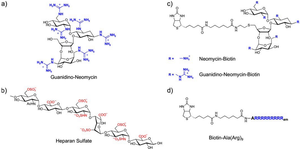
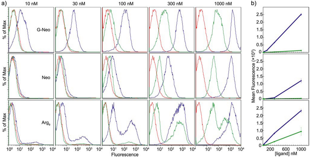
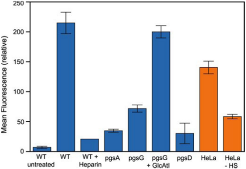
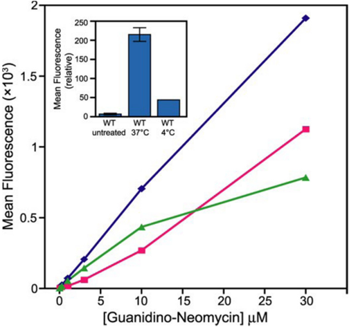
Facilitating the uptake of molecules into living cells is of substantial interest for basic research and drug delivery applications. Arginine-rich peptides have been shown to facilitate uptake of high molecular mass cargos into cells, but the mechanism of uptake is complex and may involve multiple receptors. In this report, we show that a derivative of the aminoglycoside antibiotic neomycin, in which all of the ammonium groups have been converted into guanidinium groups, can carry large (>300 kDa) bioactive molecules across cell membranes. Delivery occurs at nanomolar transporter concentrations and under these conditions depends entirely on cell surface heparan sulfate proteoglycans. Conjugation of guanidinoneomycin to the plant toxin saporin, a ribosome-inactivating agent, results in proteoglycan-dependent cell toxicity. In contrast, an arginine-rich peptide shows both heparan sulfate-dependent and -independent cellular uptake. The high selectivity of guanidinoneomycin for heparan sulfate suggests the possibility of exploiting differences in proteoglycan compositions to target delivery to different cell types.
Overview
The claims and statistics of this paper haven’t been checked yet.
Run the check — free, no account needed. It reads the open-access text and checks the sources it cites.
Integrity signals
Registry
Cites
Statistics
Figures

Verification
Claims the paper makes
The claims and statistics of this paper haven’t been checked yet.
Run the check — free, no account needed. It reads the open-access text and checks the sources it cites.
Claims other papers make about this one
Each is checked against this paper’s own text: does it say what the citing paper says it does?
The source backs part of the claim, not all of it — or backs it for a different population, condition or comparison.
In the citing paper
“Finally, a large body data indicates that proteoglycans can act as independent endocytic receptors for a variety of ligands, both natural and synthetic (5, 64, 65).”
From Syndecan-1 is the primary heparan sulfate proteoglycan mediating hepatic clearance of triglyceride-rich lipoproteins in mice (2009) · cites this paper as [65]
Experimental — accuracy not yet measured
Self-check
Percentages, sums and reported differences re-computed from the paper’s own numbers.
The self-check needs the paper’s full text.
Citations
Cites
Goodman & Gilman's The Pharmacological Basis of Therapeutics
Brunton, L. L. (1985) · Cited by 7,321
Cellular uptake of the tat protein from human immunodeficiency virus
Frankel, A. D., & Pabo, C. O. (1988) · Cell · doi:10.1016/0092-8674(88)90263-2 · Cited by 2,783
Yayon, A., Klagsbrun, M., Esko, J. D., Leder, P., & Ornitz, D. M. (1991) · Cell · doi:10.1016/0092-8674(91)90512-w · Cited by 2,440
Self-citationWender, P. A., Mitchell, D. J., Pattabiraman, K., Pelkey, E. T., Steinman, L., & Rothbard, J. B. (2000) · Proceedings of the National Academy of Sciences · doi:10.1073/pnas.97.24.13003 · Cited by 1,615
Green, M., & Loewenstein, P. M. (1988) · Cell · doi:10.1016/0092-8674(88)90262-0 · Cited by 1,553
Requirement of Heparan Sulfate for bFGF-Mediated Fibroblast Growth and Myoblast Differentiation
Rapraeger, A. C., Krufka, A., & Olwin, B. B. (1991) · Science · doi:10.1126/science.1646484 · Cited by 1,508
Order Out of Chaos: Assembly of Ligand Binding Sites in Heparan Sulfate
Esko, J. D., & Selleck, S. B. (2002) · Annual Review of Biochemistry · doi:10.1146/annurev.biochem.71.110601.135458 · Cited by 1,495
Self-citationInternalization of HIV-1 Tat Requires Cell Surface Heparan Sulfate Proteoglycans
Tyagi, M., Rusnati, M., Presta, M., & Giacca, M. (2001) · Journal of Biological Chemistry · doi:10.1074/jbc.m006701200 · Cited by 719
Cationic TAT peptide transduction domain enters cells by macropinocytosis
Kaplan, I. M., Wadia, J. S., & Dowdy, S. F. (2005) · Journal of Controlled Release · doi:10.1016/j.jconrel.2004.10.018 · Cited by 668
Rothbard, J. B., Jessop, T. C., Lewis, R. S., Murray, B. A., & Wender, P. A. (2004) · Journal of the American Chemical Society · doi:10.1021/ja0482536 · Cited by 624
Richard, J. P., Melikov, K., Brooks, H., Prevot, P., Lebleu, B., & Chernomordik, L. V. (2005) · Journal of Biological Chemistry · doi:10.1074/jbc.m401604200 · Cited by 604
Animal cell mutants defective in glycosaminoglycan biosynthesis.
Esko, J. D., Stewart, T. E., & Taylor, W. H. (1985) · Proceedings of the National Academy of Sciences · doi:10.1073/pnas.82.10.3197 · Cited by 513
Self-citationDelivery of bioactive molecules into the cell: the Trojan horse approach
Dietz, G. P. H., & Bähr, M. (2004) · Molecular and Cellular Neuroscience · doi:10.1016/j.mcn.2004.03.005 · Cited by 469
WADIA, J., & DOWDY, S. (2005) · Advanced Drug Delivery Reviews · doi:10.1016/j.addr.2004.10.005 · Cited by 428
Membrane-permeable arginine-rich peptides and the translocation mechanisms
FUTAKI, S. (2005) · Advanced Drug Delivery Reviews · doi:10.1016/j.addr.2004.10.009 · Cited by 406
Pathway for Polyarginine Entry into Mammalian Cells
Fuchs, S. M., & Raines, R. T. (2004) · Biochemistry · doi:10.1021/bi035933x · Cited by 374
Lidholt, K., Weinke, J. L., Kiser, C. S., Lugemwa, F. N., Bame, K. J., Cheifetz, S., Massagué, J., Lindahl, U., & Esko, J. D. (1992) · Proceedings of the National Academy of Sciences · doi:10.1073/pnas.89.6.2267 · Cited by 295
Self-citationRyser, H. J.-P. (1968) · Science · doi:10.1126/science.159.3813.390 · Cited by 288
ROTHBARD, J., JESSOP, T., & WENDER, P. (2005) · Advanced Drug Delivery Reviews · doi:10.1016/j.addr.2004.10.003 · Cited by 286
The emergence of clathrin-independent pinocytic pathways
Lamaze, C., & Schmid, S. L. (1995) · Current Opinion in Cell Biology · doi:10.1016/0955-0674(95)80015-8 · Cited by 283
Deutscher, S. L., Nuwayhid, N., Stanley, P., Briles, E. I. B., & Hirschberg, C. B. (1984) · Cell · doi:10.1016/0092-8674(84)90007-2 · Cited by 265
Translocation of a β-Peptide Across Cell Membranes
Umezawa, N., Gelman, M. A., Haigis, M. C., Raines, R. T., & Gellman, S. H. (2001) · Journal of the American Chemical Society · doi:10.1021/ja017283v · Cited by 226
Oligoarginine vectors for intracellular delivery: Design and cellular‐uptake mechanisms
Futaki, S. (2005) · Biopolymers · doi:10.1002/bip.20421 · Cited by 190
Cell Penetrating Agents Based on a Polyproline Helix Scaffold
Fillon, Y. A., Anderson, J. P., & Chmielewski, J. A. (2005) · Journal of the American Chemical Society · doi:10.1021/ja052377g · Cited by 164
Futaki, S., Nakase, I., Suzuki, T. W., Zhang, & Sugiura, Y. (2002) · Biochemistry · doi:10.1021/bi0256173 · Cited by 163
Cellular Uptake of Aminoglycosides, Guanidinoglycosides, and Poly-arginine
Luedtke, N. W., Carmichael, P., & Tor, Y. (2003) · Journal of the American Chemical Society · doi:10.1021/ja0360135 · Cited by 158
Self-citationFernández‐Carneado, J., Gool, M. V., Martos, V., Castel, S., Prados, P., de Mendoza, J., & Giralt, E. (2004) · Journal of the American Chemical Society · doi:10.1021/ja044006q · Cited by 125
Cell-surface heparan sulfate proteoglycans: dynamic molecules mediating ligand catabolism
Jon Williams, K., & Fuki, I. V. (1997) · Current Opinion in Lipidology · doi:10.1097/00041433-199710000-00003 · Cited by 113
Guanidinoglycosides: A Novel Family of RNA Ligands
Luedtke, N. W., Baker, T. J., Goodman, M., & Tor, Y. (2000) · Journal of the American Chemical Society · doi:10.1021/ja002421m · Cited by 105
Self-citationBai, X., Wei, G., Sinha, A., & Esko, J. D. (1999) · Journal of Biological Chemistry · doi:10.1074/jbc.274.19.13017 · Cited by 105
Self-citationCaesar, C. E. B., Esbjörner, E. K., Lincoln, P., & Nordén, B. (2006) · Biochemistry · doi:10.1021/bi052095t · Cited by 101
Hitz, T., Iten, R., Gardiner, J., Namoto, K., Walde, P., & Seebàch, D. (2006) · Biochemistry · doi:10.1021/bi060285d · Cited by 74
Wei, G., Bai, X., Sarkar, A. K., & Esko, J. D. (1999) · Journal of Biological Chemistry · doi:10.1074/jbc.274.12.7857 · Cited by 59
Self-citationDendritic oligoguanidines as intracellular translocators
Chung, H., Harms, G., Seong, C. M., Choi, B. H., Min, C., Taulane, J. P., & Goodman, M. (2004) · Biopolymers · doi:10.1002/bip.10597 · Cited by 58
Research works printed but not linked: 1
- Flavell, 1998, Curr. Top Microbiol. Immunol., 234, 57
Cited by
Stanford, K. I., Bishop, J. R., Foley, E. M., Gonzales, J. C., Niesman, I. R., Witztum, J. L., & Esko, J. D. (2009) · Journal of Clinical Investigation · doi:10.1172/jci38251 · Cited by 226
Self-citationFoley, E. M., Gordts, P. L. S. M., Stanford, K. I., Gonzales, J. C., Lawrence, R., Stoddard, N., & Esko, J. D. (2013) · Arteriosclerosis, Thrombosis, and Vascular Biology · doi:10.1161/atvbaha.113.301637 · Cited by 77
Self-citation
Metadata
- Authors
- Elson-Schwab, L., Garner, O. B., Schuksz, M., Crawford, B. E., Esko, J. D., & Tor, Y.
- Venue
- Journal of Biological Chemistry
- Publisher
- Elsevier BV
- Published
- 2007-05
- Volume
- 282
- Issue
- 18
- Pages
- 13585-13591
- ISSN
- 0021-9258
- Cited by
- 68
- References
- 34
- Language
- en
- Licence
- www.elsevier.com/tdm/userlicense/1.0/ (tdm)
- creativecommons.org/licenses/by/4.0/ (vor, 4932 d)
About these checks
Registry, retraction, Cites and statistics results are computed, not judged, with one exception: a printed reference that no index lists is read once by a model before it counts as a citation error. Claims are extracted by a model and kept only when their quote appears verbatim in the text; each cited source is then checked, with a model’s help, for whether it says what the claim relies on. Those verdicts can be wrong — read the claim’s quote and the cited source before relying on one.
Statistics and arithmetic are recomputed, not judged.
Confidence bands are shown only for fields calibrated on our annotated test set. None is calibrated yet, so no band is shown.
What counts as a claim ›How we measure confidence ›How we check this ›
Are you an author, or do you see a problem with this page? Request a review
Data sources
Bibliographic data: Semantic Scholar, OpenAlex, Crossref.
Contains information from the Semantic Scholar Academic Graph Datasets, made available under the ODC Attribution License (ODC-BY 1.0).
Semantic Scholar’s data is described in: Kinney, R., et al. (2023). The Semantic Scholar Open Data Platform. arXiv:2301.10140.
