Cited by 50

The DOI matches its Crossref record.

Active Plasma Kallikrein Localizes to Mast Cells and Regulates Epithelial Cell Apoptosis, Adipocyte Differentiation, and Stromal Remodeling during Mammary Gland Involution

Lilla, Jennifer N., Joshi, Ravi V., Craik, Charles S., Werb, Zena·2009·Journal of Biological Chemistry

Overview

The claims and statistics of this paper haven’t been checked yet.

Run the check — free, no account needed. It reads the open-access text and checks the sources it cites.

A few free checks a day · papers already verified open instantly · results are public

Integrity signals

Registry

Crossref · 10.1074/jbc.m900508200

Cites

Statistics

Figures

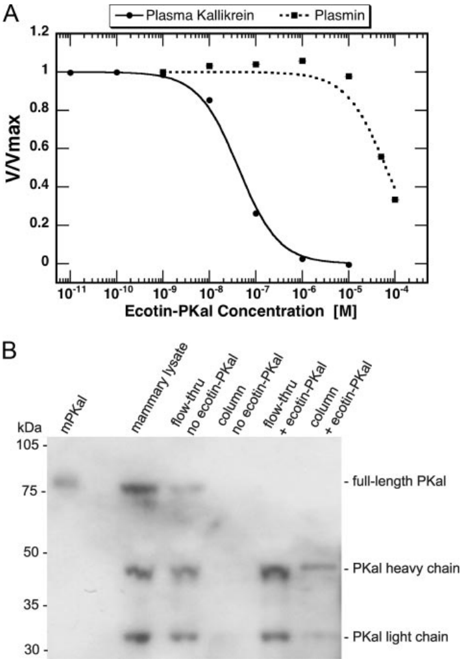
FIGURE 1 Ecotin-PKal inhibits mouse plasma kallikrein and binds PKal isolated from mammary gland lysates. A , binding assays were performed to determine Ki values for ecotin-PKal to mouse PKal or plasmin. After a 4-h incubation at 37 °C, remaining protease activity was measured using a p -nitroanilide substrate (see 'Experimental Procedures'). Reaction velocities were averaged over three trials and normalized to the baseline reaction velocity. The apparent Ki of ecotin-PKal for mouse PKal was determined to be 40nM;the Ki for mouseplasminwasdeterminedtobe60 𝛍 M. B , protein lysates from 4-day-involuting mammary glands were run over either streptavidinonly beads ( no ecotin-PKal ) or streptavidin beads bound with biotinylated ecotin-PKal ( + ecotin-PKal ). Flow-thru from the columns was collected, and bead-bound proteins were eluted by boiling the beads in sample buffer. All samples were subjected to electrophoresis and Western blotting using an antibody against mouse prekallikrein ( 𝛂 -mouse KLKB1, R&D Systems). The bands likely represent full-length PKal ( ∼ 80 kDa), its heavy chain (42 kDa, unglycosylated), and its light chain (28 kDa, unglycosylated). mPKal , mouse prekallikrein peptide, R&D Systems), against which the antibody was raised.
FIGURE 1 Ecotin-PKal inhibits mouse plasma kallikrein and binds PKal isolated from mammary gland lysates. A , binding assays were performed to determine Ki values for ecotin-PKal to mouse PKal or plasmin. After a 4-h incubation at 37 °C, remaining protease activity was measured using a p -nitroanilide substrate (see 'Experimental Procedures'). Reaction velocities were averaged over three trials and normalized to the baseline reaction velocity. The apparent Ki of ecotin-PKal for mouse PKal was determined to be 40nM;the Ki for mouseplasminwasdeterminedtobe60 𝛍 M. B , protein lysates from 4-day-involuting mammary glands were run over either streptavidinonly beads ( no ecotin-PKal ) or streptavidin beads bound with biotinylated ecotin-PKal ( + ecotin-PKal ). Flow-thru from the columns was collected, and bead-bound proteins were eluted by boiling the beads in sample buffer. All samples were subjected to electrophoresis and Western blotting using an antibody against mouse prekallikrein ( 𝛂 -mouse KLKB1, R&D Systems). The bands likely represent full-length PKal ( ∼ 80 kDa), its heavy chain (42 kDa, unglycosylated), and its light chain (28 kDa, unglycosylated). mPKal , mouse prekallikrein peptide, R&D Systems), against which the antibody was raised.CC-BY · From the PDF
Image 2
CC-BY · From the PDF
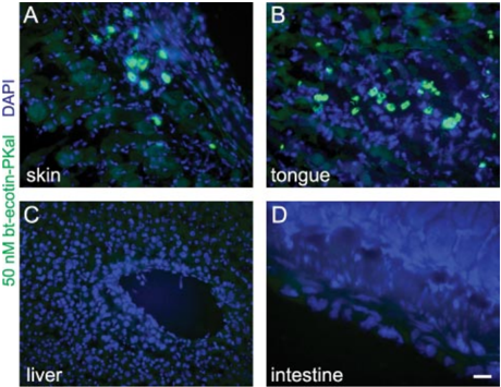
FIGURE 3 Biotinylated ecotin-PKal stains tissues with connective tissuetype mast cells but not liver. Frozen sections using 50 nM ecotin-PKal plus Alexa488-conjugatedstreptavidin,DAPIcounterstain. Scalebar for A-D is 100 𝛍 m. Active PKal is seen in mast cells of the skin ( A ) and tongue ( B ), but not in liver ( C ) or intestine ( D ). Active PKal cannot be seen in liver, where prekallikrein is synthesized but not activated, and therefore is not bound by ecotin-PKal. Active PKal is not seen in the small intestine, where mast cells are of the mucosal subtype.
FIGURE 3 Biotinylated ecotin-PKal stains tissues with connective tissuetype mast cells but not liver. Frozen sections using 50 nM ecotin-PKal plus Alexa488-conjugatedstreptavidin,DAPIcounterstain. Scalebar for A-D is 100 𝛍 m. Active PKal is seen in mast cells of the skin ( A ) and tongue ( B ), but not in liver ( C ) or intestine ( D ). Active PKal cannot be seen in liver, where prekallikrein is synthesized but not activated, and therefore is not bound by ecotin-PKal. Active PKal is not seen in the small intestine, where mast cells are of the mucosal subtype.CC-BY · From the PDF
Image 4
CC-BY · From the PDF
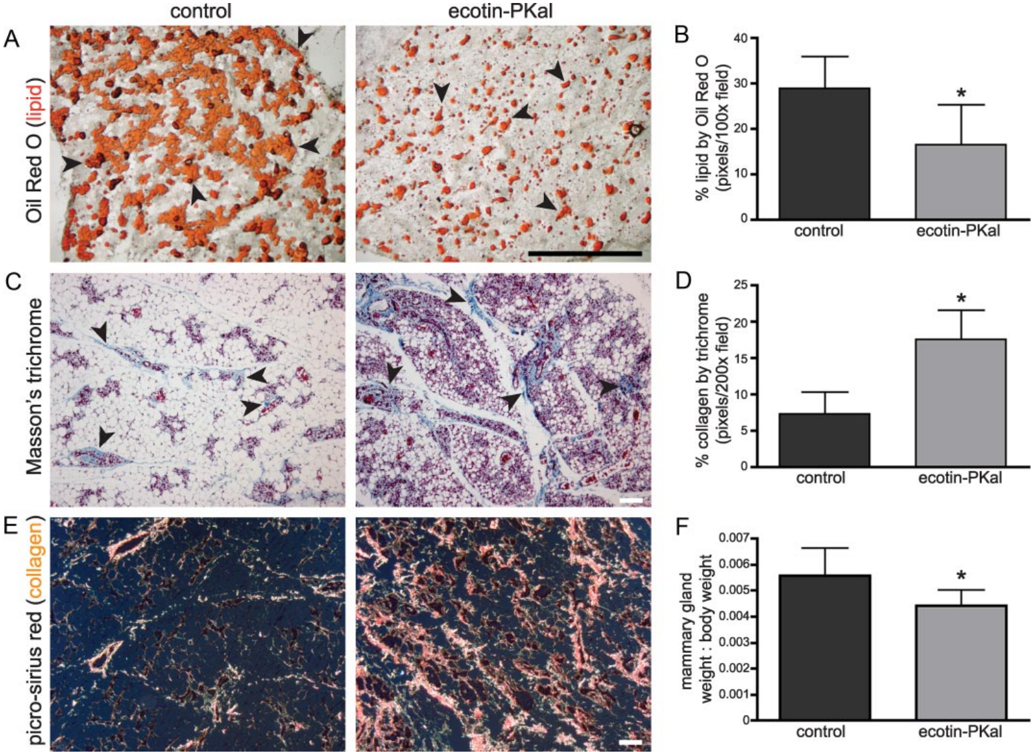
FIGURE5 Delayinmammaryglandinvolutionaftertreatmentwithecotin-PKal. Animals were treated twice daily during the first 4 days of involution with 100 𝛍 g of ecotin-PKal or with vehicle control. n = 8 mice/group. All error bars represent S.D. A , frozen sections of mammary glands stained with Oil Red O for lipid showecotin-PKal-treatedglandshavefewerandlessmatureadipocytes. Blackarrowheads indicate representative lipid staining. Scale bar is 1 mm. B , quantification of lipid contentbyOilRedOstainingshowsecotin-PKal-treatedglandshavesignificantlylesslipidafter4daysofinvolution.Student's t test: p = 0.041. C , paraffin sections of mammary glands stained with Masson's trichrome to detect collagen ( blue ) and cytoplasm ( pink ) show that ecotin-PKal-treated glands have more collagen depositionandmoreimmaturestromalcellsthancontrols. Blackarrowheads indicaterepresentativeareasofcollagendeposition. Scalebar is 100 𝛍 m. D , quantification ofcollagencontentbyMasson'strichromestainshowsecotin-PKal-treatedglandshavesignificantlymorecollagen.Student's t test: p = 0.003. All error bars represent S.D. E , paraffin sections of mammary glands stained with picro-sirius red to demonstrate by birefringence under polarized light the difference between thick ( orangeyellow ) and thin ( green ) collagen fibrils. Scale bar is 100 𝛍 m. F , abdominal mammary gland weights expressed as a ratio of overall body weight on day 5 of involution showthat ecotin-PKal-treated glands weigh less after treatment than controls. Student's t test: p = 0.029.
FIGURE5 Delayinmammaryglandinvolutionaftertreatmentwithecotin-PKal. Animals were treated twice daily during the first 4 days of involution with 100 𝛍 g of ecotin-PKal or with vehicle control. n = 8 mice/group. All error bars represent S.D. A , frozen sections of mammary glands stained with Oil Red O for lipid showecotin-PKal-treatedglandshavefewerandlessmatureadipocytes. Blackarrowheads indicate representative lipid staining. Scale bar is 1 mm. B , quantification of lipid contentbyOilRedOstainingshowsecotin-PKal-treatedglandshavesignificantlylesslipidafter4daysofinvolution.Student's t test: p = 0.041. C , paraffin sections of mammary glands stained with Masson's trichrome to detect collagen ( blue ) and cytoplasm ( pink ) show that ecotin-PKal-treated glands have more collagen depositionandmoreimmaturestromalcellsthancontrols. Blackarrowheads indicaterepresentativeareasofcollagendeposition. Scalebar is 100 𝛍 m. D , quantification ofcollagencontentbyMasson'strichromestainshowsecotin-PKal-treatedglandshavesignificantlymorecollagen.Student's t test: p = 0.003. All error bars represent S.D. E , paraffin sections of mammary glands stained with picro-sirius red to demonstrate by birefringence under polarized light the difference between thick ( orangeyellow ) and thin ( green ) collagen fibrils. Scale bar is 100 𝛍 m. F , abdominal mammary gland weights expressed as a ratio of overall body weight on day 5 of involution showthat ecotin-PKal-treated glands weigh less after treatment than controls. Student's t test: p = 0.029.CC-BY · From the PDF
Image 6
CC-BY · From the PDF
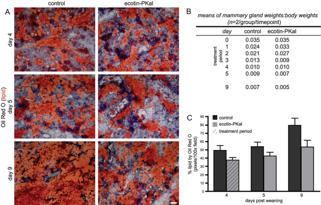
FIGURE 7 Ecotin-PKal-treated mammary glands have an altered epithelial-stromal ratio during treatment. Animals were treated as described, n = 2 mice/ group/timepoint. A , frozen sections of mammaryglandsstainedwithOilRedOforlipidshowecotin-PKal-treatedglandshavefewerandlessmatureadipocytesinthe second phase of involution, even 5 days after end of treatment. Scale bar is 100 𝛍 m. B , ecotin-PKal-treated mammary glands weigh more during the first apoptotic phaseofinvolution,thenweighlessduringthestromalremodelingphase.Abdominalmammaryglandweightsareexpressedasaratioofoverallbodyweightondays 1-5andday9ofinvolution.Ecotin-PKaltreatmentoccurredduringdays1-5. C , quantification of lipid content by Oil Red O staining shows ecotin-PKal-treated glands have less lipid even after 5 days of recovery from ecotin-PKal treatment. Shaded area marks ecotin-PKal treatment. Error bars represent S.D.
FIGURE 7 Ecotin-PKal-treated mammary glands have an altered epithelial-stromal ratio during treatment. Animals were treated as described, n = 2 mice/ group/timepoint. A , frozen sections of mammaryglandsstainedwithOilRedOforlipidshowecotin-PKal-treatedglandshavefewerandlessmatureadipocytesinthe second phase of involution, even 5 days after end of treatment. Scale bar is 100 𝛍 m. B , ecotin-PKal-treated mammary glands weigh more during the first apoptotic phaseofinvolution,thenweighlessduringthestromalremodelingphase.Abdominalmammaryglandweightsareexpressedasaratioofoverallbodyweightondays 1-5andday9ofinvolution.Ecotin-PKaltreatmentoccurredduringdays1-5. C , quantification of lipid content by Oil Red O staining shows ecotin-PKal-treated glands have less lipid even after 5 days of recovery from ecotin-PKal treatment. Shaded area marks ecotin-PKal treatment. Error bars represent S.D.CC-BY · From the PDF
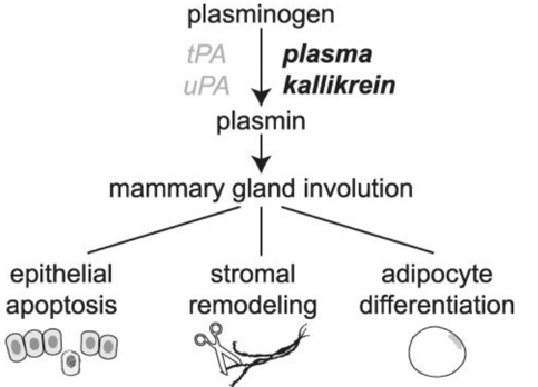
FIGURE8 Modelofthefunctionofplasmakallikreinduringmammarygland involution. As loss of tPA and uPA (in gray ) does not recapitulate the effect of plasminogendeficiencyinmammaryglandinvolution,wepositthatplasmakallikrein (in bold ) is the critical activator of plasminogen during this process. Active PKal is required for apoptosis of the secretory epithelium, subsequent stromal remodeling, and eventual adipocyte replenishment of the mammary gland.
FIGURE8 Modelofthefunctionofplasmakallikreinduringmammarygland involution. As loss of tPA and uPA (in gray ) does not recapitulate the effect of plasminogendeficiencyinmammaryglandinvolution,wepositthatplasmakallikrein (in bold ) is the critical activator of plasminogen during this process. Active PKal is required for apoptosis of the secretory epithelium, subsequent stromal remodeling, and eventual adipocyte replenishment of the mammary gland.CC-BY · From the PDF

Verification

Claims the paper makes

The claims and statistics of this paper haven’t been checked yet.

Run the check — free, no account needed. It reads the open-access text and checks the sources it cites.

A few free checks a day · papers already verified open instantly · results are public

Claims other papers make about this one

Each is checked against this paper’s own text: does it say what the citing paper says it does?

Self-check

Percentages, sums and reported differences re-computed from the paper’s own numbers.

The self-check needs the paper’s full text.

Citations

Cites

No references could be resolved yet.

Research works printed but not linked: 35

  1. Dano, 2005, Thromb. Haemost., 93, 676, 10.1160/TH05-01-0054 · DOI
  2. Lund, 2000, Development, 127, 4481, 10.1242/dev.127.20.4481 · DOI
  3. Miles, 1983, Thromb. Res., 29, 407, 10.1016/0049-3848(83)90244-X · DOI
  4. Selvarajan, 2001, Nat. Cell Biol., 3, 267, 10.1038/35060059 · DOI
  5. Baumgarten, 1986, J. Immunol., 137, 977, 10.4049/jimmunol.137.3.977 · DOI
  6. Motta, 1998, Blood, 91, 516, 10.1182/blood.V91.2.516 · DOI
  7. Rojkjaer, 1999, Immunopharmacology, 43, 109, 10.1016/S0162-3109(99)00069-7 · DOI
  8. Schmaier, 1999, Thromb. Haemost, 82, 226, 10.1055/s-0037-1615837 · DOI
  9. Lima, 2008, Biol. Chem., 389, 1487, 10.1515/BC.2008.166 · DOI
  10. Stoop, 2003, Nat. Biotechnol., 21, 1063, 10.1038/nbt860 · DOI
  11. Lund, 2006, EMBO J., 25, 2686, 10.1038/sj.emboj.7601173 · DOI
  12. Pham, 1999, Proc. Natl. Acad. Sci. U. S. A., 96, 8627, 10.1073/pnas.96.15.8627 · DOI
  13. Jamieson, 2005, Nat. Immunol., 6, 403, 10.1038/ni1182 · DOI
  14. Junqueira, 1979, Histochem. J., 11, 447, 10.1007/BF01002772 · DOI
  15. Leers, 1999, J. Pathol., 187, 567, 10.1002/(SICI)1096-9896(199904)187:5<567::AID-PATH288>3.0.CO;2-J · DOI
  16. Moloney, 1960, J. Histochem. Cytochem., 8, 200, 10.1177/8.3.200 · DOI
  17. Yang, 1998, J. Mol. Biol., 279, 1001, 10.1006/jmbi.1998.1749 · DOI
  18. Eggers, 2001, J. Mol. Biol., 308, 975, 10.1006/jmbi.2001.4754 · DOI
  19. Cerf, 2000, Metab. Brain. Dis., 15, 315, 10.1023/A:1011179426420 · DOI
  20. Ciechanowicz, 1993, Biochem. Biophys. Res. Commun., 197, 1370, 10.1006/bbrc.1993.2628 · DOI
  21. Neth, 2001, Thromb. Haemost., 85, 1043, 10.1055/s-0037-1615961 · DOI
  22. Metcalfe, 1997, Physiol. Rev., 77, 1033, 10.1152/physrev.1997.77.4.1033 · DOI
  23. Duttlinger, 1993, Development, 118, 705, 10.1242/dev.118.3.705 · DOI
  24. McIntyre, 1981, Int. Arch. Allergy Appl. Immunol., 66, 244, 10.1159/000232827 · DOI
  25. Wolters, 2001, J. Biol. Chem., 276, 18551, 10.1074/jbc.M100223200 · DOI
  26. Alexander, 2001, J. Cell Biol., 152, 693, 10.1083/jcb.152.4.693 · DOI
  27. Lund, 1996, Development, 122, 181, 10.1242/dev.122.1.181 · DOI
  28. Mandrup, 1997, J. Biol. Chem., 272, 5367, 10.1074/jbc.272.9.5367 · DOI
  29. Smas, 1995, Biochem. J., 309, 697, 10.1042/bj3090697 · DOI
  30. Spiegelman, 1983, Cell, 35, 657, 10.1016/0092-8674(83)90098-3 · DOI
  31. Strange, 1992, Development, 115, 49, 10.1242/dev.115.1.49 · DOI
  32. Ichinose, 1984, FEBS Lett., 175, 412, 10.1016/0014-5793(84)80779-6 · DOI
  33. Schmaier, 2007, J. Thromb. Haemost., 5, 2323, 10.1111/j.1538-7836.2007.02770.x · DOI
  34. Neth, 2005, Biol. Chem., 386, 101, 10.1515/BC.2005.013 · DOI
  35. Razin, 1984, J. Immunol., 132, 1479, 10.4049/jimmunol.132.3.1479 · DOI

Cited by

Sorted by citations

Metadata

Authors
Lilla, J. N., Joshi, R. V., Craik, C. S., & Werb, Z.
Venue
Journal of Biological Chemistry
Publisher
Elsevier BV
Published
2009-05
Volume
284
Issue
20
Pages
13792-13803
ISSN
0021-9258
Cited by
50
References
0
Language
en

About these checks

Registry, retraction, Cites and statistics results are computed, not judged, with one exception: a printed reference that no index lists is read once by a model before it counts as a citation error. Claims are extracted by a model and kept only when their quote appears verbatim in the text; each cited source is then checked, with a model’s help, for whether it says what the claim relies on. Those verdicts can be wrong — read the claim’s quote and the cited source before relying on one.

Statistics and arithmetic are recomputed, not judged.

Confidence bands are shown only for fields calibrated on our annotated test set. None is calibrated yet, so no band is shown.

What counts as a claim ›How we measure confidence ›How we check this ›

Are you an author, or do you see a problem with this page? Request a review