

The DOI matches its Crossref record; 251 of the 252 references it cites were found in a registry or an index; none of the 278 sources its claims cite is checked for support yet.
Mast cell ontogeny: From fetal development to life‐long health and disease
Open accessAbstract
Summary: Mast cells (MCs) are evolutionarily ancient innate immune cells with important roles in protective immunity against bacteria, parasites, and venomous animals. They can be found in most organs of the body, where they also contribute to normal tissue functioning, for example by engaging in crosstalk with nerves. Despite this, they are most widely known for their detrimental roles in allergy, anaphylaxis, and atopic disease. Just like macrophages, mast cells were conventionally thought to originate from the bone marrow. However, they are already present in fetal tissues before the onset of bone marrow hematopoiesis, questioning this dogma. In recent years, our view of myeloid cell ontogeny has been revised. We now know that the first mast cells originate from progenitors made in the extra‐embryonic yolk sac, and later get supplemented with mast cells produced from subsequent waves of hematopoiesis. In most connective tissues, sizeable populations of fetal‐derived mast cells persist into adulthood, where they self‐maintain largely independently from the bone marrow. These developmental origins are highly reminiscent of macrophages, which are known to have critical functions in development. Mast cells too may thus support healthy development. Their fetal origins and longevity also make mast cells susceptible to genetic and environmental perturbations, which may render them pathological. Here, we review our current understanding of mast cell biology from a developmental perspective. We first summarize how mast cell populations are established from distinct hematopoietic progenitor waves, and how they are subsequently maintained throughout life. We then discuss what functions mast cells may normally have at early life stages, and how they may be co‐opted to cause, worsen, or increase susceptibility to disease.
Overview
AI snapshot
Written from the paper’s full text.
TL;DR
This review argues that mast cells are not solely bone-marrow-derived: the first mast cells arise from yolk-sac erythro-myeloid progenitors (EMPs), are later supplemented by fetal-restricted hematopoietic stem cells, and persist into adulthood with tissue-specific replacement. It proposes that these layered origins shape mast cell functions in development, allergy, mastocytosis, and cancer.
Key findings
- Mast cell ontogeny is layered: the first mast cell progenitors arise from extraembryonic yolk-sac EMPs starting at E8.25 in mice, followed by a second wave predominantly from fetal-restricted HSCs, with possible perinatal contribution from adult-type HSCs. Adult connective-tissue mast cells, such as in skin and adipose tissue, remain largely fetal-derived and self-maintain, whereas mucosal mast cells receive more bone-marrow input.
- Bone-marrow contribution to mast cells is limited at steady state: after bone-marrow transfer into lethally irradiated mice, approximately 50% of resident mast cells in cecum and stomach were donor-derived after 8 months, while over 4.5 months of parabiosis only cecum, stomach, and spleen received sizeable input from the parabiotic partner.
- Fetal mast cells appear functionally mature early: granule-containing mast cells are detected in mouse cornea at E12.5 and skin at E14.5, and human fetal mast cells express tryptase (TPSAB1) and carry granules. The review links fetal mast cells to potential developmental roles in corneal vascular/nerve branching, brain sexual differentiation, mammary gland branching, and uterine spiral artery remodeling.
- Ontogeny may explain disease heterogeneity: pediatric- and adult-onset mastocytosis may have distinct cells of origin, with activating KIT mutations described in HSCs and downstream progenitors of adult systemic disease but not pediatric patients; the paper also notes D186V in 80% of adult patients and almost 40% of pediatric cases, with another 40% of pediatric patients carrying other activating mutations.
Methods
- Narrative review; no new primary data are reported. The authors synthesize mouse genetic fate-mapping models (Cdh5-CreERT2, Runx1-CreERT2, Csf1r-based), adoptive transfer, parabiosis, bone-marrow transplantation, Kit-mutant and Cre/LoxP mast cell depletion models, single-cell transcriptomics, and human fetal tissue studies.
- The main analysis is conceptual: the authors reconcile conflicting fate-mapping interpretations into an updated model of mast cell origins and discuss implications for development, allergy, mastocytosis, mast cell activation syndrome, and cancer.
Limitations
- The paper states that current fate-mapping models cannot spatially distinguish yolk-sac from intraembryonic hematopoiesis and rely on temporal distinctions; 4-hydroxy-tamoxifen remains bioavailable for at least 24 hours, causing overlap between labeled progenitor waves, and no single model is specific to each wave.
- The paper states that Kit-dependent mast cell-deficient models are not mast-cell-specific because Kit has pleiotropic effects, and that many Cre-based models also target basophils, can show off-target effects or Cre leakiness, and vary in depletion efficiency; it recommends using complementary models.
- Evident from the text: as a review, it does not perform a systematic search or meta-analysis, and several proposed developmental and pathological roles for mast cells remain speculative or based largely on mouse studies, with limited direct human evidence.
Model-written summary. Check it against the paper before relying on it.
Integrity signals
Registry
Cites
Statistics
Figures
Verification
Claims the paper makes
94 Check pending · 86 Own interpretation · 26 No full text of the sources · 23 Not checked yet · 5 Sources not found · 3 Own method · 2 No basis given · 1 Own result
In the paper
“Moreover, mast cell phenotypes are not rigid, but can change in inflammatory conditions. 16 , 17 , 18 , 19 , 20 , 21”
§ Mast cell ontogeny: From fetal development to life‐long health and disease > MAST CELL ONTOGENY
What the sources say
- Check pendingcites [16] Abonia JP, Blanchard C, Butz BB, et al. Involvement of mast cells in eosinophilic esophagitis. J Allergy Clin Immunol. 2010; 126(1): 140‐ 149. doi: 10.1016/J.JACI.2010.04.009 20538331 PMC2902643 source
- Not checked — no full text of the sourcecites [17] Andersson CK, Mori M, Bjermer L, Löfdahl CG, Erjefält JS. Alterations in lung mast cell populations in patients with chronic obstructive pulmonary disease. Am J Respir Crit Care Med. 2010; 181(3): 206‐ 217. doi: 10.1164/RCCM.200906-0932OC 19926870 source
- Check pendingcites [18] Dougherty RH, Sidhu SS, Raman K, et al. Accumulation of intraepithelial mast cells with a unique protease phenotype in T(H)2‐high asthma. J Allergy Clin Immunol. 2010; 125(5): 1046‐ 1053.e8. doi: 10.1016/J.JACI.2010.03.003 20451039 PMC2918406 source
plainstates a possible cause- Asserted: passes — The authors put it forward as true.
- Checkable: passes — It can be checked against evidence.
- Load-bearing: unclear — It is unclear whether the paper’s conclusions depend on it.
- Based: passes — The paper gives a reason why it holds.
In the paper
“They also mediate protection from venoms produced by a range of animals, including snakes and bees. 5 , 6 , 7”
§ Mast cell ontogeny: From fetal development to life‐long health and disease > MAST CELL ONTOGENY
What the sources say
- Not checked — no full text of the sourcecites [5] Metz M, Piliponsky AM, Chan CC, et al. Mast cells can enhance resistance to snake and honeybee venoms. Science (80‐). 2006; 313(5786): 526‐ 530. doi: 10.1126/SCIENCE.1128877/SUPPL_FILE/METZ_SOM.PDF 16873664 source
- Check pendingcites [6] Marichal T, Starkl P, Reber LL, et al. A beneficial role for immunoglobulin E in host defense against honeybee venom. Immunity. 2013; 39(5): 963‐ 975. doi: 10.1016/j.immuni.2013.10.005 24210352 PMC4164235 source
- Check pendingcites [7] Starkl P, Gaudenzio N, Marichal T, et al. IgE antibodies increase honeybee venom responsiveness and detoxification efficiency of mast cells. Allergy. 2022; 77(2): 499‐ 512. doi: 10.1111/all.14852 33840121 PMC8502784 source
plainstates a cause- Asserted: passes — The authors put it forward as true.
- Checkable: passes — It can be checked against evidence.
- Load-bearing: unclear — It is unclear whether the paper’s conclusions depend on it.
- Based: passes — The paper gives a reason why it holds.
None of the sources cited for this claim have been checked yet.
In the paper
“With this in mind, it is perhaps not surprising that mast cells are evolutionarily conserved.”
§ Mast cell ontogeny: From fetal development to life‐long health and disease > MAST CELL ONTOGENY
What the sources say
- Not checked yetcites [8] Cavalcante MCM, de Andrade LR, Du Bocage Santos‐Pinto C, et al. Colocalization of heparin and histamine in the intracellular granules of test cells from the invertebrate Styela plicata (Chordata‐Tunicata). J Struct Biol. 2002; 137(3): 313‐ 321. doi: 10.1016/s1047-8477(02)00007-2 12096899
- Not checked — no full text of the sourcecites [9] de Barros CM, Andrade LR, Allodi S, et al. The hemolymph of the ascidian Styela plicata (Chordata‐Tunicata) contains heparin inside basophil‐like cells and a unique sulfated galactoglucan in the plasma. J Biol Chem. 2007; 282(3): 1615‐ 1626. doi: 10.1074/jbc.M604056200 17114184 source
- Not checked — no full text of the sourcecites [10] St John AL, Rathore APS, Ginhoux F. New perspectives on the origins and heterogeneity of mast cells. Nat Rev Immunol. 2022; 23: 55‐ 68. doi: 10.1038/s41577-022-00731-2 35610312 source
hedged- Asserted: passes — The authors put it forward as true.
- Checkable: passes — It can be checked against evidence.
- Load-bearing: unclear — It is unclear whether the paper’s conclusions depend on it.
- Based: passes — The paper gives a reason why it holds.
A statement the paper makes itself — it cites no source for it, so there is none to check. The quote shows it is in the paper.
In the paper
“The first hematopoietic progenitors are produced in the extraembryonic yolk sac (YS).”
§ Mast cell ontogeny: From fetal development to life‐long health and disease > MAST CELL ONTOGENY > Layered hematopoiesis
plainstates a cause- Asserted: passes — The authors put it forward as true.
- Checkable: passes — It can be checked against evidence.
- Load-bearing: unclear — It is unclear whether the paper’s conclusions depend on it.
- Based: passes — The paper gives a reason why it holds.
In the paper
“Recognized already in the 1970s, there is now growing appreciation that mast cell heterogeneity extends beyond this dichotomy.”
§ Mast cell ontogeny: From fetal development to life‐long health and disease > MAST CELL ONTOGENY
What the sources say
- Not checked yetcites [15] Dwyer DF, Barrett NA, Austen KF, Immunological Genome Project Consortium. Expression profiling of constitutive mast cells reveals a unique identity within the immune system. Nat Immunol. 2016; 17(7): 878‐ 887. doi: 10.1038/ni.3445 27135604 PMC5045264
plain- Asserted: passes — The authors put it forward as true.
- Checkable: passes — It can be checked against evidence.
- Load-bearing: unclear — It is unclear whether the paper’s conclusions depend on it.
- Based: passes — The paper gives a reason why it holds.
A conclusion the paper draws from its own results — it cites no other source for it, so there is none to check. The quote shows it is in the paper.
In the paper
“As we will discuss below, this developmental pattern also applies to mast cells.”
§ Mast cell ontogeny: From fetal development to life‐long health and disease > MAST CELL ONTOGENY
plainstates an association- Asserted: passes — The authors put it forward as true.
- Checkable: passes — It can be checked against evidence.
- Load-bearing: passes — The paper’s conclusions depend on it.
- Based: unclear — It is unclear why the paper says it holds.
In the paper
“These findings were the first indication that mast cells are indeed hematopoietic in nature.”
- Also stated in: § MAST CELL ONTOGENY · boosted
“This was confirmed in in vitro studies and parabiosis, an experimental approach in which two animals are surgically joined, resulting in a shared blood circulation. 24 , 25 , 26”
§ Mast cell ontogeny: From fetal development to life‐long health and disease > MAST CELL ONTOGENY
What the sources say
- Not matchedcites [23] Kitamura Y, Shimada M, Hatanaka K, Miyano Y. Development of mast cells from grafted bone marrow cells in irradiated mice. Nature. 1977; 268(5619): 442‐ 443. http://www.ncbi.nlm.nih.gov/pubmed/331117 331117
Couldn’t be matched to a paper in Crossref, so it can’t be checked.
- Not checked — no full text of the sourcecites [24] Kitamura V, Hatanaka K, Murakami M, Shibata H. Presence of mast cell precursors in peripheral blood of mice demonstrated by parabiosis. Blood. 1979; 53(6): 1085‐ 1088. 444650 source
- Check pendingcites [25] Schrader JW, Lewis SJ, Clark‐Lewis I, Culvenor JG. The persisting (P) cell: histamine content, regulation by a T cell‐derived factor, origin from a bone marrow precursor, and relationship to mast cells. Proc Natl Acad Sci USA. 1981; 78(1): 323‐ 327. doi: 10.1073/PNAS.78.1.323 6972532 PMC319045 source
boosted- Asserted: passes — The authors put it forward as true.
- Checkable: passes — It can be checked against evidence.
- Load-bearing: passes — The paper’s conclusions depend on it.
- Based: passes — The paper gives a reason why it holds.
- Also stated in: § MAST CELL ONTOGENY · boosted
A statement the paper makes itself — it cites no source for it, so there is none to check. The quote shows it is in the paper.
In the paper
“In the mouse, so‐called primitive progenitors are made as early as day 7 of embryonic development (E7.0).”
§ Mast cell ontogeny: From fetal development to life‐long health and disease > MAST CELL ONTOGENY > Layered hematopoiesis
plain- Asserted: passes — The authors put it forward as true.
- Checkable: passes — It can be checked against evidence.
- Load-bearing: unclear — It is unclear whether the paper’s conclusions depend on it.
- Based: passes — The paper gives a reason why it holds.
In the paper
“This notion was first challenged 40 years ago with the demonstration that bone marrow (BM) transplanted into irradiated hosts can generate mast cells. 23”
§ Mast cell ontogeny: From fetal development to life‐long health and disease > MAST CELL ONTOGENY
What the sources say
- Not matchedcites [23] Kitamura Y, Shimada M, Hatanaka K, Miyano Y. Development of mast cells from grafted bone marrow cells in irradiated mice. Nature. 1977; 268(5619): 442‐ 443. http://www.ncbi.nlm.nih.gov/pubmed/331117 331117
Couldn’t be matched to a paper in Crossref, so it can’t be checked.
boostedstates a cause- Asserted: passes — The authors put it forward as true.
- Checkable: passes — It can be checked against evidence.
- Load-bearing: passes — The paper’s conclusions depend on it.
- Based: passes — The paper gives a reason why it holds.
- Not matched
A statement the paper makes itself — it cites no source for it, so there is none to check. The quote shows it is in the paper.
In the paper
“However, it is now abundantly clear that many tissue‐resident immune cell lineages, including macrophages and certain innate lymphocytes, first originate from fetal‐restricted progenitors.”
§ Mast cell ontogeny: From fetal development to life‐long health and disease > MAST CELL ONTOGENY
boostedstates a cause- Asserted: passes — The authors put it forward as true.
- Checkable: passes — It can be checked against evidence.
- Load-bearing: unclear — It is unclear whether the paper’s conclusions depend on it.
- Based: passes — The paper gives a reason why it holds.
- It may state two separate claims as one. 113
- Probably not a substantial claim. 26
In the paper
“EMPs can still differentiate into cells of erythroid, megakaryocytic lineages, but also more readily produce different types of myeloid cells. 33 , 34”
§ Mast cell ontogeny: From fetal development to life‐long health and disease > MAST CELL ONTOGENY > Layered hematopoiesis
What the sources say
- Check pendingcites [33] McGrath KE, Frame JM, Fegan KH, et al. Distinct sources of hematopoietic progenitors emerge before HSCs and provide functional blood cells in the mammalian embryo. Cell Rep. 2015; 11(12): 1892‐ 1904. doi: 10.1016/j.celrep.2015.05.036 26095363 PMC4490098 source
- Not checked — no full text of the sourcecites [34] Palis J, Robertson S, Kennedy M, Wall C, Keller G. Development of erythroid and myeloid progenitors in the yolk sac and embryo proper of the mouse. Development. 1999; 126: 5073‐ 5084. 10529424 source
plainstates a cause- Asserted: passes — The authors put it forward as true.
- Checkable: passes — It can be checked against evidence.
- Load-bearing: unclear — It is unclear whether the paper’s conclusions depend on it.
- Based: passes — The paper gives a reason why it holds.
It may state two separate claims as one.
In the paper
“Based on their localization and granule content, mast cells are typically classified as either mucosal (MTMCs) or connective tissue‐type mast cells (CTMCs), which in humans, respectively, express tryptase or tryptase and chymase. 12 , 13 , 14”
§ Mast cell ontogeny: From fetal development to life‐long health and disease > MAST CELL ONTOGENY
What the sources say
- Not checked — no full text of the sourcecites [12] He D, Esquenazi‐Behar S, Soter NA, Lim HW. Mast‐cell heterogeneity: functional comparison of purified mouse cutaneous and peritoneal mast cells. J Invest Dermatol. 1990; 95(2): 178‐ 185. doi: 10.1111/1523-1747.ep12477951 1696300 source
- Not checked — no full text of the sourcecites [13] Irani AM, Schwartz LB. Human mast cell heterogeneity. Allergy Proc. 1994; 15(6): 303‐ 308. doi: 10.2500/108854194778816472 7721078 source
- Check pendingcites [14] Levi‐Schaffer F, Gibbs BF, Hallgren J, et al. Selected recent advances in understanding the role of human mast cells in health and disease. J Allergy Clin Immunol. 2022; 149(6): 1833‐ 1844. doi: 10.1016/j.jaci.2022.01.030 35276243 source
plain- Asserted: passes — The authors put it forward as true.
- Checkable: passes — It can be checked against evidence.
- Load-bearing: fails — The paper’s conclusions may not depend on it.
- Based: passes — The paper gives a reason why it holds.
Probably not a substantial claim. The paper’s conclusions may not depend on it. It may state two separate claims as one.
None of the sources cited for this claim have been checked yet.
In the paper
“These give rise primarily to erythrocytes and megakaryocytes, but also contribute to the very first macrophages. 27 , 28 , 29 , 30 , 31”
§ Mast cell ontogeny: From fetal development to life‐long health and disease > MAST CELL ONTOGENY > Layered hematopoiesis
What the sources say
- Not checked yetcites [27] Moore MAS, Metcalf D. Ontogeny of the haemopoietic system: yolk sac origin of in vivo and in vitro colony forming cells in the developing mouse embryo. Br J Haematol. 1970; 18(3): 279‐ 296. doi: 10.1111/j.1365-2141.1970.tb01443.x 5491581
- Not checked yetcites [28] Wong PMC, Chung SW, Chui DHK, Eaves CJ. Properties of the earliest clonogenic hemopoietic precursors to appear in the developing murine yolk sac. Proc Natl Acad Sci USA. 1986; 83(11): 3851‐ 3854. doi: 10.1073/pnas.83.11.3851 3012535 PMC323622
- Not checked — no full text of the sourcecites [29] Alliot F, Godin I, Pessac B. Microglia derive from progenitors, originating from the yolk sac, and which proliferate in the brain. Dev Brain Res. 1999; 117(2): 145‐ 152. doi: 10.1016/S0165-3806(99)00113-3 10567732 source
plainstates a cause- Asserted: passes — The authors put it forward as true.
- Checkable: passes — It can be checked against evidence.
- Load-bearing: fails — The paper’s conclusions may not depend on it.
- Based: passes — The paper gives a reason why it holds.
Probably not a substantial claim. The paper’s conclusions may not depend on it. It may state two separate claims as one.
In the paper
“From E10.5 onwards, HSCs arise in the main arteries of the embryo properly. This occurs at several sites including the head, heart, placenta and vitelline, and umbilical veins, 35 , 36 , 37 , 38 , 39 , 40 , 41 , 42 though most notably, the aorta‐gonad‐mesonephros (AGM) region. 43”
§ Mast cell ontogeny: From fetal development to life‐long health and disease > MAST CELL ONTOGENY > Layered hematopoiesis
What the sources say
- Not checked — no full text of the sourcecites [35] Alvarez‐Silva M, Belo‐Diabangouaya P, Salaün J, Dieterlen‐Lièvre F. Mouse placenta is a major hematopoietic organ. Development. 2003; 130: 5437‐ 5444. doi: 10.1242/dev.00755 14507780 source
- Check pendingcites [36] Gekas C, Dieterlen‐Lièvre F, Orkin SH, Mikkola HKA. The placenta is a niche for hematopoietic stem cells. Dev Cell. 2005; 8(3): 365‐ 375. doi: 10.1016/j.devcel.2004.12.016 15737932 source
- Not checked — no full text of the sourcecites [43] Boisset J‐CC, van Cappellen W, Andrieu‐Soler C, Galjart N, Dzierzak E, Robin C. In vivo imaging of haematopoietic cells emerging from the mouse aortic endothelium. Nature. 2010; 464(7285): 116‐ 120. doi: 10.1038/nature08764 20154729 source
plain- Asserted: passes — The authors put it forward as true.
- Checkable: passes — It can be checked against evidence.
- Load-bearing: unclear — It is unclear whether the paper’s conclusions depend on it.
- Based: passes — The paper gives a reason why it holds.
It may state two separate claims as one.
In the paper
“For example, connective tissue‐type mast cells share a core transcriptional program but differ between the skin, peritoneal cavity, and the esophagus, trachea, and tongue. 15”
§ Mast cell ontogeny: From fetal development to life‐long health and disease > MAST CELL ONTOGENY
What the sources say
- Not checked yetcites [15] Dwyer DF, Barrett NA, Austen KF, Immunological Genome Project Consortium. Expression profiling of constitutive mast cells reveals a unique identity within the immune system. Nat Immunol. 2016; 17(7): 878‐ 887. doi: 10.1038/ni.3445 27135604 PMC5045264
plainstates an association- Asserted: passes — The authors put it forward as true.
- Checkable: passes — It can be checked against evidence.
- Load-bearing: unclear — It is unclear whether the paper’s conclusions depend on it.
- Based: passes — The paper gives a reason why it holds.
It may state two separate claims as one.
None of the sources cited for this claim have been checked yet.
In the paper
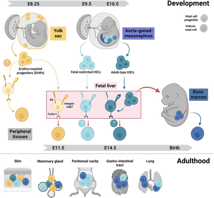
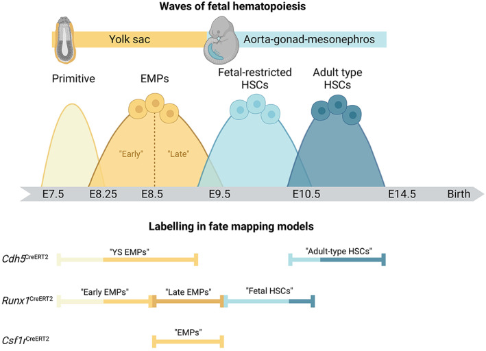
“This is followed by the production of erythro‐myeloid progenitors (EMPs) starting at E8.25. 32 , 33”
§ Mast cell ontogeny: From fetal development to life‐long health and disease > MAST CELL ONTOGENY > Layered hematopoiesis
What the sources say
- Not checked yetcites [32] Gomez Perdiguero E, Klapproth K, Schulz C, et al. Tissue‐resident macrophages originate from yolk‐sac‐derived erythro‐myeloid progenitors. Nature. 2015; 518(7540): 547‐ 551. doi: 10.1038/nature13989 25470051 PMC5997177 source
- Not checked yetcites [33] McGrath KE, Frame JM, Fegan KH, et al. Distinct sources of hematopoietic progenitors emerge before HSCs and provide functional blood cells in the mammalian embryo. Cell Rep. 2015; 11(12): 1892‐ 1904. doi: 10.1016/j.celrep.2015.05.036 26095363 PMC4490098 source
plain- Asserted: passes — The authors put it forward as true.
- Checkable: passes — It can be checked against evidence.
- Load-bearing: fails — The paper’s conclusions may not depend on it.
- Based: passes — The paper gives a reason why it holds.
Probably not a substantial claim. The paper’s conclusions may not depend on it.
A statement the paper makes itself — it cites no source for it, so there is none to check. The quote shows it is in the paper.
In the paper
“Hematopoiesis is a complex process that is initiated early in fetal development and involves several spatio‐temporally distinct (but overlapping) progenitor waves (Figure 1).”
§ Mast cell ontogeny: From fetal development to life‐long health and disease > MAST CELL ONTOGENY > Layered hematopoiesis
plain- Asserted: passes — The authors put it forward as true.
- Checkable: passes — It can be checked against evidence.
- Load-bearing: fails — The paper’s conclusions may not depend on it.
- Based: passes — The paper gives a reason why it holds.
Probably not a substantial claim. The paper’s conclusions may not depend on it. It may state two separate claims as one.
In the paper
“They are critical for host defense against bacteria such as Staphylococcus aureus 1 and other pathogens like helminths. 2 , 3 , 4”
§ Mast cell ontogeny: From fetal development to life‐long health and disease > MAST CELL ONTOGENY
What the sources say
- Check pendingcites [1] Starkl P, Watzenboeck ML, Popov LM, et al. IgE effector mechanisms, in concert with mast cells, contribute to acquired host defense against Staphylococcus aureus. Immunity. 2020; 53(4): 793‐ 804.e9. doi: 10.1016/J.IMMUNI.2020.08.002 32910906 PMC7572876 source
- Check pendingcites [2] Hepworth MR, Daniłowicz‐Luebert E, Rausch S, et al. Mast cells orchestrate type 2 immunity to helminths through regulation of tissue‐derived cytokines. Proc Natl Acad Sci USA. 2012; 109(17): 6644‐ 6649. doi: 10.1073/PNAS.1112268109/SUPPL_FILE/PNAS.201112268SI.PDF 22493240 PMC3340035 source
- Check pendingcites [3] Reitz M, Brunn ML, Rodewald HR, et al. Mucosal mast cells are indispensable for the timely termination of Strongyloides ratti infection. Mucosal Immunol. 2017; 10(2): 481‐ 492. doi: 10.1038/MI.2016.56 27381924 source
plainstates a cause- Asserted: passes — The authors put it forward as true.
- Checkable: passes — It can be checked against evidence.
- Load-bearing: unclear — It is unclear whether the paper’s conclusions depend on it.
- Based: passes — The paper gives a reason why it holds.
It may state two separate claims as one.
In the paper
“SCF can be produced by a range of non‐immune cells including fibroblasts and keratinocytes, and dermal mast cells are absent in mice in which SCF is specifically ablated in keratinocytes. 83”
§ Mast cell ontogeny: From fetal development to life‐long health and disease > MAST CELL ONTOGENY > How are mast cells maintained postnatally?
What the sources say
- Check pendingcites [83] Wang Z, Mascarenhas N, Eckmann L, et al. Skin microbiome promotes mast cell maturation by triggering stem cell factor production in keratinocytes. J Allergy Clin Immunol. 2017; 139(4): 1205‐ 1216.e6. doi: 10.1016/j.jaci.2016.09.019 27746235 PMC5385284 source
plainstates a cause- Asserted: passes — The authors put it forward as true.
- Checkable: passes — It can be checked against evidence.
- Load-bearing: unclear — It is unclear whether the paper’s conclusions depend on it.
- Based: passes — The paper gives a reason why it holds.
It may state two separate claims as one.
In the paper
“An interesting explanation for the low degree of BM turnover has been put forward in the 1980‐1990s, according to which mature mast cells may (actively) inhibit progenitor recruitment. 84 , 85”
§ Mast cell ontogeny: From fetal development to life‐long health and disease > MAST CELL ONTOGENY > How are mast cells maintained postnatally?
What the sources say
- Check pendingcites [84] Kanakura Y, Kuriu A, Waki N, et al. Changes in numbers and types of mast cell colony‐forming cells in the peritoneal cavity of mice after injection of distilled water: evidence that mast cells suppress differentiation of bone marrow‐derived precursors. Blood. 1988; 71(3): 573‐ 580. doi: 10.1182/BLOOD.V71.3.573.573 3345338 source
- Not checked — no full text of the sourcecites [85] Waki N, Kitamura Y, Kanakura Y, Asai H, Nakano T. Intraperitoneally injected cultured mast cells suppress recruitment and differentiation of bone marrow‐derived mast cell precursors in the peritoneal cavity of W/Wv mice. Exp Hematol. 1990; 18(3): 243‐ 247. 1968010
hedgedstates a possible cause- Asserted: passes — The authors put it forward as true.
- Checkable: passes — It can be checked against evidence.
- Load-bearing: fails — The paper’s conclusions may not depend on it.
- Based: passes — The paper gives a reason why it holds.
Probably not a substantial claim. The paper’s conclusions may not depend on it.
In the paper
“However, integrin α4β7 is also expressed on progenitors for other immune cells, including BM‐derived progenitors for T cells and innate lymphoid cells, and is often thought of as a more general tissue homing marker. 67 , 68”
§ Mast cell ontogeny: From fetal development to life‐long health and disease > MAST CELL ONTOGENY > How do fetal progenitors generate mast cells?
What the sources say
- Check pendingcites [67] Cimbro R, Vassena L, Arthos J, et al. IL‐7 induces expression and activation of integrin α4β7 promoting naive T‐cell homing to the intestinal mucosa. Blood. 2012; 120(13): 2610‐ 2619. doi: 10.1182/BLOOD-2012-06-434779 22896005 PMC3460683 source
- Not checked yetcites [68] Klose CSN, Flach M, Möhle L, et al. Differentiation of type 1 ILCs from a common progenitor to all helper‐like innate lymphoid cell lineages. Cell. 2014; 157(2): 340‐ 356. doi: 10.1016/j.cell.2014.03.030 24725403
plainstates an association- Asserted: passes — The authors put it forward as true.
- Checkable: passes — It can be checked against evidence.
- Load-bearing: unclear — It is unclear whether the paper’s conclusions depend on it.
- Based: passes — The paper gives a reason why it holds.
It may state two separate claims as one.
In the paper
“These do not yet have granules but express cardinal markers of mast cells such as Kit, as well as several mast cell proteases, at least on transcriptional level. 54 , 59 , 60”
§ Mast cell ontogeny: From fetal development to life‐long health and disease > MAST CELL ONTOGENY > How do fetal progenitors generate mast cells?
What the sources say
- Not checked — no full text of the sourcecites [54] Li Z, Liu S, Xu J, et al. Adult connective tissue‐resident mast cells originate from late erythro‐myeloid progenitors. Immunity. 2018; 49(4): 640‐ 653.e5. doi: 10.1016/j.immuni.2018.09.023 30332630 source
- Not checked yetcites [59] Popescu D‐M, Botting RA, Stephenson E, et al. Decoding human fetal liver haematopoiesis. Nature. 2019; 574(7778): 365‐ 371. doi: 10.1038/s41586-019-1652-y 31597962 PMC6861135 source
- Check pendingcites [60] Suo C, Dann E, Goh I, et al. Mapping the developing human immune system across organs. Science. 2022; 376(6597): eabo0510. doi: 10.1126/science.abo0510 35549310 PMC7612819 source
plain- Asserted: passes — The authors put it forward as true.
- Checkable: passes — It can be checked against evidence.
- Load-bearing: passes — The paper’s conclusions depend on it.
- Based: passes — The paper gives a reason why it holds.
It may state two separate claims as one.
In the paper
“Protein expression of FcεR1, on the other hand, appears to be variable for committed progenitors in peripheral tissues of mice and is also not found on human peripheral blood progenitors. 79”
§ Mast cell ontogeny: From fetal development to life‐long health and disease > MAST CELL ONTOGENY > How are mast cells maintained postnatally?
What the sources say
- Check pendingcites [79] Rottem M, Okada T, Goff JP, Metcalfe DD. Mast cells cultured from the peripheral blood of normal donors and patients with mastocytosis originate from a CD34+/fc epsilon RI‐ cell population. Blood. 1994; 84(8): 2489‐ 2496. doi: 10.1182/BLOOD.V84.8.2489.2489 7522630 source
hedged- Asserted: passes — The authors put it forward as true.
- Checkable: passes — It can be checked against evidence.
- Load-bearing: unclear — It is unclear whether the paper’s conclusions depend on it.
- Based: passes — The paper gives a reason why it holds.
It may state two separate claims as one.
In the paper
“This revealed that between E9.5 and E11.5, the highest mast cell potential is found in the YS, followed by the fetal liver that predominates from E13.5 onwards. 51”
§ Mast cell ontogeny: From fetal development to life‐long health and disease > MAST CELL ONTOGENY > Current understanding of mast cell origins
What the sources say
- Not checked — no full text of the sourcecites [51] Sonoda T, Hayashi C, Kitamura Y. Presence of mast cell precursors in the yolk sac of mice. Dev Biol. 1983; 97: 89‐ 94. doi: 10.1016/0012-1606(83)90066-0 6341120 source
plainstates an association- Asserted: passes — The authors put it forward as true.
- Checkable: passes — It can be checked against evidence.
- Load-bearing: passes — The paper’s conclusions depend on it.
- Based: passes — The paper gives a reason why it holds.
It may state two separate claims as one.
In the paper
“HSCs transiently colonize and are thought to expand in the fetal liver, although this view has recently been challenged 44 and hence, remains an area of ongoing debate.”
§ Mast cell ontogeny: From fetal development to life‐long health and disease > MAST CELL ONTOGENY > Layered hematopoiesis
What the sources say
- Check pendingcites [44] Ganuza M, Hall T, Myers J, et al. Murine foetal liver supports limited detectable expansion of life‐long haematopoietic progenitors. Nat Cell Biol. 2022; 24(10): 1475‐ 1486. doi: 10.1038/s41556-022-00999-5 36202972 PMC10026622 source
hedgedstates a possible cause- Asserted: passes — The authors put it forward as true.
- Checkable: passes — It can be checked against evidence.
- Load-bearing: fails — The paper’s conclusions may not depend on it.
- Based: passes — The paper gives a reason why it holds.
Probably not a substantial claim. The paper’s conclusions may not depend on it. It may state two separate claims as one.
In the paper
“SCF is required for mast cell (progenitor) chemotaxis, 87 , 88 differentiation, adhesion, 89 , 90 proliferation, and survival by blocking apoptosis. 91 , 92 , 93”
§ Mast cell ontogeny: From fetal development to life‐long health and disease > MAST CELL ONTOGENY > How are mast cells maintained postnatally?
What the sources say
- Check pendingcites [87] Meininger CJ, Yano H, Rottapel R, Bernstein A, Zsebo KM, Zetter BR. The c‐kit receptor ligand functions as a mast cell chemoattractant. Blood. 1992; 79(4): 958‐ 963. doi: 10.1182/BLOOD.V79.4.958.958 1371080 source
- Not checked — no full text of the sourcecites [88] Nilsson G, Butterfield J, Nilsson K, Siegbahn A. Stem cell factor is a chemotactic factor for human mast cells. J Immunol. 1994; 153(8): 3717‐ 3723. 7523504 source
- Not checked — no full text of the sourcecites [91] Mekori Y, Oh C, Metcalfe D. IL‐3‐dependent murine mast cells undergo apoptosis on removal of IL‐3. Prevention of apoptosis by c‐kit ligand. J Immunol. 1993; 151(7): 3775‐ 3784. 7690814 source
plainstates a cause- Asserted: passes — The authors put it forward as true.
- Checkable: passes — It can be checked against evidence.
- Load-bearing: passes — The paper’s conclusions depend on it.
- Based: passes — The paper gives a reason why it holds.
It may state two separate claims as one.
In the paper
“Integrin α4β7 specifically is expressed by adult BM progenitors with mast cell potential 64 and required for gut homing of mast cell progenitors. 65”
§ Mast cell ontogeny: From fetal development to life‐long health and disease > MAST CELL ONTOGENY > How do fetal progenitors generate mast cells?
What the sources say
- Check pendingcites [64] Chen C‐C, Grimbaldeston MA, Tsai M, Weissman IL, Galli SJ. Identification of mast cell progenitors in adult mice. Proc Natl Acad Sci USA. 2005; 102(32): 11408‐ 11413. doi: 10.1073/pnas.0504197102 16006518 PMC1183570 source
- Check pendingcites [65] Gurish MF, Tao H, Abonia JP, et al. Intestinal mast cell progenitors require CD49dbeta7 (alpha4beta7 integrin) for tissue‐specific homing. J Exp Med. 2001; 194(9): 1243‐ 1252. doi: 10.1084/JEM.194.9.1243 11696590 PMC2195984 source
plainstates a cause- Asserted: passes — The authors put it forward as true.
- Checkable: passes — It can be checked against evidence.
- Load-bearing: unclear — It is unclear whether the paper’s conclusions depend on it.
- Based: passes — The paper gives a reason why it holds.
It may state two separate claims as one.
In the paper
“In support of this notion, progenitors with mast cell potential can be found in the fetal liver from E11.5 onwards, and these progenitors give rise to mast cells when transferred into fetal or newborn recipients. 54 , 69”
§ Mast cell ontogeny: From fetal development to life‐long health and disease > MAST CELL ONTOGENY > How do fetal progenitors generate mast cells?
What the sources say
- Not checked — no full text of the sourcecites [54] Li Z, Liu S, Xu J, et al. Adult connective tissue‐resident mast cells originate from late erythro‐myeloid progenitors. Immunity. 2018; 49(4): 640‐ 653.e5. doi: 10.1016/j.immuni.2018.09.023 30332630 source
- Check pendingcites [69] Yoshimoto M, Kosters A, Cornelius S, et al. Mast cell repopulating ability is lost during the transition from pre‐HSCs to FL HSC. Front Immunol Accept. 2022; 13(July): 1‐ 11. doi: 10.3389/fimmu.2022.896396 PMC9309215 35898504 source
plainstates a cause- Asserted: passes — The authors put it forward as true.
- Checkable: passes — It can be checked against evidence.
- Load-bearing: passes — The paper’s conclusions depend on it.
- Based: passes — The paper gives a reason why it holds.
It may state two separate claims as one.
A statement the paper makes itself — it cites no source for it, so there is none to check. The quote shows it is in the paper.
In the paper
“On the other hand, mice with hypomorphic alleles of Kit are profoundly deficient in mast cells and have therefore long been used as models to study their functions.”
§ Mast cell ontogeny: From fetal development to life‐long health and disease > MAST CELL ONTOGENY > How are mast cells maintained postnatally?
plainstates a cause- Asserted: passes — The authors put it forward as true.
- Checkable: passes — It can be checked against evidence.
- Load-bearing: unclear — It is unclear whether the paper’s conclusions depend on it.
- Based: passes — The paper gives a reason why it holds.
It may state two separate claims as one.
A conclusion the paper draws from its own results — it cites no other source for it, so there is none to check. The quote shows it is in the paper.
In the paper
“EMPs differentiate into mast cells via committed progenitors that may seed peripheral tissues directly from the yolk sac and/or through fetal liver intermediates.”
§ Mast cell ontogeny: From fetal development to life‐long health and disease > MAST CELL ONTOGENY > Layered hematopoiesis
hedgedstates a possible cause- Asserted: passes — The authors put it forward as true.
- Checkable: passes — It can be checked against evidence.
- Load-bearing: passes — The paper’s conclusions depend on it.
- Based: passes — The paper gives a reason why it holds.
It may state two separate claims as one.
A statement the paper makes itself — it cites no source for it, so there is none to check. The quote shows it is in the paper.
In the paper
“HSCs ultimately settle in the BM, where they persist lifelong.”
§ Mast cell ontogeny: From fetal development to life‐long health and disease > MAST CELL ONTOGENY > Layered hematopoiesis
plain- Asserted: passes — The authors put it forward as true.
- Checkable: passes — It can be checked against evidence.
- Load-bearing: fails — The paper’s conclusions may not depend on it.
- Based: passes — The paper gives a reason why it holds.
Probably not a substantial claim. The paper’s conclusions may not depend on it. It may state two separate claims as one.
In the paper
“At steady state, these clusters remain stable in size and distribution and are independent from BM input. 86”
§ Mast cell ontogeny: From fetal development to life‐long health and disease > MAST CELL ONTOGENY > How are mast cells maintained postnatally?
What the sources say
- Not checked — no full text of the sourcecites [86] Weitzmann A, Naumann R, Dudeck A, Zerjatke T, Gerbaulet A, Roers A. Mast cells occupy stable clonal territories in adult steady‐state skin. J Invest Dermatol. 2020; 140(12): 2433‐ 2441.e5. doi: 10.1016/J.JID.2020.03.963 32311397 source
plain- Asserted: passes — The authors put it forward as true.
- Checkable: passes — It can be checked against evidence.
- Load-bearing: passes — The paper’s conclusions depend on it.
- Based: passes — The paper gives a reason why it holds.
It may state two separate claims as one.
In the paper
“In vitro, they efficiently produce mast cell colonies, but not any other lineages.”
§ Mast cell ontogeny: From fetal development to life‐long health and disease > MAST CELL ONTOGENY > Current understanding of mast cell origins
What the sources say
- Not checked — no full text of the sourcecites [52] Rodewald HR, Dessing M, Dvorak AM, Galli SJ. Identification of a committed precursor for the mast cell lineage. Science (80‐). 1996; 271(5250): 818‐ 822. doi: 10.1126/science.271.5250.818 8629001 source
plainstates a cause- Asserted: passes — The authors put it forward as true.
- Checkable: passes — It can be checked against evidence.
- Load-bearing: passes — The paper’s conclusions depend on it.
- Based: passes — The paper gives a reason why it holds.
It may state two separate claims as one.
A conclusion the paper draws from its own results — it cites no other source for it, so there is none to check. The quote shows it is in the paper.
In the paper
“These EMP‐derived mast cells are then supplemented with a second wave that originates predominantly from fetal‐restricted HSCs, but may also receive contribution from adult‐type HSCs.”
§ Mast cell ontogeny: From fetal development to life‐long health and disease > MAST CELL ONTOGENY > Current understanding of mast cell origins
hedgedstates a cause- Asserted: passes — The authors put it forward as true.
- Checkable: passes — It can be checked against evidence.
- Load-bearing: passes — The paper’s conclusions depend on it.
- Based: passes — The paper gives a reason why it holds.
It may state two separate claims as one.
None of the sources cited for this claim have been checked yet.
In the paper
“Considering that turnover from the BM is very low for mast cells in connective tissues populations, 23 , 53 such HSC‐derived mast cell progenitors might be recruited in the perinatal window prior to settling in the BM.”
§ Mast cell ontogeny: From fetal development to life‐long health and disease > MAST CELL ONTOGENY > Current understanding of mast cell origins
What the sources say
- Not matchedcites [23] Kitamura Y, Shimada M, Hatanaka K, Miyano Y. Development of mast cells from grafted bone marrow cells in irradiated mice. Nature. 1977; 268(5619): 442‐ 443. http://www.ncbi.nlm.nih.gov/pubmed/331117 331117
Couldn’t be matched to a paper in Crossref, so it can’t be checked.
- Not checked yetcites [53] Gentek R, Ghigo C, Hoeffel G, et al. Hemogenic endothelial fate mapping reveals dual developmental origin of mast cells. Immunity. 2018; 48(6): 1160‐ 1171.e5. doi: 10.1016/j.immuni.2018.04.025 29858009 source
hedgedstates a possible cause- Asserted: passes — The authors put it forward as true.
- Checkable: passes — It can be checked against evidence.
- Load-bearing: fails — The paper’s conclusions may not depend on it.
- Based: passes — The paper gives a reason why it holds.
Probably not a substantial claim. The paper’s conclusions may not depend on it. It may state two separate claims as one.
- Not matched
A conclusion the paper draws from its own results — it cites no other source for it, so there is none to check. The quote shows it is in the paper.
In the paper
“While mast cells in connective tissues largely self‐maintain thereafter, mucosal mast cells receive further input from the BM after birth.”
§ Mast cell ontogeny: From fetal development to life‐long health and disease > MAST CELL ONTOGENY > Current understanding of mast cell origins
plainstates a cause- Asserted: passes — The authors put it forward as true.
- Checkable: passes — It can be checked against evidence.
- Load-bearing: unclear — It is unclear whether the paper’s conclusions depend on it.
- Based: unclear — It is unclear why the paper says it holds.
It may state two separate claims as one.
A conclusion the paper draws from its own results — it cites no other source for it, so there is none to check. The quote shows it is in the paper.
In the paper
“This indicates that lineage commitment indeed takes place prior to tissue colonization, while final maturation of fetal mast cells occurs in tissues.”
§ Mast cell ontogeny: From fetal development to life‐long health and disease > MAST CELL ONTOGENY > How do fetal progenitors generate mast cells?
hedgedstates a cause- Asserted: passes — The authors put it forward as true.
- Checkable: passes — It can be checked against evidence.
- Load-bearing: passes — The paper’s conclusions depend on it.
- Based: passes — The paper gives a reason why it holds.
It may state two separate claims as one.
In the paper
“These Kithigh Thy1low cells are first detected at E14.5, peak in numbers at E15.5 and decline towards birth.”
§ Mast cell ontogeny: From fetal development to life‐long health and disease > MAST CELL ONTOGENY > Current understanding of mast cell origins
What the sources say
- Not checked — no full text of the sourcecites [52] Rodewald HR, Dessing M, Dvorak AM, Galli SJ. Identification of a committed precursor for the mast cell lineage. Science (80‐). 1996; 271(5250): 818‐ 822. doi: 10.1126/science.271.5250.818 8629001 source
plain- Asserted: passes — The authors put it forward as true.
- Checkable: passes — It can be checked against evidence.
- Load-bearing: unclear — It is unclear whether the paper’s conclusions depend on it.
- Based: passes — The paper gives a reason why it holds.
It may state two separate claims as one.
In the paper
“SCF exists in soluble and membrane‐bound forms, which are the result of alternative splicing of the same transcript. 94”
§ Mast cell ontogeny: From fetal development to life‐long health and disease > MAST CELL ONTOGENY > How are mast cells maintained postnatally?
What the sources say
- Not checked — no full text of the sourcecites [94] Flanagan JG, Chan DC, Leder P. Transmembrane form of the kit ligand growth factor is determined by alternative splicing and is missing in the Sld mutant. Cell. 1991; 64(5): 1025‐ 1035. doi: 10.1016/0092-8674(91)90326-T 1705866 source
plainstates a cause- Asserted: passes — The authors put it forward as true.
- Checkable: passes — It can be checked against evidence.
- Load-bearing: unclear — It is unclear whether the paper’s conclusions depend on it.
- Based: passes — The paper gives a reason why it holds.
It may state two separate claims as one.
The paper states this without a source or data behind it here, so there is nothing to check it against. The quote shows it is in the paper.
In the paper
“Mast cell‐committed progenitors express integrin α4β7, Cx3cr1, and Kit and undergo terminal maturation following recruitment into tissues.”
§ Mast cell ontogeny: From fetal development to life‐long health and disease > MAST CELL ONTOGENY > Layered hematopoiesis
plainstates a cause- Asserted: passes — The authors put it forward as true.
- Checkable: passes — It can be checked against evidence.
- Load-bearing: passes — The paper’s conclusions depend on it.
- Based: unclear — It is unclear why the paper says it holds.
It may state two separate claims as one.
In the paper
“Indeed, based on transplantation assays, mast cell potential is present in the YS until E13.5, and mast cells are in fact generated more efficiently from the YS than the fetal liver until E11.5. 51”
§ Mast cell ontogeny: From fetal development to life‐long health and disease > MAST CELL ONTOGENY > How do fetal progenitors generate mast cells?
What the sources say
- Not checked — no full text of the sourcecites [51] Sonoda T, Hayashi C, Kitamura Y. Presence of mast cell precursors in the yolk sac of mice. Dev Biol. 1983; 97: 89‐ 94. doi: 10.1016/0012-1606(83)90066-0 6341120 source
plainstates a cause- Asserted: passes — The authors put it forward as true.
- Checkable: passes — It can be checked against evidence.
- Load-bearing: unclear — It is unclear whether the paper’s conclusions depend on it.
- Based: passes — The paper gives a reason why it holds.
It may state two separate claims as one.
A conclusion the paper draws from its own results — it cites no other source for it, so there is none to check. The quote shows it is in the paper.
In the paper
“Interestingly, those found in the limbs are transcriptomically very similar to those in the fetal liver at the same stage, yet do show some differences in gene expression, suggesting they have either started differentiating in situ and must have been recruited earlier, and/or they may have originated elsewhere.”
§ Mast cell ontogeny: From fetal development to life‐long health and disease > MAST CELL ONTOGENY > How do fetal progenitors generate mast cells?
hedgedstates a possible cause- Asserted: passes — The authors put it forward as true.
- Checkable: passes — It can be checked against evidence.
- Load-bearing: fails — The paper’s conclusions may not depend on it.
- Based: passes — The paper gives a reason why it holds.
Probably not a substantial claim. The paper’s conclusions may not depend on it. It may state two separate claims as one.
In the paper
“From a combination of genetic fate mapping, adoptive transfer, and in vitro differentiation assays, Yoshimoto and colleagues conclude that only the earliest HSCs found in the fetal liver give rise to mast cells, but not those present at a time when adult‐type HSCs dominate prior to BM colonization. 69”
§ Mast cell ontogeny: From fetal development to life‐long health and disease > MAST CELL ONTOGENY > Current understanding of mast cell origins
What the sources say
- Check pendingcites [69] Yoshimoto M, Kosters A, Cornelius S, et al. Mast cell repopulating ability is lost during the transition from pre‐HSCs to FL HSC. Front Immunol Accept. 2022; 13(July): 1‐ 11. doi: 10.3389/fimmu.2022.896396 PMC9309215 35898504 source
boostedstates a cause- Asserted: passes — The authors put it forward as true.
- Checkable: passes — It can be checked against evidence.
- Load-bearing: passes — The paper’s conclusions depend on it.
- Based: passes — The paper gives a reason why it holds.
It may state two separate claims as one.
What the paper did — it cites no source for this, so there is none to check. The quote shows it is in the paper.
In the paper
“Kit‐dependent mice are therefore by no means mast cell‐specific and findings obtained in these models should be interpreted with caution (see Box 2).”
§ Mast cell ontogeny: From fetal development to life‐long health and disease > MAST CELL ONTOGENY > How are mast cells maintained postnatally?
plain- Asserted: passes — The authors put it forward as true.
- Checkable: passes — It can be checked against evidence.
- Load-bearing: passes — The paper’s conclusions depend on it.
- Based: passes — The paper gives a reason why it holds.
It may state two separate claims as one.
A statement the paper makes itself — it cites no source for it, so there is none to check. The quote shows it is in the paper.
In the paper
“The intra‐embryonic aorta‐gonad‐mesonephros (AGM) region generates fetal‐restricted hematopoietic stem cells (HSCs) between E9.0 and E10.0 and adult‐type HSCs from E10.5 onwards.”
§ Mast cell ontogeny: From fetal development to life‐long health and disease > MAST CELL ONTOGENY > Layered hematopoiesis
plainstates a cause- Asserted: passes — The authors put it forward as true.
- Checkable: passes — It can be checked against evidence.
- Load-bearing: unclear — It is unclear whether the paper’s conclusions depend on it.
- Based: passes — The paper gives a reason why it holds.
It may state two separate claims as one.
In the paper
“It was observed that the rate of mast cell reconstitution from the BM is tissue‐dependent, but overall low.”
§ Mast cell ontogeny: From fetal development to life‐long health and disease > MAST CELL ONTOGENY > Current understanding of mast cell origins
What the sources say
- Not matchedcites [23] Kitamura Y, Shimada M, Hatanaka K, Miyano Y. Development of mast cells from grafted bone marrow cells in irradiated mice. Nature. 1977; 268(5619): 442‐ 443. http://www.ncbi.nlm.nih.gov/pubmed/331117 331117
Couldn’t be matched to a paper in Crossref, so it can’t be checked.
plainstates an association- Asserted: passes — The authors put it forward as true.
- Checkable: passes — It can be checked against evidence.
- Load-bearing: passes — The paper’s conclusions depend on it.
- Based: passes — The paper gives a reason why it holds.
It may state two separate claims as one.
- Not matched
None of the sources cited for this claim have been checked yet.
In the paper
“Replenishment from grafted wild‐type BM is not only very slow, it also does not restore wild‐type numbers. 86 , 97 , 98”
§ Mast cell ontogeny: From fetal development to life‐long health and disease > MAST CELL ONTOGENY > How are mast cells maintained postnatally?
What the sources say
- Not checked — no full text of the sourcecites [86] Weitzmann A, Naumann R, Dudeck A, Zerjatke T, Gerbaulet A, Roers A. Mast cells occupy stable clonal territories in adult steady‐state skin. J Invest Dermatol. 2020; 140(12): 2433‐ 2441.e5. doi: 10.1016/J.JID.2020.03.963 32311397 source
- Not matchedcites [97] Kitamura Y, Go S, Hatanaka K. Decrease of mast cells in W/Wv mice and their increase by bone marrow transplantation. Blood. 1978; 52(2): 447‐ 452. 352443
Couldn’t be matched to a paper in Crossref, so it can’t be checked.
- Not checked yetcites [98] Du T, Friend DS, Austen KF, Katz HR. Tissue‐dependent differences in the asynchronous appearance of mast cells in normal mice and in congenic mast cell‐deficient mice after infusion of normal bone marrow cells. Clin Exp Immunol. 1996; 103(2): 316‐ 321. doi: 10.1046/J.1365-2249.1996.D01-610.X 8565318 PMC2200335
plainstates a cause- Asserted: passes — The authors put it forward as true.
- Checkable: passes — It can be checked against evidence.
- Load-bearing: passes — The paper’s conclusions depend on it.
- Based: passes — The paper gives a reason why it holds.
It may state two separate claims as one.
In the paper
“The frequency of EMP‐derived mast cells gradually declines over time, although sizeable populations are retained in adult connective tissues, such as the body skin, as well as adipose tissues.”
§ Mast cell ontogeny: From fetal development to life‐long health and disease > MAST CELL ONTOGENY > Current understanding of mast cell origins
What the sources say
- Check pendingcites [53] Gentek R, Ghigo C, Hoeffel G, et al. Hemogenic endothelial fate mapping reveals dual developmental origin of mast cells. Immunity. 2018; 48(6): 1160‐ 1171.e5. doi: 10.1016/j.immuni.2018.04.025 29858009 source
- Not checked — no full text of the sourcecites [54] Li Z, Liu S, Xu J, et al. Adult connective tissue‐resident mast cells originate from late erythro‐myeloid progenitors. Immunity. 2018; 49(4): 640‐ 653.e5. doi: 10.1016/j.immuni.2018.09.023 30332630 source
plainstates a cause- Asserted: passes — The authors put it forward as true.
- Checkable: passes — It can be checked against evidence.
- Load-bearing: passes — The paper’s conclusions depend on it.
- Based: passes — The paper gives a reason why it holds.
It may state two separate claims as one.
A statement the paper makes itself — it cites no source for it, so there is none to check. The quote shows it is in the paper.
In the paper
“In adulthood, progenitors first commit to the mast cell lineage and then undergo terminal maturation in peripheral tissues.”
§ Mast cell ontogeny: From fetal development to life‐long health and disease > MAST CELL ONTOGENY > How do fetal progenitors generate mast cells?
plainstates a cause- Asserted: passes — The authors put it forward as true.
- Checkable: passes — It can be checked against evidence.
- Load-bearing: passes — The paper’s conclusions depend on it.
- Based: passes — The paper gives a reason why it holds.
It may state two separate claims as one.
In the paper
“They are, however, also reduced and appear less mature in the skin of germ‐free mice. 83”
§ Mast cell ontogeny: From fetal development to life‐long health and disease > MAST CELL ONTOGENY > How are mast cells maintained postnatally?
What the sources say
- Check pendingcites [83] Wang Z, Mascarenhas N, Eckmann L, et al. Skin microbiome promotes mast cell maturation by triggering stem cell factor production in keratinocytes. J Allergy Clin Immunol. 2017; 139(4): 1205‐ 1216.e6. doi: 10.1016/j.jaci.2016.09.019 27746235 PMC5385284 source
hedgedstates a cause- Asserted: passes — The authors put it forward as true.
- Checkable: passes — It can be checked against evidence.
- Load-bearing: unclear — It is unclear whether the paper’s conclusions depend on it.
- Based: passes — The paper gives a reason why it holds.
It may state two separate claims as one.
A conclusion the paper draws from its own results — it cites no other source for it, so there is none to check. The quote shows it is in the paper.
In the paper
“It would follow that mast cells are established from YS EMPs, followed by fetal‐restricted HSCs.”
§ Mast cell ontogeny: From fetal development to life‐long health and disease > MAST CELL ONTOGENY > Current understanding of mast cell origins
hedgedstates a cause- Asserted: passes — The authors put it forward as true.
- Checkable: passes — It can be checked against evidence.
- Load-bearing: passes — The paper’s conclusions depend on it.
- Based: passes — The paper gives a reason why it holds.
It may state two separate claims as one.
None of the sources cited for this claim have been checked yet.
In the paper
“This is because unlike primitive progenitors, they express the transcription factor c‐Myb, 47 , 48 but they cease to exist perinatally and are thus transient in nature.”
§ Mast cell ontogeny: From fetal development to life‐long health and disease > MAST CELL ONTOGENY > Layered hematopoiesis
What the sources say
- Not checked — no full text of the sourcecites [47] Mukouyama YS, Chiba N, Mucenski ML, et al. Hematopoietic cells in cultures of the murine embryonic aorta‐gonad‐mesonephros region are induced by c‐Myb. Curr Biol. 1999; 9(15): 833‐ 836. doi: 10.1016/S0960-9822(99)80368-6 10469571 source
- Not checked yetcites [48] Hoeffel G, Chen J, Lavin Y, et al. C‐Myb+ erythro‐myeloid progenitor‐derived fetal monocytes give rise to adult tissue‐resident macrophages. Immunity. 2015; 42(4): 665‐ 678. doi: 10.1016/j.immuni.2015.03.011 25902481 PMC4545768 source
plainstates a cause- Asserted: passes — The authors put it forward as true.
- Checkable: passes — It can be checked against evidence.
- Load-bearing: unclear — It is unclear whether the paper’s conclusions depend on it.
- Based: passes — The paper gives a reason why it holds.
It may state two separate claims as one.
What the paper did — it cites no source for this, so there is none to check. The quote shows it is in the paper.
In the paper
“However, they were based on either in vitro differentiation assays or adoptive transfer into hosts that were not age‐matched and either pre‐conditioned by irradiation or mast cell‐deficient.”
§ Mast cell ontogeny: From fetal development to life‐long health and disease > MAST CELL ONTOGENY > Current understanding of mast cell origins
plain- Asserted: passes — The authors put it forward as true.
- Checkable: passes — It can be checked against evidence.
- Load-bearing: passes — The paper’s conclusions depend on it.
- Based: passes — The paper gives a reason why it holds.
It may state two separate claims as one.
What the paper did — it cites no source for this, so there is none to check. The quote shows it is in the paper.
In the paper
“None of the currently available models can spatially distinguish YS from intra‐embryonic hematopoiesis, and all models available to study mast cell origins rely on a temporally defined distinction of different progenitors.”
§ Mast cell ontogeny: From fetal development to life‐long health and disease > MAST CELL ONTOGENY > Current understanding of mast cell origins
plain- Asserted: passes — The authors put it forward as true.
- Checkable: passes — It can be checked against evidence.
- Load-bearing: passes — The paper’s conclusions depend on it.
- Based: passes — The paper gives a reason why it holds.
It may state two separate claims as one.
In the paper
“These were phenotypically identified by high expression of Kit, low‐level expression of Thy1 and a low density of granules. They also express transcripts for three different mast cell proteases, Cpa3, Mcpt2, and Mcpt4.”
§ Mast cell ontogeny: From fetal development to life‐long health and disease > MAST CELL ONTOGENY > Current understanding of mast cell origins
What the sources say
- Not checked — no full text of the sourcecites [52] Rodewald HR, Dessing M, Dvorak AM, Galli SJ. Identification of a committed precursor for the mast cell lineage. Science (80‐). 1996; 271(5250): 818‐ 822. doi: 10.1126/science.271.5250.818 8629001 source
plain- Asserted: passes — The authors put it forward as true.
- Checkable: passes — It can be checked against evidence.
- Load-bearing: passes — The paper’s conclusions depend on it.
- Based: passes — The paper gives a reason why it holds.
It may state two separate claims as one.
None of the sources cited for this claim have been checked yet.
In the paper
“This heterogeneous disease is characterized by recurring episodes of allergic and anaphylactic symptoms, which typically involve multiple organ systems, such as the skin, lung, heart, and gastrointestinal tract. Common symptoms include skin itching, swelling, and flushing, wheezing, abdominal pain, cramping, and diarrhea. These symptoms are driven by high levels of potent mast cell mediators, 163”
§ Mast cell ontogeny: From fetal development to life‐long health and disease > IMPLICATIONS OF MAST CELL ONTOGENY FOR LIFE‐LONG HEALTH AND DISEASE > Abnormal mast cell development and pathology
What the sources say
- Not checked — no full text of the sourcecites [163] Afrin LB, Butterfield JJ, Raithel M, Molderings GG. Often seen, rarely recognized: mast cell activation disease – a guide to diagnosis and therapeutic options. Ann Med. 2016; 48(3): 190‐ 201. doi: 10.3109/07853890.2016.1161231 27012973 source
- Not checked yetcites [164] Valent P, Akin C, Nedoszytko B, et al. Diagnosis, classification and Management of Mast Cell Activation Syndromes (MCAS) in the era of personalized medicine. Int J Mol Sci. 2020; 21(23): 9030. doi: 10.3390/IJMS21239030 33261124 PMC7731385 source
plainstates a cause- Asserted: passes — The authors put it forward as true.
- Checkable: passes — It can be checked against evidence.
- Load-bearing: unclear — It is unclear whether the paper’s conclusions depend on it.
- Based: passes — The paper gives a reason why it holds.
It may state two separate claims as one.
In the paper
“While surface expression of FcεR1 is only upregulated perinatally, 53 , 63 fetal mast cells express other receptors through which they can sense the environment, such as the IL‐33 receptor. 53 , 54 , 63 They can also respond to environmental signals and stressors already in utero. 63”
§ Mast cell ontogeny: From fetal development to life‐long health and disease > IMPLICATIONS OF MAST CELL ONTOGENY FOR LIFE‐LONG HEALTH AND DISEASE > (Potential) functions of mast cells in early life
What the sources say
- Check pendingcites [53] Gentek R, Ghigo C, Hoeffel G, et al. Hemogenic endothelial fate mapping reveals dual developmental origin of mast cells. Immunity. 2018; 48(6): 1160‐ 1171.e5. doi: 10.1016/j.immuni.2018.04.025 29858009 source
- Not checked — no full text of the sourcecites [63] Msallam R, Balla J, Rathore APS, et al. Fetal mast cells mediate postnatal allergic responses dependent on maternal IgE. Science. 2020; 370: 941‐ 950. doi: 10.1126/science.aba0864 33122426 source
- Not checked — no full text of the sourcecites [54] Li Z, Liu S, Xu J, et al. Adult connective tissue‐resident mast cells originate from late erythro‐myeloid progenitors. Immunity. 2018; 49(4): 640‐ 653.e5. doi: 10.1016/j.immuni.2018.09.023 30332630 source
plainstates an association- Asserted: passes — The authors put it forward as true.
- Checkable: passes — It can be checked against evidence.
- Load-bearing: passes — The paper’s conclusions depend on it.
- Based: passes — The paper gives a reason why it holds.
It may state two separate claims as one.
In the paper
“Specifically, maternal separation results in reduced sucrose preference, a symptom of depression in rodents. 197 These effects are more pronounced in females than in males. 197”
§ Mast cell ontogeny: From fetal development to life‐long health and disease > IMPLICATIONS OF MAST CELL ONTOGENY FOR LIFE‐LONG HEALTH AND DISEASE > Abnormal mast cell development and pathology
What the sources say
- Check pendingcites [197] Duque‐Wilckens N, Teis R, Sarno E, et al. Early life adversity drives sex‐specific anhedonia and meningeal immune gene expression through mast cell activation. Brain Behav Immun. 2022; 103: 73‐ 84. doi: 10.1016/j.bbi.2022.03.009 35339629 PMC9149134 source
plainstates a cause- Asserted: passes — The authors put it forward as true.
- Checkable: passes — It can be checked against evidence.
- Load-bearing: unclear — It is unclear whether the paper’s conclusions depend on it.
- Based: passes — The paper gives a reason why it holds.
It may state two separate claims as one.
In the paper
“On the other hand, mast cells can also be protective, for example against Staphylococcus, honeybee, and snake venom. 1 , 5 , 6 , 7”
§ Mast cell ontogeny: From fetal development to life‐long health and disease > IMPLICATIONS OF MAST CELL ONTOGENY FOR LIFE‐LONG HEALTH AND DISEASE
What the sources say
- Check pendingcites [1] Starkl P, Watzenboeck ML, Popov LM, et al. IgE effector mechanisms, in concert with mast cells, contribute to acquired host defense against Staphylococcus aureus. Immunity. 2020; 53(4): 793‐ 804.e9. doi: 10.1016/J.IMMUNI.2020.08.002 32910906 PMC7572876 source
- Not checked — no full text of the sourcecites [5] Metz M, Piliponsky AM, Chan CC, et al. Mast cells can enhance resistance to snake and honeybee venoms. Science (80‐). 2006; 313(5786): 526‐ 530. doi: 10.1126/SCIENCE.1128877/SUPPL_FILE/METZ_SOM.PDF 16873664 source
- Check pendingcites [6] Marichal T, Starkl P, Reber LL, et al. A beneficial role for immunoglobulin E in host defense against honeybee venom. Immunity. 2013; 39(5): 963‐ 975. doi: 10.1016/j.immuni.2013.10.005 24210352 PMC4164235 source
- Check pendingcites [7] Starkl P, Gaudenzio N, Marichal T, et al. IgE antibodies increase honeybee venom responsiveness and detoxification efficiency of mast cells. Allergy. 2022; 77(2): 499‐ 512. doi: 10.1111/all.14852 33840121 PMC8502784 source
plainstates a cause- Asserted: passes — The authors put it forward as true.
- Checkable: passes — It can be checked against evidence.
- Load-bearing: unclear — It is unclear whether the paper’s conclusions depend on it.
- Based: passes — The paper gives a reason why it holds.
It may state two separate claims as one.
In the paper
“Indeed, mast cells have been linked to anxiety, 194 and MCAS patients frequently report suffering from psychiatric symptoms. 195 , 196”
§ Mast cell ontogeny: From fetal development to life‐long health and disease > IMPLICATIONS OF MAST CELL ONTOGENY FOR LIFE‐LONG HEALTH AND DISEASE > Abnormal mast cell development and pathology
What the sources say
- Check pendingcites [194] Nautiyal KM, Ribeiro AC, Pfaff DW, Silver R. Brain mast cells link the immune system to anxiety‐like behavior. Proc Natl Acad Sci USA. 2008; 105(46): 18053‐ 18057. doi: 10.1073/PNAS.0809479105/SUPPL_FILE/0809479105SI.PDF 19004805 PMC2584714 source
- Not checked — no full text of the sourcecites [195] Afrin LB, Pöhlau D, Raithel M, et al. Mast cell activation disease: an underappreciated cause of neurologic and psychiatric symptoms and diseases. Brain Behav Immun. 2015; 50: 314‐ 321. doi: 10.1016/J.BBI.2015.07.002 26162709 source
- Not checked — no full text of the sourcecites [196] Rosenberg D, Mathur S, Viswanathan R. Cognitive dysfunction and co‐morbid psychiatric disturbances in chronic allergic conditions. J Allergy Clin Immunol. 2021; 147(2): AB137. doi: 10.1016/j.jaci.2020.12.499 source
plainstates an association- Asserted: passes — The authors put it forward as true.
- Checkable: passes — It can be checked against evidence.
- Load-bearing: unclear — It is unclear whether the paper’s conclusions depend on it.
- Based: passes — The paper gives a reason why it holds.
It may state two separate claims as one.
In the paper
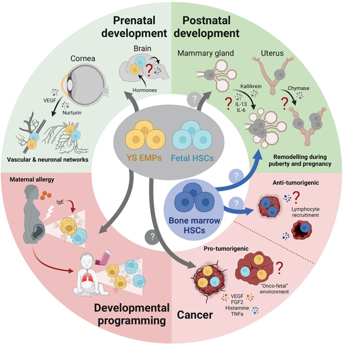
“Mast cells localize to the terminal end buds of the mammary gland ducts during puberty, lactation, and subsequent involution. 126 , 127 Mast cell‐deficient KitWsh/Wsh mice show a delay in pubertal ductal branching. 128”
§ Mast cell ontogeny: From fetal development to life‐long health and disease > IMPLICATIONS OF MAST CELL ONTOGENY FOR LIFE‐LONG HEALTH AND DISEASE > (Potential) functions of mast cells in early life
What the sources say
- Not checked — no full text of the sourcecites [126] Szewczyk G, Szukiewicz D, Zaczek R, Maślińska D. Mast cells in the mouse mammary gland – correlation with the development of lactiferous structures. Folia Biolotgica. 2000; 48(1–2): 13‐ 17. 11080912
- Check pendingcites [127] Lilla JN, Joshi RV, Craik CS, Werb Z. Active plasma kallikrein localizes to mast cells and regulates epithelial cell apoptosis, adipocyte differentiation, and stromal remodeling during mammary gland involution. J Biol Chem. 2009; 284(20): 13792‐ 13803. doi: 10.1074/jbc.M900508200 19297327 PMC2679480 source
- Check pendingcites [128] Lilla JN, Werb Z. Mast cells contribute to the stromal microenvironment in mammary gland branching morphogenesis. Dev Biol. 2010; 337: 124‐ 133. doi: 10.1016/j.ydbio.2009.10.021 19850030 PMC2787992 source
plainstates a cause- Asserted: passes — The authors put it forward as true.
- Checkable: passes — It can be checked against evidence.
- Load-bearing: passes — The paper’s conclusions depend on it.
- Based: passes — The paper gives a reason why it holds.
It may state two separate claims as one.
In the paper
“The cause of mastocytosis is mutational hyperactivation of KIT, the tyrosine kinase receptor for stem cell factor, which mast cell development and survival depend on. These mutations are typically somatic, and germline mutations are very rare. 149 , 150 , 151 , 152 , 153 , 154 , 155”
§ Mast cell ontogeny: From fetal development to life‐long health and disease > IMPLICATIONS OF MAST CELL ONTOGENY FOR LIFE‐LONG HEALTH AND DISEASE > Abnormal mast cell development and pathology
What the sources say
- Not checked — no full text of the sourcecites [149] Beghini A, Tibiletti MG, Roversi G, et al. Germline mutation in the juxtamembrane domain of the kit gene in a family with gastrointestinal stromal tumors and urticaria pigmentosa. Cancer. 2001; 92(3): 657‐ 662. doi: 10.1002/1097-0142(20010801)92:3<657::aid-cncr1367>3.0.co;2-d 11505412 source
- Not checked — no full text of the sourcecites [150] Akin C, Fumo G, Yavuz AS, Lipsky PE, Neckers L, Metcalfe DD. A novel form of mastocytosis associated with a transmembrane c‐kit mutation and response to imatinib. Blood. 2004; 103(8): 3222‐ 3225. doi: 10.1182/blood-2003-11-3816 15070706 source
- Check pendingcites [151] Tang X, Boxer M, Drummond A, Ogston P, Hodgins M, Burden AD. A germline mutation in KIT in familial diffuse cutaneous mastocytosis. J Med Genet. 2004; 41(6): e88. doi: 10.1136/jmg.2003.015156 15173254 PMC1735799 source
- Check pendingcites [152] Hartmann K, Wardelmann E, Ma Y, et al. Novel germline mutation of KIT associated with familial gastrointestinal stromal tumors and mastocytosis. Gastroenterology. 2005; 129(3): 1042‐ 1046. doi: 10.1053/j.gastro.2005.06.060 16143141 source
- Not checked — no full text of the sourcecites [153] Zhang LY, Smith ML, Schultheis B, et al. A novel K509I mutation of KIT identified in familial mastocytosis‐in vitro and in vivo responsiveness to imatinib therapy. Leuk Res. 2006; 30(4): 373‐ 378. doi: 10.1016/j.leukres.2005.08.015 16183119 source
- Check pendingcites [154] Wasag B, Niedoszytko M, Piskorz A, et al. Novel, activating KIT‐N822I mutation in familial cutaneous mastocytosis. Exp Hematol. 2011; 39(8): 859‐ 65.e2. doi: 10.1016/j.exphem.2011.05.009 21689725 source
- Not checked — no full text of the sourcecites [155] Speight RA, Nicolle A, Needham SJ, Verrill MW, Bryon J, Panter S. Rare, germline mutation of KIT with imatinib‐resistant multiple GI stromal tumors and mastocytosis. J Clin Oncol. 2013; 31(16): e245‐ e247. doi: 10.1200/JCO.2012.42.0133 23610110 source
plainstates a cause- Asserted: passes — The authors put it forward as true.
- Checkable: passes — It can be checked against evidence.
- Load-bearing: passes — The paper’s conclusions depend on it.
- Based: passes — The paper gives a reason why it holds.
It may state two separate claims as one.
In the paper
“At least on transcript level, fetal mast cells also express key effectors like proteases. 53 , 54 , 63 This is also true for human fetal mast cells, which express, for example, TPSAB1, the gene encoding tryptase, and carry granules. 59 , 60 , 63”
§ Mast cell ontogeny: From fetal development to life‐long health and disease > IMPLICATIONS OF MAST CELL ONTOGENY FOR LIFE‐LONG HEALTH AND DISEASE > (Potential) functions of mast cells in early life
What the sources say
- Check pendingcites [53] Gentek R, Ghigo C, Hoeffel G, et al. Hemogenic endothelial fate mapping reveals dual developmental origin of mast cells. Immunity. 2018; 48(6): 1160‐ 1171.e5. doi: 10.1016/j.immuni.2018.04.025 29858009 source
- Not checked — no full text of the sourcecites [54] Li Z, Liu S, Xu J, et al. Adult connective tissue‐resident mast cells originate from late erythro‐myeloid progenitors. Immunity. 2018; 49(4): 640‐ 653.e5. doi: 10.1016/j.immuni.2018.09.023 30332630 source
- Not checked — no full text of the sourcecites [63] Msallam R, Balla J, Rathore APS, et al. Fetal mast cells mediate postnatal allergic responses dependent on maternal IgE. Science. 2020; 370: 941‐ 950. doi: 10.1126/science.aba0864 33122426 source
- Not checked yetcites [59] Popescu D‐M, Botting RA, Stephenson E, et al. Decoding human fetal liver haematopoiesis. Nature. 2019; 574(7778): 365‐ 371. doi: 10.1038/s41586-019-1652-y 31597962 PMC6861135 source
- Check pendingcites [60] Suo C, Dann E, Goh I, et al. Mapping the developing human immune system across organs. Science. 2022; 376(6597): eabo0510. doi: 10.1126/science.abo0510 35549310 PMC7612819 source
plain- Asserted: passes — The authors put it forward as true.
- Checkable: passes — It can be checked against evidence.
- Load-bearing: passes — The paper’s conclusions depend on it.
- Based: passes — The paper gives a reason why it holds.
It may state two separate claims as one.
In the paper
“This view may be oversimplified, however, and it originally did not take into account the prenatal period, although prenatal exposure to microorganisms inversely correlates with the risk of developing atopy. 184 It has since been proposed that restricted maternal microbial exposure also renders their offspring hypersensitive, and that this is at least in part mediated by effects on fetal immune”
§ Mast cell ontogeny: From fetal development to life‐long health and disease > IMPLICATIONS OF MAST CELL ONTOGENY FOR LIFE‐LONG HEALTH AND DISEASE > Abnormal mast cell development and pathology
What the sources say
- Check pendingcites [184] Ege MJ, Herzum I, Büchele G, et al. Prenatal exposure to a farm environment modifies atopic sensitization at birth. J Allergy Clin Immunol. 2008; 122(2): 412.e4. doi: 10.1016/J.JACI.2008.06.011 18678343 source
- Check pendingcites [185] Apostol AC, Jensen KDC, Beaudin AE. Training the fetal immune system through maternal inflammation—a layered hygiene hypothesis. Front Immunol. 2020; 11: 1‐ 14. doi: 10.3389/fimmu.2020.00123 32117273 PMC7026678 source
hedgedstates a possible cause- Asserted: passes — The authors put it forward as true.
- Checkable: passes — It can be checked against evidence.
- Load-bearing: passes — The paper’s conclusions depend on it.
- Based: passes — The paper gives a reason why it holds.
Probably not a substantial claim. It may state two separate claims as one.
In the paper
“A firm link with early life adversity has also been established for neuro‐developmental and psychiatric disorders, 191 and immune dysregulation is involved in driving these pathologies. 192 , 193”
§ Mast cell ontogeny: From fetal development to life‐long health and disease > IMPLICATIONS OF MAST CELL ONTOGENY FOR LIFE‐LONG HEALTH AND DISEASE > Abnormal mast cell development and pathology
What the sources say
- Not checked — no full text of the sourcecites [191] Maccari S, Polese D, Reynaert ML, Amici T, Morley‐Fletcher S, Fagioli F. Early‐life experiences and the development of adult diseases with a focus on mental illness: the human birth theory. Neuroscience. 2017; 342: 232‐ 251. doi: 10.1016/J.NEUROSCIENCE.2016.05.042 27235745 source
- Check pendingcites [192] Bennett FC, Molofsky AV. The immune system and psychiatric disease: a basic science perspective. Clin Exp Immunol. 2019; 197(3): 294‐ 307. doi: 10.1111/CEI.13334 31125426 PMC6693968 source
- Not checked yetcites [193] Yuan N, Chen Y, Xia Y, Dai J, Liu C. Inflammation‐related biomarkers in major psychiatric disorders: a cross‐disorder assessment of reproducibility and specificity in 43 meta‐analyses. Transl Psychiatry. 2019; 9(1): 1‐ 13. doi: 10.1038/S41398-019-0570-Y 31534116 PMC6751188 source
boostedstates a cause- Asserted: passes — The authors put it forward as true.
- Checkable: passes — It can be checked against evidence.
- Load-bearing: passes — The paper’s conclusions depend on it.
- Based: passes — The paper gives a reason why it holds.
It may state two separate claims as one.
A conclusion the paper draws from its own results — it cites no other source for it, so there is none to check. The quote shows it is in the paper.
In the paper
“Overall, available data thus indicate that following perturbations, replenishment, or expansion of tissue‐resident mast cell populations can be achieved via several, complementary mechanisms. Progenitors can be mobilized from the BM or alternative sources, and/or recruited progenitors and (remaining) mature mast cells can (further) expand in the affected tissue by proliferation.”
§ Mast cell ontogeny: From fetal development to life‐long health and disease > MAST CELL ONTOGENY > Non‐homeostatic mast cell replenishment
hedgedstates a cause- Asserted: passes — The authors put it forward as true.
- Checkable: passes — It can be checked against evidence.
- Load-bearing: passes — The paper’s conclusions depend on it.
- Based: passes — The paper gives a reason why it holds.
It may state two separate claims as one.
In the paper
“Similarly, Dwyer and colleagues identified a population of progenitor‐like immature mast cells in nasal polyps of patients with chronic rhinosinusitis. 102 These are distinct from circulating mast cell progenitors and appear highly proliferative.”
§ Mast cell ontogeny: From fetal development to life‐long health and disease > MAST CELL ONTOGENY > Non‐homeostatic mast cell replenishment
What the sources say
- Check pendingcites [102] Dwyer DF, Ordovas‐Montanes J, Allon SJ, et al. Human airway mast cells proliferate and acquire distinct inflammation‐driven phenotypes during type 2 inflammation. Sci Immunol. 2021; 6(56): 1‐ 31. doi: 10.1126/SCIIMMUNOL.ABB7221 PMC8362933 33637594 source
hedgedstates an association- Asserted: passes — The authors put it forward as true.
- Checkable: passes — It can be checked against evidence.
- Load-bearing: unclear — It is unclear whether the paper’s conclusions depend on it.
- Based: passes — The paper gives a reason why it holds.
It may state two separate claims as one.
A statement the paper makes itself — it cites no source for it, so there is none to check. The quote shows it is in the paper.
In the paper
“Historically, mice were used that carry hypomorphic alleles of Kit, such as KitW/Wv and KitWsh/Wsh mice. In these mice, mast cells are absent or reduced because they depend on Kit signaling for their growth and survival.”
§ Mast cell ontogeny: From fetal development to life‐long health and disease > MAST CELL ONTOGENY > How are mast cells maintained postnatally?
plainstates a cause- Asserted: passes — The authors put it forward as true.
- Checkable: passes — It can be checked against evidence.
- Load-bearing: unclear — It is unclear whether the paper’s conclusions depend on it.
- Based: passes — The paper gives a reason why it holds.
It may state two separate claims as one.
A conclusion the paper draws from its own results — it cites no other source for it, so there is none to check. The quote shows it is in the paper.
In the paper
“Connective tissue‐resident mast cells originate from YS EMPs and likely later fetal‐restricted progenitors. Once established, they self‐maintain independently from the BM.”
§ Mast cell ontogeny: From fetal development to life‐long health and disease > MAST CELL ONTOGENY > Non‐homeostatic mast cell replenishment
hedgedstates a cause- Asserted: passes — The authors put it forward as true.
- Checkable: passes — It can be checked against evidence.
- Load-bearing: passes — The paper’s conclusions depend on it.
- Based: unclear — It is unclear why the paper says it holds.
It may state two separate claims as one.
In the paper
“Substantial influx of BM progenitors to the skin also occurs following application of 12‐O‐tetradecanoylphorbol‐13‐acetate (TPA), a small molecule drug originally derived from a plant that induces skin rashes. 86 Within TPA‐treated inflamed skin, BM progenitors clonally expand and invade the territories of pre‐existing mast cells. 86”
§ Mast cell ontogeny: From fetal development to life‐long health and disease > MAST CELL ONTOGENY > Non‐homeostatic mast cell replenishment
What the sources say
- Not checked — no full text of the sourcecites [86] Weitzmann A, Naumann R, Dudeck A, Zerjatke T, Gerbaulet A, Roers A. Mast cells occupy stable clonal territories in adult steady‐state skin. J Invest Dermatol. 2020; 140(12): 2433‐ 2441.e5. doi: 10.1016/J.JID.2020.03.963 32311397 source
plainstates a cause- Asserted: passes — The authors put it forward as true.
- Checkable: passes — It can be checked against evidence.
- Load-bearing: unclear — It is unclear whether the paper’s conclusions depend on it.
- Based: passes — The paper gives a reason why it holds.
It may state two separate claims as one.
In the paper
“Mast cell‐deficient female mice display defective spiral artery remodeling and impaired implantation, collectively resulting in fetal growth restriction. 122 , 124 , 125 Interestingly, mast cells appear to mediate these processes cooperatively with uterine NK cells, since both can (partially) compensate in the absence of the other. 125”
§ Mast cell ontogeny: From fetal development to life‐long health and disease > IMPLICATIONS OF MAST CELL ONTOGENY FOR LIFE‐LONG HEALTH AND DISEASE > (Potential) functions of mast cells in early life
What the sources say
- Check pendingcites [122] Woidacki K, Popovic M, Metz M, et al. Mast cells rescue implantation defects caused by c‐kit deficiency. Cell Death Dis. 2013; 4: e462. doi: 10.1038/cddis.2012.214 23328669 PMC3564001 source
- Check pendingcites [124] Meyer N, Woidacki K, Knöfler M, et al. Chymase‐producing cells of the innate immune system are required for decidual vascular remodeling and fetal growth. Sci Rep. 2017; 7: 45106. doi: 10.1038/srep45106 28327604 PMC5361184 source
- Check pendingcites [125] Meyer N, Woidacki K, Maurer M, Zenclussen AC. Safeguarding of fetal growth by mast cells and natural killer cells: deficiency of one is counterbalanced by the other. Front Immunol. 2017; 8(JUN): 711. doi: 10.3389/fimmu.2017.00711 28670317 PMC5472686 source
hedgedstates a cause- Asserted: passes — The authors put it forward as true.
- Checkable: passes — It can be checked against evidence.
- Load-bearing: passes — The paper’s conclusions depend on it.
- Based: passes — The paper gives a reason why it holds.
It may state two separate claims as one.
A statement the paper makes itself — it cites no source for it, so there is none to check. The quote shows it is in the paper.
In the paper
“Additionally, Kit‐dependent models lack interstitial cells of Cajal and show abnormal pacemaker activity in the gastrointestinal tract.”
§ Mast cell ontogeny: From fetal development to life‐long health and disease > MAST CELL ONTOGENY > How are mast cells maintained postnatally?
plainstates a cause- Asserted: passes — The authors put it forward as true.
- Checkable: passes — It can be checked against evidence.
- Load-bearing: passes — The paper’s conclusions depend on it.
- Based: passes — The paper gives a reason why it holds.
It may state two separate claims as one.
A claim is checked only against the cited paper’s full text, never its abstract, and none is available for its sources.
In the paper
“Their ontogeny may make mast cells particularly susceptible to developmental programming: They are already present and phenotypically plastic at fetal stages and can sense environmental perturbations. 63 , 170 They are also long‐lived and many fetal‐derived mast cells persist postnatally.”
§ Mast cell ontogeny: From fetal development to life‐long health and disease > IMPLICATIONS OF MAST CELL ONTOGENY FOR LIFE‐LONG HEALTH AND DISEASE > Abnormal mast cell development and pathology
What the sources say
- Not checked — no full text of the sourcecites [63] Msallam R, Balla J, Rathore APS, et al. Fetal mast cells mediate postnatal allergic responses dependent on maternal IgE. Science. 2020; 370: 941‐ 950. doi: 10.1126/science.aba0864 33122426 source
- Not checked — no full text of the sourcecites [170] Balistreri CR, Garagnani P, Madonna R, Vaiserman A, Melino G. Developmental programming of adult haematopoiesis system. Ageing Res Rev. 2019; 54: 100918. doi: 10.1016/j.arr.2019.100918 31226498 source
hedgedstates a possible cause- Asserted: passes — The authors put it forward as true.
- Checkable: passes — It can be checked against evidence.
- Load-bearing: passes — The paper’s conclusions depend on it.
- Based: passes — The paper gives a reason why it holds.
It may state two separate claims as one.
A conclusion the paper draws from its own results — it cites no other source for it, so there is none to check. The quote shows it is in the paper.
In the paper
“Rather than waiting for adult‐type HSCs that become the dominant source of immune cells perinatally, fetal tissues are thus seeded with mast cells from the earliest possible source, suggesting they are required before birth.”
§ Mast cell ontogeny: From fetal development to life‐long health and disease > IMPLICATIONS OF MAST CELL ONTOGENY FOR LIFE‐LONG HEALTH AND DISEASE > (Potential) functions of mast cells in early life
hedgedstates a cause- Asserted: passes — The authors put it forward as true.
- Checkable: passes — It can be checked against evidence.
- Load-bearing: passes — The paper’s conclusions depend on it.
- Based: passes — The paper gives a reason why it holds.
It may state two separate claims as one.
A conclusion the paper draws from its own results — it cites no other source for it, so there is none to check. The quote shows it is in the paper.
In the paper
“This supports the hypothesis that pediatric‐ and adult‐onset mastocytosis may have distinct cells of origin.”
§ Mast cell ontogeny: From fetal development to life‐long health and disease > IMPLICATIONS OF MAST CELL ONTOGENY FOR LIFE‐LONG HEALTH AND DISEASE > Abnormal mast cell development and pathology
hedgedstates a possible cause- Asserted: fails — The authors may not put it forward as true.
- Checkable: passes — It can be checked against evidence.
- Load-bearing: passes — The paper’s conclusions depend on it.
- Based: passes — The paper gives a reason why it holds.
Probably not a substantial claim. The authors may not put it forward as true.
- Rests on an assumption: The normal developmental kinetics of mast cells in mice, in which YS EMP-derived mast cells are gradually replaced by later progenitor waves, are similar enough in humans to explain pediatric- versus adult-onset mastocytosis.
In the paper
“Of note, the area covered by blood vessels and the density of nerve fibers are both significantly reduced in newborn KitWsh/Wsh mice that do not have corneal mast cells. 55 This indicates that fetal mast cells mediate vascular and nerve branching in the developing cornea.”
§ Mast cell ontogeny: From fetal development to life‐long health and disease > IMPLICATIONS OF MAST CELL ONTOGENY FOR LIFE‐LONG HEALTH AND DISEASE > (Potential) functions of mast cells in early life
What the sources say
- Check pendingcites [55] Liu J, Fu T, Song F, et al. Mast cells participate in corneal development in mice. Sci Rep. 2015; 5: 17569. doi: 10.1038/srep17569 26627131 PMC4667177 source
hedgedstates a cause- Asserted: passes — The authors put it forward as true.
- Checkable: passes — It can be checked against evidence.
- Load-bearing: passes — The paper’s conclusions depend on it.
- Based: passes — The paper gives a reason why it holds.
It may state two separate claims as one.
In the paper
“Indeed, mast cells appear to be functionally mature at fetal stages: In mice, mast cells detected as early as E12.5 in the cornea and E14.5 in the skin already contain characteristic granules. 53 , 55”
§ Mast cell ontogeny: From fetal development to life‐long health and disease > IMPLICATIONS OF MAST CELL ONTOGENY FOR LIFE‐LONG HEALTH AND DISEASE > (Potential) functions of mast cells in early life
What the sources say
- Check pendingcites [53] Gentek R, Ghigo C, Hoeffel G, et al. Hemogenic endothelial fate mapping reveals dual developmental origin of mast cells. Immunity. 2018; 48(6): 1160‐ 1171.e5. doi: 10.1016/j.immuni.2018.04.025 29858009 source
- Check pendingcites [55] Liu J, Fu T, Song F, et al. Mast cells participate in corneal development in mice. Sci Rep. 2015; 5: 17569. doi: 10.1038/srep17569 26627131 PMC4667177 source
hedgedstates an association- Asserted: passes — The authors put it forward as true.
- Checkable: passes — It can be checked against evidence.
- Load-bearing: passes — The paper’s conclusions depend on it.
- Based: passes — The paper gives a reason why it holds.
It may state two separate claims as one.
None of the sources cited for this claim have been checked yet.
In the paper
“Sensitization to allergens may begin before birth. 176 , 177 , 178 , 179 Indeed, heredity of allergy is more strongly associated with maternal IgE compared to paternal IgE. 180 , 181”
§ Mast cell ontogeny: From fetal development to life‐long health and disease > IMPLICATIONS OF MAST CELL ONTOGENY FOR LIFE‐LONG HEALTH AND DISEASE > Abnormal mast cell development and pathology
What the sources say
- Not checked yetcites [176] Piccinni MP, Mecacci F, Sampognaro S, et al. Aeroallergen sensitization can occur during fetal life. Int Arch Allergy Immunol. 1993; 102(3): 301‐ 303. doi: 10.1159/000236541 8219784
- Not checked — no full text of the sourcecites [177] Kihlström A, Lilja G, Pershagen G, Hedlin G. Exposure to high doses of birch pollen during pregnancy, and risk of sensitization and atopic disease in the child. Allergy Eur J Allergy Clin Immunol. 2003; 58: 871‐ 877. doi: 10.1034/j.1398-9995.2003.00232.x 12911415 source
- Not checked — no full text of the sourcecites [178] Boyle RJ, Tang MLK. Can allergic diseases be prevented prenatally? Allergy Eur J Allergy Clin Immunol. 2006; 61: 1423‐ 1431. doi: 10.1111/j.1398-9995.2006.01113.x 17073873 source
- Not checked yetcites [179] Warner JA, Jones AC, Miles EA, Colwell BM, Warner JO. Prenatal origins of asthma and allergy. Ciba Found Symp. 1997; 206: 220‐ 232. doi: 10.1002/9780470515334.CH14 9257015
- Not checked — no full text of the sourcecites [180] Liu CA, Wang CL, Chuang H, Ou CY, Hsu TY, Yang KD. Prenatal prediction of infant atopy by maternal but not paternal total IgE levels. J Allergy Clin Immunol. 2003; 112: 899‐ 904. doi: 10.1016/j.jaci.2003.08.030 14610477 source
- Not checked yetcites [181] Wu CC, Chen RF, Kuo HC. Different implications of paternal and maternal atopy for perinatal IgE production and asthma development. Clin Dev Immunol. 2012; 2012: 1‐ 10. doi: 10.1155/2012/132142 PMC3261469 22272211
hedgedstates an association- Asserted: passes — The authors put it forward as true.
- Checkable: passes — It can be checked against evidence.
- Load-bearing: passes — The paper’s conclusions depend on it.
- Based: passes — The paper gives a reason why it holds.
It may state two separate claims as one.
A claim is checked only against the cited paper’s full text, never its abstract, and none is available for its sources.
In the paper
“It has been proposed that BM progenitors are a primary source of mast cells following environmental perturbations, such as inflammatory insults. 99 , 100”
§ Mast cell ontogeny: From fetal development to life‐long health and disease > MAST CELL ONTOGENY > Non‐homeostatic mast cell replenishment
What the sources say
- Not checked — no full text of the sourcecites [99] Grootens J, Ungerstedt JS, Nilsson G, Dahlin JS. Deciphering the differentiation trajectory from hematopoietic stem cells to mast cells. Blood Adv. 2018; 2(17): 2273‐ 2281. doi: 10.1182/BLOODADVANCES.2018019539 30206100 PMC6134220 source
- Not checked — no full text of the sourcecites [100] Nilsson G, Dahlin JS. New insights into the origin of mast cells. Allergy. 2019; 74(4): 844‐ 845. doi: 10.1111/ALL.13668 30427067 source
hedgedstates a possible cause- Asserted: passes — The authors put it forward as true.
- Checkable: passes — It can be checked against evidence.
- Load-bearing: fails — The paper’s conclusions may not depend on it.
- Based: passes — The paper gives a reason why it holds.
Probably not a substantial claim. The paper’s conclusions may not depend on it.
In the paper
“Mast cells colonize the developing cornea in two waves. The first mast cells concentrate around the corneal stroma from E12.5 onwards and disappear by the time of eyelid opening. A second wave of mast cells settles in the corneal limbus within the first postnatal week and stabilizes thereafter. These are recruited to the corneal limbus via a mechanism that depends on integrin α4β7 and Cxcr2. 55”
§ Mast cell ontogeny: From fetal development to life‐long health and disease > IMPLICATIONS OF MAST CELL ONTOGENY FOR LIFE‐LONG HEALTH AND DISEASE > (Potential) functions of mast cells in early life
What the sources say
- Check pendingcites [55] Liu J, Fu T, Song F, et al. Mast cells participate in corneal development in mice. Sci Rep. 2015; 5: 17569. doi: 10.1038/srep17569 26627131 PMC4667177 source
plainstates a cause- Asserted: passes — The authors put it forward as true.
- Checkable: passes — It can be checked against evidence.
- Load-bearing: unclear — It is unclear whether the paper’s conclusions depend on it.
- Based: passes — The paper gives a reason why it holds.
It may state two separate claims as one.
A conclusion the paper draws from its own results — it cites no other source for it, so there is none to check. The quote shows it is in the paper.
In the paper
“This mechanism thus propagates maternal disease to the next generation and could explain the rapidly rising incidence of allergic disease.”
§ Mast cell ontogeny: From fetal development to life‐long health and disease > IMPLICATIONS OF MAST CELL ONTOGENY FOR LIFE‐LONG HEALTH AND DISEASE > Abnormal mast cell development and pathology
hedgedstates a cause- Asserted: passes — The authors put it forward as true.
- Checkable: passes — It can be checked against evidence.
- Load-bearing: passes — The paper’s conclusions depend on it.
- Based: passes — The paper gives a reason why it holds.
It may state two separate claims as one.
In the paper
“In keeping with the hygiene hypothesis, germ‐free mice have reduced mast cell numbers in the intestinal mucosa and are resistant to food allergy models. 82”
§ Mast cell ontogeny: From fetal development to life‐long health and disease > IMPLICATIONS OF MAST CELL ONTOGENY FOR LIFE‐LONG HEALTH AND DISEASE > Abnormal mast cell development and pathology
What the sources say
- Check pendingcites [82] Schwarzer M, Hermanova P, Srutkova D, et al. Germ‐free mice exhibit mast cells with impaired functionality and gut homing and do not develop food allergy. Front Immunol. 2019; 10: 205. doi: 10.3389/fimmu.2019.00205 30809227 PMC6379318 source
plainstates a cause- Asserted: passes — The authors put it forward as true.
- Checkable: passes — It can be checked against evidence.
- Load-bearing: passes — The paper’s conclusions depend on it.
- Based: passes — The paper gives a reason why it holds.
It may state two separate claims as one.
In the paper
“Pharmacological inhibition of mast cells at late fetal stages dampens copulation behavior in adult male offspring, suggesting that masculinization is normally mediated by mast cells. Conversely, perinatal administration of exogenous estradiol or mast cell activation using the compound 48/80 both partially masculinize the copulation behavior of female mice.”
§ Mast cell ontogeny: From fetal development to life‐long health and disease > IMPLICATIONS OF MAST CELL ONTOGENY FOR LIFE‐LONG HEALTH AND DISEASE > (Potential) functions of mast cells in early life
What the sources say
- Check pendingcites [120] Lenz KM, Pickett LA, Wright CL, Davis KT, Joshi A, McCarthy MM. Mast cells in the developing brain determine adult sexual behavior. J Neurosci. 2018; 38(37): 8044‐ 8059. doi: 10.1523/JNEUROSCI.1176-18.2018 30093566 PMC6136154 source
hedgedstates a cause- Asserted: passes — The authors put it forward as true.
- Checkable: passes — It can be checked against evidence.
- Load-bearing: passes — The paper’s conclusions depend on it.
- Based: passes — The paper gives a reason why it holds.
It may state two separate claims as one.
A statement the paper makes itself — it cites no source for it, so there is none to check. The quote shows it is in the paper.
In the paper
“For example, KitW/Wv mice have fewer neutrophils and basophils, are anemic and sterile.”
§ Mast cell ontogeny: From fetal development to life‐long health and disease > MAST CELL ONTOGENY > How are mast cells maintained postnatally?
plainstates an association- Asserted: passes — The authors put it forward as true.
- Checkable: passes — It can be checked against evidence.
- Load-bearing: unclear — It is unclear whether the paper’s conclusions depend on it.
- Based: passes — The paper gives a reason why it holds.
It may state two separate claims as one.
A conclusion the paper draws from its own results — it cites no other source for it, so there is none to check. The quote shows it is in the paper.
In the paper
“As we will discuss in the following section, their functions may be adapted to the challenges of distinct life stages, and can potentially be co‐opted in pathological settings.”
§ Mast cell ontogeny: From fetal development to life‐long health and disease > MAST CELL ONTOGENY > Non‐homeostatic mast cell replenishment
hedgedstates a possible cause- Asserted: passes — The authors put it forward as true.
- Checkable: passes — It can be checked against evidence.
- Load-bearing: passes — The paper’s conclusions depend on it.
- Based: passes — The paper gives a reason why it holds.
Probably not a substantial claim. It may state two separate claims as one.
A statement the paper makes itself — it cites no source for it, so there is none to check. The quote shows it is in the paper.
In the paper
“Other potential issues include off‐target effects or “Cre leakiness,” which can in principle be a concern for all Cre‐based approaches, as well as varying levels of depletion, certainly in inducible models.”
§ Mast cell ontogeny: From fetal development to life‐long health and disease > MAST CELL ONTOGENY > How are mast cells maintained postnatally?
hedged- Asserted: passes — The authors put it forward as true.
- Checkable: passes — It can be checked against evidence.
- Load-bearing: unclear — It is unclear whether the paper’s conclusions depend on it.
- Based: passes — The paper gives a reason why it holds.
It may state two separate claims as one.
None of the sources cited for this claim have been checked yet.
In the paper
“Fetal mast cells share their YS EMP origin with macrophages, which are known to have pivotal roles in development. 31 , 32 , 48”
§ Mast cell ontogeny: From fetal development to life‐long health and disease > IMPLICATIONS OF MAST CELL ONTOGENY FOR LIFE‐LONG HEALTH AND DISEASE > (Potential) functions of mast cells in early life
What the sources say
- Not checked yetcites [31] Ginhoux F, Greter M, Leboeuf M, et al. Fate mapping analysis reveals that adult microglia derive from primitive macrophages. Science. 2010; 330(6005): 841‐ 845. doi: 10.1126/science.1194637 20966214 PMC3719181 source
- Not checked yetcites [32] Gomez Perdiguero E, Klapproth K, Schulz C, et al. Tissue‐resident macrophages originate from yolk‐sac‐derived erythro‐myeloid progenitors. Nature. 2015; 518(7540): 547‐ 551. doi: 10.1038/nature13989 25470051 PMC5997177 source
- Not checked yetcites [48] Hoeffel G, Chen J, Lavin Y, et al. C‐Myb+ erythro‐myeloid progenitor‐derived fetal monocytes give rise to adult tissue‐resident macrophages. Immunity. 2015; 42(4): 665‐ 678. doi: 10.1016/j.immuni.2015.03.011 25902481 PMC4545768 source
boostedstates an association- Asserted: passes — The authors put it forward as true.
- Checkable: passes — It can be checked against evidence.
- Load-bearing: passes — The paper’s conclusions depend on it.
- Based: passes — The paper gives a reason why it holds.
It may state two separate claims as one.
In the paper
“Based on numerous epidemiological studies, it is now firmly established that adult health and disease have developmental origins. 167 Exposure to adverse environments in utero or at early postnatal stages is associated with a wide range of non‐communicable diseases, 168 many of which are characterized by inflammation and aberrant immune responses. 169”
§ Mast cell ontogeny: From fetal development to life‐long health and disease > IMPLICATIONS OF MAST CELL ONTOGENY FOR LIFE‐LONG HEALTH AND DISEASE > Abnormal mast cell development and pathology
What the sources say
- Not checked yetcites [167] Barker DJP. Developmental origins of adult health and disease. J Epidemiol Community Health. 2004; 58(2): 114‐ 115. doi: 10.1136/JECH.58.2.114 14729887 PMC1732687
- Check pendingcites [168] Padmanabhan V, Cardoso RC, Puttabyatappa M. Developmental programming, a pathway to disease. Endocrinology. 2016; 157(4): 1328‐ 1340. doi: 10.1210/en.2016-1003 26859334 PMC4816734 source
- Check pendingcites [169] Chen T, Liu HX, Yan HY, Wu DM, Ping J. Developmental origins of inflammatory and immune diseases. Mol Hum Reprod. 2016; 22(8): 558‐ 565. doi: 10.1093/molehr/gaw036 PMC4986419 27226490 source
boostedstates an association- Asserted: passes — The authors put it forward as true.
- Checkable: passes — It can be checked against evidence.
- Load-bearing: passes — The paper’s conclusions depend on it.
- Based: passes — The paper gives a reason why it holds.
It may state two separate claims as one.
In the paper
“Indeed, mast cell progenitors are also present in the spleen 104 and murine adipose tissues, 105 to which the latter are mobilized upon experimental myocardial infarction. 106”
§ Mast cell ontogeny: From fetal development to life‐long health and disease > MAST CELL ONTOGENY > Non‐homeostatic mast cell replenishment
What the sources say
- Check pendingcites [104] Arinobu Y, Iwasaki H, Gurish MF, et al. Developmental checkpoints of the basophil/mast cell lineages in adult murine hematopoiesis. Proc Natl Acad Sci USA. 2005; 102(50): 18105‐ 18110. doi: 10.1073/PNAS.0509148102 16330751 PMC1312421 source
- Not checked — no full text of the sourcecites [105] Poglio S, De Toni‐Costes F, Arnaud E, et al. Adipose tissue as a dedicated reservoir of functional mast cell progenitors. Stem Cells. 2010; 28(11): 2065‐ 2072. doi: 10.1002/stem.523 20845475 source
- Check pendingcites [106] Gomez I, Robert V, Alayrac P, et al. Identification of adipose tissue as a reservoir of macrophages after acute myocardial infarction. Int J Mol Sci. 2022; 23(18): 1‐ 15. doi: 10.3390/IJMS231810498 PMC9499676 36142416 source
plainstates a cause- Asserted: passes — The authors put it forward as true.
- Checkable: passes — It can be checked against evidence.
- Load-bearing: unclear — It is unclear whether the paper’s conclusions depend on it.
- Based: passes — The paper gives a reason why it holds.
It may state two separate claims as one.
In the paper
“Mast cells can also be found in the developing brain of mice 54 and rats, 119 including the pre‐optic area. 120 This brain region is essential for male copulatory behavior, which is marked by a higher synaptic density compared to females. Late fetal and early postnatal stages are critical for sexual differentiation in this region. At these stages, mast cell numbers are higher in the pre‐optic area”
§ Mast cell ontogeny: From fetal development to life‐long health and disease > IMPLICATIONS OF MAST CELL ONTOGENY FOR LIFE‐LONG HEALTH AND DISEASE > (Potential) functions of mast cells in early life
What the sources say
- Not checked — no full text of the sourcecites [54] Li Z, Liu S, Xu J, et al. Adult connective tissue‐resident mast cells originate from late erythro‐myeloid progenitors. Immunity. 2018; 49(4): 640‐ 653.e5. doi: 10.1016/j.immuni.2018.09.023 30332630 source
- Not checked — no full text of the sourcecites [119] Lambracht‐Hall M, Dimitriadou V, Theoharides TC. Migration of mast cells in the developing rat brain. Dev Brain Res. 1990; 56(2): 151‐ 159. doi: 10.1016/0165-3806(90)90077-C 2261679 source
- Check pendingcites [120] Lenz KM, Pickett LA, Wright CL, Davis KT, Joshi A, McCarthy MM. Mast cells in the developing brain determine adult sexual behavior. J Neurosci. 2018; 38(37): 8044‐ 8059. doi: 10.1523/JNEUROSCI.1176-18.2018 30093566 PMC6136154 source
plainstates a cause- Asserted: passes — The authors put it forward as true.
- Checkable: passes — It can be checked against evidence.
- Load-bearing: passes — The paper’s conclusions depend on it.
- Based: passes — The paper gives a reason why it holds.
It may state two separate claims as one.
A conclusion the paper draws from its own results — it cites no other source for it, so there is none to check. The quote shows it is in the paper.
In the paper
“Mast cells thus regulate sexual differentiation in the brain during development in a hormone‐responsive manner and via mechanisms that involve other immune cells.”
§ Mast cell ontogeny: From fetal development to life‐long health and disease > IMPLICATIONS OF MAST CELL ONTOGENY FOR LIFE‐LONG HEALTH AND DISEASE > (Potential) functions of mast cells in early life
plainstates a cause- Asserted: passes — The authors put it forward as true.
- Checkable: passes — It can be checked against evidence.
- Load-bearing: passes — The paper’s conclusions depend on it.
- Based: passes — The paper gives a reason why it holds.
It may state two separate claims as one.
A conclusion the paper draws from its own results — it cites no other source for it, so there is none to check. The quote shows it is in the paper.
In the paper
“In summary, mast cells are versatile immune cells that arise during fetal development and show considerable heterogeneity between and within tissues and in response to environmental perturbations.”
§ Mast cell ontogeny: From fetal development to life‐long health and disease > MAST CELL ONTOGENY > Non‐homeostatic mast cell replenishment
plainstates an association- Asserted: passes — The authors put it forward as true.
- Checkable: passes — It can be checked against evidence.
- Load-bearing: passes — The paper’s conclusions depend on it.
- Based: passes — The paper gives a reason why it holds.
It may state two separate claims as one.
A statement the paper makes itself — it cites no source for it, so there is none to check. The quote shows it is in the paper.
In the paper
“They are potent immune effectors that can rapidly respond to harmful stimuli owing to their intracellular granules loaded with pre‐formed effectors. Furthermore, mast cells precede the adaptive immune system both ontogenetically and phylogenetically.”
§ Mast cell ontogeny: From fetal development to life‐long health and disease > IMPLICATIONS OF MAST CELL ONTOGENY FOR LIFE‐LONG HEALTH AND DISEASE > (Potential) functions of mast cells in early life
plainstates a cause- Asserted: passes — The authors put it forward as true.
- Checkable: passes — It can be checked against evidence.
- Load-bearing: passes — The paper’s conclusions depend on it.
- Based: passes — The paper gives a reason why it holds.
It may state two separate claims as one.
In the paper
“The underlying mechanisms have not been explored in detail, but it was reported that corneal mast cells produce vascular endothelial growth factor (VEGF) and neurturin, which, respectively, have angiogenic and neurotrophic actions. In agreement with this, inappropriately high levels of mast cell‐derived VEGF promote neovascularization in the adult cornea, that is, excessive angiogenesis that can”
§ Mast cell ontogeny: From fetal development to life‐long health and disease > IMPLICATIONS OF MAST CELL ONTOGENY FOR LIFE‐LONG HEALTH AND DISEASE > (Potential) functions of mast cells in early life
What the sources say
- Check pendingcites [118] Cho WK, Mittal SK, Elbasiony E, Chauhan SK. Activation of ocular surface mast cells promotes corneal neovascularization. Ocul Surf. 2020; 18(4): 857‐ 864. doi: 10.1016/J.JTOS.2020.09.002 32916251 PMC7686271 source
plainstates a cause- Asserted: passes — The authors put it forward as true.
- Checkable: passes — It can be checked against evidence.
- Load-bearing: unclear — It is unclear whether the paper’s conclusions depend on it.
- Based: passes — The paper gives a reason why it holds.
It may state two separate claims as one.
A statement the paper makes itself — it cites no source for it, so there is none to check. The quote shows it is in the paper.
In the paper
“Pediatric‐onset disease is predominantly cutaneous, tends to follow a milder clinical course and usually regresses spontaneously by adolescence. Adult‐onset disease, however, is more frequently systemic and does not normally regress.”
§ Mast cell ontogeny: From fetal development to life‐long health and disease > IMPLICATIONS OF MAST CELL ONTOGENY FOR LIFE‐LONG HEALTH AND DISEASE > Abnormal mast cell development and pathology
plainstates an association- Asserted: passes — The authors put it forward as true.
- Checkable: passes — It can be checked against evidence.
- Load-bearing: passes — The paper’s conclusions depend on it.
- Based: passes — The paper gives a reason why it holds.
It may state two separate claims as one.
In the paper
“Symptomatic MCAS episodes appear to be triggered by environmental exposures, for example to psychological stress, 165 sudden temperature changes or odors and chemicals. 166 In addition to triggering symptoms, environmental exposures may also contribute to MCAS etiology: A recent study has explored a possible link between MCAS and (acquired) chemical intolerance, and proposed that xenobiotic”
§ Mast cell ontogeny: From fetal development to life‐long health and disease > IMPLICATIONS OF MAST CELL ONTOGENY FOR LIFE‐LONG HEALTH AND DISEASE > Abnormal mast cell development and pathology
What the sources say
- Check pendingcites [165] Theoharides TC, Alysandratos KD, Angelidou A, et al. Mast cells and inflammation. Biochim Biophys Acta. 2012; 1822(1): 21‐ 33. doi: 10.1016/J.BBADIS.2010.12.014 21185371 PMC3318920 source
- Check pendingcites [166] Miller CS, Palmer RF, Dempsey TT, Ashford NA, Afrin LB. Mast cell activation may explain many cases of chemical intolerance. Environ Sci Eur. 2021; 33(1): 1‐ 15. doi: 10.1186/S12302-021-00570-3/FIGURES/7 source
hedgedstates a possible cause- Asserted: passes — The authors put it forward as true.
- Checkable: passes — It can be checked against evidence.
- Load-bearing: unclear — It is unclear whether the paper’s conclusions depend on it.
- Based: passes — The paper gives a reason why it holds.
It may state two separate claims as one.
A statement the paper makes itself — it cites no source for it, so there is none to check. The quote shows it is in the paper.
In the paper
“RMB mice are an interesting model in this regard, since basophils recover much more quickly from Diphtheria toxin‐induced depletion than mast cells. This makes this model more mast cell‐specific at later stages.”
§ Mast cell ontogeny: From fetal development to life‐long health and disease > MAST CELL ONTOGENY > How are mast cells maintained postnatally?
plainstates a cause- Asserted: passes — The authors put it forward as true.
- Checkable: passes — It can be checked against evidence.
- Load-bearing: passes — The paper’s conclusions depend on it.
- Based: passes — The paper gives a reason why it holds.
It may state two separate claims as one.
In the paper
“As in mice, committed mast cell progenitors (or immature mast cells) have also been found at extra‐medullary sites in humans, including the spleen and a range of non‐lymphoid tissues, and they too can be recruited in inflammatory conditions. 10”
§ Mast cell ontogeny: From fetal development to life‐long health and disease > MAST CELL ONTOGENY > Non‐homeostatic mast cell replenishment
What the sources say
- Not checked — no full text of the sourcecites [10] St John AL, Rathore APS, Ginhoux F. New perspectives on the origins and heterogeneity of mast cells. Nat Rev Immunol. 2022; 23: 55‐ 68. doi: 10.1038/s41577-022-00731-2 35610312 source
plainstates an association- Asserted: passes — The authors put it forward as true.
- Checkable: passes — It can be checked against evidence.
- Load-bearing: passes — The paper’s conclusions depend on it.
- Based: passes — The paper gives a reason why it holds.
It may state two separate claims as one.
In the paper
“Auto‐activating KIT mutations are a shared feature of different types of mastocytosis. 156 Most common is a substituting point mutation in codon 186 (D186V), which is found in 80% of adult patients. 157 Although less abundant in these patients, this mutation still accounts for almost 40% of pediatric cases, and of the remaining pediatric patients, another 40% carry activating mutations in other”
§ Mast cell ontogeny: From fetal development to life‐long health and disease > IMPLICATIONS OF MAST CELL ONTOGENY FOR LIFE‐LONG HEALTH AND DISEASE > Abnormal mast cell development and pathology
What the sources say
- Check pendingcites [156] Frieri M, Quershi M. Pediatric mastocytosis: a review of the literature. Pediatr Allergy Immunol Pulmonol. 2013; 26(4): 175‐ 180. doi: 10.1089/ped.2013.0275 24380017 PMC3869446 source
- Not checked — no full text of the sourcecites [157] Bodemer C, Hermine O, Palmérini F, et al. Pediatric mastocytosis is a clonal disease associated with D816V and other activating c‐KIT mutations. J Invest Dermatol. 2010; 130(3): 804‐ 815. doi: 10.1038/jid.2009.281 19865100 source
plainstates an association- Asserted: passes — The authors put it forward as true.
- Checkable: passes — It can be checked against evidence.
- Load-bearing: passes — The paper’s conclusions depend on it.
- Based: passes — The paper gives a reason why it holds.
It may state two separate claims as one.
In the paper
“In the pubertal and post‐lactational mammary gland, mast cells also produce the protease plasma kallikrein, which activates the plasminogen cascade. 127 Mast cell proteases may also enhance IL‐33 signaling 133 and promote stromal remodeling via secretion of pro‐inflammatory cytokines like IL‐6 134 , 135 and IL‐13. 136 , 137”
§ Mast cell ontogeny: From fetal development to life‐long health and disease > IMPLICATIONS OF MAST CELL ONTOGENY FOR LIFE‐LONG HEALTH AND DISEASE > (Potential) functions of mast cells in early life
What the sources say
- Check pendingcites [127] Lilla JN, Joshi RV, Craik CS, Werb Z. Active plasma kallikrein localizes to mast cells and regulates epithelial cell apoptosis, adipocyte differentiation, and stromal remodeling during mammary gland involution. J Biol Chem. 2009; 284(20): 13792‐ 13803. doi: 10.1074/jbc.M900508200 19297327 PMC2679480 source
- Not checked yetcites [133] Lefranҫais E, Duval A, Mirey E, et al. Central domain of IL‐33 is cleaved by mast cell proteases for potent activation of group‐2 innate lymphoid cells. Proc Natl Acad Sci USA. 2014; 111(43): 15502‐ 15507. doi: 10.1073/PNAS.1410700111/SUPPL_FILE/PNAS.201410700SI.PDF 25313073 PMC4217470
- Check pendingcites [134] Zhao L, Melenhorst JJ, Hennighausen L. Loss of interleukin 6 results in delayed mammary gland involution: a possible role for mitogen‐activated protein kinase and not signal transducer and activator of transcription 3. Mol Endocrinol. 2002; 16(12): 2902‐ 2912. doi: 10.1210/ME.2001-0330 12456808 source
- Check pendingcites [135] Johnson AL, Zinser GM, Waltz SE. Loss of vitamin D receptor signaling from the mammary epithelium or adipose tissue alters pubertal glandular development. Am J Physiol – Endocrinol Metab. 2014; 307(8): E674‐ E685. doi: 10.1152/AJPENDO.00200.2014/ASSET/IMAGES/LARGE/ZH10201472200006.JPEG 25139050 PMC4200307 source
hedgedstates a possible cause- Asserted: passes — The authors put it forward as true.
- Checkable: passes — It can be checked against evidence.
- Load-bearing: unclear — It is unclear whether the paper’s conclusions depend on it.
- Based: passes — The paper gives a reason why it holds.
It may state two separate claims as one.
In the paper
“This is evidenced by a growing body of literature reporting protective effects of maternal helminth infections on offspring susceptibility to allergic disease. While seemingly at odds with potentiating postnatal immunity, the effects of maternal infections appear to be dependent on the phase of infection experienced during pregnancy, which for example for Schistosoma mansoni ranges from a predomin”
§ Mast cell ontogeny: From fetal development to life‐long health and disease > IMPLICATIONS OF MAST CELL ONTOGENY FOR LIFE‐LONG HEALTH AND DISEASE > (Potential) functions of mast cells in early life
What the sources say
- Check pendingcites [112] Straubinger K, Paul S, Prazeres Da Costa O, et al. Maternal immune response to helminth infection during pregnancy determines offspring susceptibility to allergic airway inflammation. J Allergy Clin Immunol. 2014; 134(6): 1271‐ 1279.e10. doi: 10.1016/j.jaci.2014.05.034 25042744 source
hedgedstates a cause- Asserted: passes — The authors put it forward as true.
- Checkable: passes — It can be checked against evidence.
- Load-bearing: passes — The paper’s conclusions depend on it.
- Based: passes — The paper gives a reason why it holds.
It may state two separate claims as one.
In the paper
“In the resting, non‐pregnant uterus, the blood vessels are thick‐walled. Early during pregnancy, these are converted into thin‐walled vessels with large lumens. 121 This allows for effective exchange of oxygen and nutrients to meet the increased demands to the developing fetus.”
§ Mast cell ontogeny: From fetal development to life‐long health and disease > IMPLICATIONS OF MAST CELL ONTOGENY FOR LIFE‐LONG HEALTH AND DISEASE > (Potential) functions of mast cells in early life
What the sources say
- Check pendingcites [121] Meyer N, Zenclussen AC. Immune cells in the uterine remodeling: are they the target of endocrine disrupting chemicals? Front Immunol. 2020; 11: 246. doi: 10.3389/FIMMU.2020.00246 32140155 PMC7043066 source
plainstates a cause- Asserted: passes — The authors put it forward as true.
- Checkable: passes — It can be checked against evidence.
- Load-bearing: unclear — It is unclear whether the paper’s conclusions depend on it.
- Based: passes — The paper gives a reason why it holds.
It may state two separate claims as one.
None of the sources cited for this claim have been checked yet.
In the paper
“Helminth infections are not commonly thought to be vertically transmitted to the fetus, however, transplacental infection has been described. 110 Moreover, transmission through the mammary glands is a major route of infection after birth. 111”
§ Mast cell ontogeny: From fetal development to life‐long health and disease > IMPLICATIONS OF MAST CELL ONTOGENY FOR LIFE‐LONG HEALTH AND DISEASE > (Potential) functions of mast cells in early life
What the sources say
- Not checked yetcites [110] Shoop WL. Vertical transmission of helminths: Hypobiosis and amphiparatenesis. Parasitol Today. 1991; 7(2): 51‐ 54. doi: 10.1016/0169-4758(91)90189-U 15463421
- Not checked yetcites [111] Miller GC. Helminths and the transmammary route of infection. Parasitology. 1981; 82(Pt 2): 335‐ 342. doi: 10.1017/S0031182000057073 7220091
plainstates a cause- Asserted: passes — The authors put it forward as true.
- Checkable: passes — It can be checked against evidence.
- Load-bearing: unclear — It is unclear whether the paper’s conclusions depend on it.
- Based: passes — The paper gives a reason why it holds.
It may state two separate claims as one.
None of the sources cited for this claim have been checked yet.
In the paper
“One main issue is that many of these also target basophils, albeit to varying degrees. Reduced basophil numbers are observed, for example, in Mas‐TRECK mice.”
§ Mast cell ontogeny: From fetal development to life‐long health and disease > MAST CELL ONTOGENY > How are mast cells maintained postnatally?
plainstates a cause- Asserted: passes — The authors put it forward as true.
- Checkable: passes — It can be checked against evidence.
- Load-bearing: unclear — It is unclear whether the paper’s conclusions depend on it.
- Based: passes — The paper gives a reason why it holds.
It may state two separate claims as one.
A conclusion the paper draws from its own results — it cites no other source for it, so there is none to check. The quote shows it is in the paper.
In the paper
“Since mast cells are long‐lived, both scenarios could induce long‐lasting changes, for example, through epigenetic mechanisms, thereby causing MCAS.”
§ Mast cell ontogeny: From fetal development to life‐long health and disease > IMPLICATIONS OF MAST CELL ONTOGENY FOR LIFE‐LONG HEALTH AND DISEASE > Abnormal mast cell development and pathology
hedgedstates a possible cause- Asserted: passes — The authors put it forward as true.
- Checkable: passes — It can be checked against evidence.
- Load-bearing: passes — The paper’s conclusions depend on it.
- Based: passes — The paper gives a reason why it holds.
It may state two separate claims as one.
In the paper
“Most cases are benign, but mastocytosis can also be associated with other hematological malignancies, 138 or can itself become malignant.”
§ Mast cell ontogeny: From fetal development to life‐long health and disease > IMPLICATIONS OF MAST CELL ONTOGENY FOR LIFE‐LONG HEALTH AND DISEASE > Abnormal mast cell development and pathology
What the sources say
- Check pendingcites [138] Valent P, Akin C, Metcalfe DD. Mastocytosis: 2016 updated WHO classification and novel emerging treatment concepts. Blood. 2017; 129(11): 1420‐ 1427. doi: 10.1182/blood-2016-09-731893 28031180 PMC5356454 source
plainstates an association- Asserted: passes — The authors put it forward as true.
- Checkable: passes — It can be checked against evidence.
- Load-bearing: unclear — It is unclear whether the paper’s conclusions depend on it.
- Based: passes — The paper gives a reason why it holds.
It may state two separate claims as one.
In the paper
“Collectively, these Kit‐independent models have allowed researchers to assess mast cell functions in a more specific manner. Indeed, using such models, many of the previous findings from Kit‐dependent models have been challenged. 254”
§ Mast cell ontogeny: From fetal development to life‐long health and disease > MAST CELL ONTOGENY > How are mast cells maintained postnatally?
What the sources say
- Check pendingcites [254] Dahlin JS, Maurer M, Metcalfe DD, Pejler G, Sagi‐Eisenberg R, Nilsson G. The ingenious mast cell: contemporary insights into mast cell behavior and function. Allergy Eur J Allergy Clin Immunol. 2022; 77(1): 83‐ 99. doi: 10.1111/all.14881 33955017 source
plainstates a cause- Asserted: passes — The authors put it forward as true.
- Checkable: passes — It can be checked against evidence.
- Load-bearing: unclear — It is unclear whether the paper’s conclusions depend on it.
- Based: passes — The paper gives a reason why it holds.
It may state two separate claims as one.
A statement the paper makes itself — it cites no source for it, so there is none to check. The quote shows it is in the paper.
In the paper
“The KitWsh/Wsh model, on the other hand, is neither anemic nor sterile but has a higher number of neutrophils and basophils.”
§ Mast cell ontogeny: From fetal development to life‐long health and disease > MAST CELL ONTOGENY > How are mast cells maintained postnatally?
plain- Asserted: passes — The authors put it forward as true.
- Checkable: passes — It can be checked against evidence.
- Load-bearing: unclear — It is unclear whether the paper’s conclusions depend on it.
- Based: passes — The paper gives a reason why it holds.
It may state two separate claims as one.
In the paper
“Although the idea that mast cells support developmental processes has gained traction, 10 , 11 , 60 , 117 mice that lack mast cells develop normally without obvious defects, and mast cell deficiencies have not been reported in humans. The current consensus therefore is that mast cells are dispensable for development.”
§ Mast cell ontogeny: From fetal development to life‐long health and disease > IMPLICATIONS OF MAST CELL ONTOGENY FOR LIFE‐LONG HEALTH AND DISEASE > (Potential) functions of mast cells in early life
What the sources say
- Not checked — no full text of the sourcecites [10] St John AL, Rathore APS, Ginhoux F. New perspectives on the origins and heterogeneity of mast cells. Nat Rev Immunol. 2022; 23: 55‐ 68. doi: 10.1038/s41577-022-00731-2 35610312 source
- Not checked — no full text of the sourcecites [11] Ribatti D, Crivellato E. Mast cell ontogeny: an historical overview. Immunol Lett. 2014; 159(1–2): 11‐ 14. doi: 10.1016/j.imlet.2014.02.003 24534641 source
- Check pendingcites [60] Suo C, Dann E, Goh I, et al. Mapping the developing human immune system across organs. Science. 2022; 376(6597): eabo0510. doi: 10.1126/science.abo0510 35549310 PMC7612819 source
- Not checked yetcites [117] Norrby K. Do mast cells contribute to the continued survival of vertebrates? APMIS. 2022; 130(10): 618‐ 624. doi: 10.1111/apm.13264 35869669 PMC9545593
plain- Asserted: passes — The authors put it forward as true.
- Checkable: passes — It can be checked against evidence.
- Load-bearing: unclear — It is unclear whether the paper’s conclusions depend on it.
- Based: passes — The paper gives a reason why it holds.
It may state two separate claims as one.
A conclusion the paper draws from its own results — it cites no other source for it, so there is none to check. The quote shows it is in the paper.
In the paper
“As the examples of the cornea, brain, and mammary gland illustrate, mast cells are likely involved in fine‐tuning such processes. The consequences of lacking mast cells may therefore be difficult to detect, in keeping with the overall normal appearance of mast cell‐deficient mice and the apparent absence of (symptomatic) human mast cell deficiencies. Moreover, even where mast cells did normally”
§ Mast cell ontogeny: From fetal development to life‐long health and disease > IMPLICATIONS OF MAST CELL ONTOGENY FOR LIFE‐LONG HEALTH AND DISEASE > (Potential) functions of mast cells in early life
hedgedstates a possible cause- Asserted: passes — The authors put it forward as true.
- Checkable: passes — It can be checked against evidence.
- Load-bearing: passes — The paper’s conclusions depend on it.
- Based: passes — The paper gives a reason why it holds.
It may state two separate claims as one.
- Rests on an assumption: If mast cells were essential for development, human mast cell deficiencies would be symptomatic or detectable.
In the paper
“Allergic symptoms are caused by aberrant activation and degranulation of mast cells in response to otherwise innocuous stimuli and range from localized swelling and itching to life‐threatening anaphylaxis. 172”
§ Mast cell ontogeny: From fetal development to life‐long health and disease > IMPLICATIONS OF MAST CELL ONTOGENY FOR LIFE‐LONG HEALTH AND DISEASE > Abnormal mast cell development and pathology
What the sources say
- Check pendingcites [172] Brown JM, Wilson TM, Metcalfe DD. The mast cell and allergic diseases: role in pathogenesis and implications for therapy. Clin Exp Allergy. 2008; 38(1): 4‐ 18. doi: 10.1111/J.1365-2222.2007.02886.X 18031566 source
plainstates a cause- Asserted: passes — The authors put it forward as true.
- Checkable: passes — It can be checked against evidence.
- Load-bearing: passes — The paper’s conclusions depend on it.
- Based: passes — The paper gives a reason why it holds.
It may state two separate claims as one.
In the paper
“Helminth infections, for example, affect approximately one‐third of the total population, with strong regional bias towards tropic regions. 109”
§ Mast cell ontogeny: From fetal development to life‐long health and disease > IMPLICATIONS OF MAST CELL ONTOGENY FOR LIFE‐LONG HEALTH AND DISEASE > (Potential) functions of mast cells in early life
What the sources say
- Check pendingcites [109] McSorley HJ, Maizels RM. Helminth infections and host immune regulation. Clin Microbiol Rev. 2012; 25(4): 585‐ 608. doi: 10.1128/CMR.05040-11 23034321 PMC3485755 source
plain- Asserted: passes — The authors put it forward as true.
- Checkable: passes — It can be checked against evidence.
- Load-bearing: fails — The paper’s conclusions may not depend on it.
- Based: passes — The paper gives a reason why it holds.
Probably not a substantial claim. The paper’s conclusions may not depend on it. It may state two separate claims as one.
In the paper
“The combination of maternal separation and early weaning induces symptoms of IBS in pigs, and these correlate with increased numbers and activation of intestinal mast cells. 203”
§ Mast cell ontogeny: From fetal development to life‐long health and disease > IMPLICATIONS OF MAST CELL ONTOGENY FOR LIFE‐LONG HEALTH AND DISEASE > Abnormal mast cell development and pathology
What the sources say
- Check pendingcites [203] Pohl CS, Medland JE, Mackey E, et al. Early weaning stress induces chronic functional diarrhea, intestinal barrier defects, and increased mast cell activity in a porcine model of early life adversity. Neurogastroenterol Motil. 2017; 29(11): e13118. doi: 10.1111/nmo.13118 PMC5650513 28573751 source
plainstates a cause- Asserted: passes — The authors put it forward as true.
- Checkable: passes — It can be checked against evidence.
- Load-bearing: unclear — It is unclear whether the paper’s conclusions depend on it.
- Based: passes — The paper gives a reason why it holds.
It may state two separate claims as one.
In the paper
“Of note, cleavage of histone H3 and loss of lamin B1 also occur during cellular senescence, 237 , 238 , 239”
§ Mast cell ontogeny: From fetal development to life‐long health and disease > IMPLICATIONS OF MAST CELL ONTOGENY FOR LIFE‐LONG HEALTH AND DISEASE > Abnormal mast cell development and pathology
What the sources say
- Not checked yetcites [237] Shimi T, Butin‐Israeli V, Adam SA, et al. The role of nuclear lamin B1 in cell proliferation and senescence. Genes Dev. 2011; 25(24): 2579‐ 2593. doi: 10.1101/GAD.179515.111 22155925 PMC3248680 source
- Not checked yetcites [238] Freund A, Laberge RM, Demaria M, Campisi J. Lamin B1 loss is a senescence‐associated biomarker. Mol Biol Cell. 2012; 23(11): 2066‐ 2075. doi: 10.1091/MBC.E11-10-0884 22496421 PMC3364172 source
- Check pendingcites [239] Duarte LF, Young ARJ, Wang Z, et al. Histone H3.3 and its proteolytically processed form drive a cellular senescence program. Nat Commun. 2014; 5: 5210. doi: 10.1038/NCOMMS6210 25394905 PMC4235654 source
plainstates an association- Asserted: passes — The authors put it forward as true.
- Checkable: passes — It can be checked against evidence.
- Load-bearing: unclear — It is unclear whether the paper’s conclusions depend on it.
- Based: passes — The paper gives a reason why it holds.
It may state two separate claims as one.
A conclusion the paper draws from its own results — it cites no other source for it, so there is none to check. The quote shows it is in the paper.
In the paper
“Considering that the abundance of BM‐derived mast cells is highest in the gut and lung mucosa, the microbiome may be a factor.”
§ Mast cell ontogeny: From fetal development to life‐long health and disease > OPEN QUESTIONS AND FUTURE DIRECTIONS > What is the relative contribution of different progenitors to mast cell (sub)populations?
hedgedstates a possible cause- Asserted: fails — The authors may not put it forward as true.
- Checkable: passes — It can be checked against evidence.
- Load-bearing: fails — The paper’s conclusions may not depend on it.
- Based: passes — The paper gives a reason why it holds.
Probably not a substantial claim. The authors may not put it forward as true. The paper’s conclusions may not depend on it.
- Rests on an assumption: The abundance of BM-derived mast cells is highest in the gut and lung mucosa.
In the paper
“In clinical and experimental studies alike, mast cells have been associated with both, beneficial and detrimental outcomes. This is true often even for the same tumor types, including pancreatic, 217 , 218 lung, 219 , 220 colorectal 221 , 222 and prostate cancer 223 , 224 , 225 as well as melanoma. 220 , 226 , 227 , 228 , 229 , 230 Other reports concluded they have no role at all. 231 , 232 , 233”
§ Mast cell ontogeny: From fetal development to life‐long health and disease > IMPLICATIONS OF MAST CELL ONTOGENY FOR LIFE‐LONG HEALTH AND DISEASE > Abnormal mast cell development and pathology
What the sources say
- Check pendingcites [217] Ma Y, Hwang RF, Logsdon CD, Ullrich SE. Dynamic mast cell‐stromal cell interactions promote growth of pancreatic cancer. Cancer Res. 2013; 73(13): 3927‐ 3937. doi: 10.1158/0008-5472.CAN-12-4479/651090/AM/DYNAMIC-MAST-CELL-STROMAL-CELL-INTERACTIONS 23633481 PMC3702652 source
- Check pendingcites [218] Wang S, Li L, Shi R, et al. Mast cell targeted chimeric toxin can Be developed as an adjunctive therapy in colon cancer treatment. Toxins. 2016; 8(3): 71. doi: 10.3390/TOXINS8030071 26978404 PMC4810216 source
- Not checked — no full text of the sourcecites [219] Carlini MJ, Dalurzo MCL, Lastiri JM, et al. Mast cell phenotypes and microvessels in non‐small cell lung cancer and its prognostic significance. Hum Pathol. 2010; 41(5): 697‐ 705. doi: 10.1016/J.HUMPATH.2009.04.029 20040391 source
- Check pendingcites [220] Banat GA, Tretyn A, Pullamsetti SS, et al. Immune and inflammatory cell composition of human lung cancer stroma. PLoS One. 2015; 10(9): e0139073. doi: 10.1371/JOURNAL.PONE.0139073 26413839 PMC4587668 source
- Check pendingcites [221] Suzuki S, Ichikawa Y, Nakagawa K, et al. High infiltration of mast cells positive to tryptase predicts worse outcome following resection of colorectal liver metastases. BMC Cancer. 2015; 15(1): 1‐ 8. doi: 10.1186/S12885-015-1863-Z/TABLES/2 26530140 PMC4632336 source
- Check pendingcites [222] Tan SY, Fan Y, Luo HS, Shen ZX, Guo Y, Zhao LJ. Prognostic significance of cell infiltrations of immunosurveillance in colorectal cancer. World J Gastroenterol. 2005; 11(8): 1210‐ 1214. doi: 10.3748/wjg.v11.i8.1210 15754407 PMC4250716 source
plainstates an association- Asserted: passes — The authors put it forward as true.
- Checkable: passes — It can be checked against evidence.
- Load-bearing: unclear — It is unclear whether the paper’s conclusions depend on it.
- Based: passes — The paper gives a reason why it holds.
It may state two separate claims as one.
A conclusion the paper draws from its own results — it cites no other source for it, so there is none to check. The quote shows it is in the paper.
In the paper
“These kinetics could potentially reflecting stage‐specific demands for mast cells.”
§ Mast cell ontogeny: From fetal development to life‐long health and disease > OPEN QUESTIONS AND FUTURE DIRECTIONS > When, where, and how are mast cells specified from fetal progenitors?
hedgedstates a possible cause- Asserted: passes — The authors put it forward as true.
- Checkable: fails — It may be too vague to check against evidence.
- Load-bearing: fails — The paper’s conclusions may not depend on it.
- Based: fails — The paper may give no reason why it holds.
Probably not a substantial claim. It may be too vague to check against evidence. The paper’s conclusions may not depend on it. The paper may give no reason why it holds.
None of the sources cited for this claim have been checked yet.
In the paper
“They can promote angiogenesis through production of VEGF and Fibroblast Growth Factor 2 (FGF2), and indirectly through degradation of the extracellular matrix, which results in release of additional pro‐angiogenic factors. 212 Matrix degradation also supports malignant tissue remodeling and tumor invasiveness. 213”
§ Mast cell ontogeny: From fetal development to life‐long health and disease > IMPLICATIONS OF MAST CELL ONTOGENY FOR LIFE‐LONG HEALTH AND DISEASE > Abnormal mast cell development and pathology
What the sources say
- Not checked yetcites [212] Chng S, Wallis RA, Yuan L, Davis PF, Tan ST. Mast cells and cutaneous malignancies. Mod Pathol. 2006; 19(1): 149‐ 159. doi: 10.1038/modpathol.3800474 16258517
- Not checked — no full text of the sourcecites [213] Hempel HA, Cuka NS, Kulac I, et al. Low Intratumoral mast cells are associated with a higher risk of prostate cancer recurrence. Prostate. 2017; 77(4): 412‐ 424. doi: 10.1002/pros.23280 27868214 source
plainstates a cause- Asserted: passes — The authors put it forward as true.
- Checkable: passes — It can be checked against evidence.
- Load-bearing: passes — The paper’s conclusions depend on it.
- Based: passes — The paper gives a reason why it holds.
It may state two separate claims as one.
A conclusion the paper draws from its own results — it cites no other source for it, so there is none to check. The quote shows it is in the paper.
In the paper
“This exaggerated responsiveness represents a case of developmental programming”
§ Mast cell ontogeny: From fetal development to life‐long health and disease > OPEN QUESTIONS AND FUTURE DIRECTIONS > Do mast cells have developmental functions? Are these co‐opted in pathology?
plainstates a cause- Asserted: passes — The authors put it forward as true.
- Checkable: passes — It can be checked against evidence.
- Load-bearing: fails — The paper’s conclusions may not depend on it.
- Based: passes — The paper gives a reason why it holds.
Probably not a substantial claim. The paper’s conclusions may not depend on it.
- Rests on an assumption: Sensitization of fetal mast cells by maternal IgE results in increased allergen sensitivity postnatally.
A conclusion the paper draws from its own results — it cites no other source for it, so there is none to check. The quote shows it is in the paper.
In the paper
“Their early life origins and longevity also make mast cells highly susceptible to genetic and environmental insults, which may render them pathological.”
§ Mast cell ontogeny: From fetal development to life‐long health and disease > OPEN QUESTIONS AND FUTURE DIRECTIONS
hedgedstates a possible cause- Asserted: passes — The authors put it forward as true.
- Checkable: passes — It can be checked against evidence.
- Load-bearing: unclear — It is unclear whether the paper’s conclusions depend on it.
- Based: passes — The paper gives a reason why it holds.
It may state two separate claims as one.
- Rests on an assumption: Mast cells are long-lived.
- Rests on an assumption: Most mast cell populations in healthy connective tissues are established in the pre- or early postnatal periods and subsequently self-maintain with site-specific, but overall low input from the BM.
In the paper
“Intriguingly however, B16 metastases are drastically reduced in the lung, 232 where only connective‐type mast cells are absent, but the mucosal‐type population marked by expression of integrin α4β7 is retained. 21”
§ Mast cell ontogeny: From fetal development to life‐long health and disease > IMPLICATIONS OF MAST CELL ONTOGENY FOR LIFE‐LONG HEALTH AND DISEASE > Abnormal mast cell development and pathology
What the sources say
- Check pendingcites [232] Öhrvik H, Grujic M, Waern I, et al. Mast cells promote melanoma colonization of lungs. Oncotarget. 2016; 7(42): 68990‐ 69001. doi: 10.18632/oncotarget.11837 27602499 PMC5356606 source
- Check pendingcites [21] Derakhshan T, Samuchiwal SK, Hallen N, et al. Lineage‐specific regulation of inducible and constitutive mast cells in allergic airway inflammation. J Exp Med. 2021; 218(1): 1‐ 15. doi: 10.1084/JEM.20200321 PMC7953627 32946563 source
plainstates a cause- Asserted: passes — The authors put it forward as true.
- Checkable: passes — It can be checked against evidence.
- Load-bearing: passes — The paper’s conclusions depend on it.
- Based: passes — The paper gives a reason why it holds.
It may state two separate claims as one.
A conclusion the paper draws from its own results — it cites no other source for it, so there is none to check. The quote shows it is in the paper.
In the paper
“Mast cells with different developmental origins and microenvironmental imprinting could mediate their pro‐ and anti‐tumorigenic effects in cancer.”
§ Mast cell ontogeny: From fetal development to life‐long health and disease > OPEN QUESTIONS AND FUTURE DIRECTIONS
hedgedstates a possible cause- Asserted: fails — The authors may not put it forward as true.
- Checkable: passes — It can be checked against evidence.
- Load-bearing: fails — The paper’s conclusions may not depend on it.
- Based: fails — The paper may give no reason why it holds.
Probably not a substantial claim. The authors may not put it forward as true. The paper’s conclusions may not depend on it. The paper may give no reason why it holds.
In the paper
“For example, mast cell‐derived histamine and TNF‐α regulate cytokine production by tumor cells, and tryptase can inhibit melanoma proliferation. 214”
§ Mast cell ontogeny: From fetal development to life‐long health and disease > IMPLICATIONS OF MAST CELL ONTOGENY FOR LIFE‐LONG HEALTH AND DISEASE > Abnormal mast cell development and pathology
What the sources say
- Check pendingcites [214] Rabelo Melo F, Santosh Martin S, Sommerhoff CP, Pejler G. Exosome‐mediated uptake of mast cell tryptase into the nucleus of melanoma cells: a novel axis for regulating tumor cell proliferation and gene expression. Cell Death Dis. 2019; 10(9): 659. doi: 10.1038/s41419-019-1879-4 31506436 PMC6736983 source
plainstates a cause- Asserted: passes — The authors put it forward as true.
- Checkable: passes — It can be checked against evidence.
- Load-bearing: unclear — It is unclear whether the paper’s conclusions depend on it.
- Based: passes — The paper gives a reason why it holds.
It may state two separate claims as one.
In the paper
“This led to cleavage of histone H3 and lamin B1 and extensive nuclear remodeling, ultimately leading to reduced tumor growth.”
§ Mast cell ontogeny: From fetal development to life‐long health and disease > IMPLICATIONS OF MAST CELL ONTOGENY FOR LIFE‐LONG HEALTH AND DISEASE > Abnormal mast cell development and pathology
What the sources say
- Check pendingcites [214] Rabelo Melo F, Santosh Martin S, Sommerhoff CP, Pejler G. Exosome‐mediated uptake of mast cell tryptase into the nucleus of melanoma cells: a novel axis for regulating tumor cell proliferation and gene expression. Cell Death Dis. 2019; 10(9): 659. doi: 10.1038/s41419-019-1879-4 31506436 PMC6736983 source
plainstates a cause- Asserted: passes — The authors put it forward as true.
- Checkable: passes — It can be checked against evidence.
- Load-bearing: passes — The paper’s conclusions depend on it.
- Based: passes — The paper gives a reason why it holds.
It may state two separate claims as one.
A statement the paper makes itself — it cites no source for it, so there is none to check. The quote shows it is in the paper.
In the paper
“They are present in most solid tumors, where they may shape the tumor microenvironment and determine therapy response like other immune cells.”
§ Mast cell ontogeny: From fetal development to life‐long health and disease > IMPLICATIONS OF MAST CELL ONTOGENY FOR LIFE‐LONG HEALTH AND DISEASE > Abnormal mast cell development and pathology
hedgedstates a possible cause- Asserted: passes — The authors put it forward as true.
- Checkable: passes — It can be checked against evidence.
- Load-bearing: unclear — It is unclear whether the paper’s conclusions depend on it.
- Based: passes — The paper gives a reason why it holds.
It may state two separate claims as one.
A conclusion the paper draws from its own results — it cites no other source for it, so there is none to check. The quote shows it is in the paper.
In the paper
“An alternative explanation is that melanoma‐associated mast cells comprise different populations with distinct and potentially antagonistic effects on tumor growth.”
§ Mast cell ontogeny: From fetal development to life‐long health and disease > IMPLICATIONS OF MAST CELL ONTOGENY FOR LIFE‐LONG HEALTH AND DISEASE > Abnormal mast cell development and pathology
hedgedstates a possible cause- Asserted: passes — The authors put it forward as true.
- Checkable: passes — It can be checked against evidence.
- Load-bearing: passes — The paper’s conclusions depend on it.
- Based: passes — The paper gives a reason why it holds.
Probably not a substantial claim.
A claim is checked only against the cited paper’s full text, never its abstract, and none is available for its sources.
In the paper
“Similar mechanisms may be at play in other tumor types. 244 , 245”
§ Mast cell ontogeny: From fetal development to life‐long health and disease > IMPLICATIONS OF MAST CELL ONTOGENY FOR LIFE‐LONG HEALTH AND DISEASE > Abnormal mast cell development and pathology
What the sources say
- Not checked — no full text of the sourcecites [244] Sharma A, Seow JJW, Dutertre C‐A, et al. Onco‐fetal reprogramming of endothelial cells drives immunosuppressive macrophages in hepatocellular carcinoma. Cell. 2020; 183(2): 377‐ 394.e21. doi: 10.1016/j.cell.2020.08.040 32976798 source
- Not checked — no full text of the sourcecites [245] Sharma A, Blériot C, Currenti J, Ginhoux F. Oncofetal reprogramming in tumour development and progression. Nat Rev Cancer. 2022; 22(10): 593‐ 602. doi: 10.1038/s41568-022-00497-8 35999292 source
hedgedstates a possible cause- Asserted: passes — The authors put it forward as true.
- Checkable: fails — It may be too vague to check against evidence.
- Load-bearing: fails — The paper’s conclusions may not depend on it.
- Based: passes — The paper gives a reason why it holds.
Probably not a substantial claim. It may be too vague to check against evidence. The paper’s conclusions may not depend on it.
A conclusion the paper draws from its own results — it cites no other source for it, so there is none to check. The quote shows it is in the paper.
In the paper
“Existing data from several fate mapping systems are best reconciled in a model in which most mast cells found in adult connective tissues originate from fetal‐restricted progenitors, with a sizeable contribution of YS EMPs.”
§ Mast cell ontogeny: From fetal development to life‐long health and disease > OPEN QUESTIONS AND FUTURE DIRECTIONS > What is the relative contribution of different progenitors to mast cell (sub)populations?
plainstates a cause- Asserted: passes — The authors put it forward as true.
- Checkable: passes — It can be checked against evidence.
- Load-bearing: passes — The paper’s conclusions depend on it.
- Based: passes — The paper gives a reason why it holds.
It may state two separate claims as one.
- Rests on an assumption: The first mast cells originate from EMPs produced in the extra-embryonic YS.
In the paper
“Finally, several mouse studies have found that the absence of mast cells has no effect on skin carcinogenesis or the growth of subcutaneously inoculated melanoma. 231 , 232 , 233”
§ Mast cell ontogeny: From fetal development to life‐long health and disease > IMPLICATIONS OF MAST CELL ONTOGENY FOR LIFE‐LONG HEALTH AND DISEASE > Abnormal mast cell development and pathology
What the sources say
- Not checked — no full text of the sourcecites [231] Ghouse SM, Polikarpova A, Muhandes L, et al. Although abundant in tumor tissue, mast cells have no effect on immunological micro‐milieu or growth of HPV‐induced or transplanted tumors. Cell Rep. 2018; 22(1): 27‐ 35. doi: 10.1016/j.celrep.2017.12.010 29298428 source
- Check pendingcites [232] Öhrvik H, Grujic M, Waern I, et al. Mast cells promote melanoma colonization of lungs. Oncotarget. 2016; 7(42): 68990‐ 69001. doi: 10.18632/oncotarget.11837 27602499 PMC5356606 source
- Not checked — no full text of the sourcecites [233] Antsiferova M, Martin C, Huber M, et al. Mast cells are dispensable for normal and activin‐promoted wound healing and skin carcinogenesis. J Immunol. 2013; 191(12): 6147‐ 6155. doi: 10.4049/jimmunol.1301350 24227781 source
plain- Asserted: passes — The authors put it forward as true.
- Checkable: passes — It can be checked against evidence.
- Load-bearing: passes — The paper’s conclusions depend on it.
- Based: passes — The paper gives a reason why it holds.
It may state two separate claims as one.
A conclusion the paper draws from its own results — it cites no other source for it, so there is none to check. The quote shows it is in the paper.
In the paper
“Much like for macrophages, mast cell identity, and functions are thus likely shaped by their ontogeny, that is, the combination of their cellular sources and differentiation trajectories, the time they have spent in the tissue, the cumulative (micro)environmental signals they are exposed to during their lifespan, and any (epi)genetic changes resulting from these exposures.”
§ Mast cell ontogeny: From fetal development to life‐long health and disease > OPEN QUESTIONS AND FUTURE DIRECTIONS
hedgedstates a possible cause- Asserted: passes — The authors put it forward as true.
- Checkable: fails — It may be too vague to check against evidence.
- Load-bearing: passes — The paper’s conclusions depend on it.
- Based: fails — The paper may give no reason why it holds.
Probably not a substantial claim. It may be too vague to check against evidence. The paper may give no reason why it holds.
A conclusion the paper draws from its own results — it cites no other source for it, so there is none to check. The quote shows it is in the paper.
In the paper
“A key determinant of the heterogeneous mast cell functions in the tumor microenvironment may be their ontogeny.”
§ Mast cell ontogeny: From fetal development to life‐long health and disease > IMPLICATIONS OF MAST CELL ONTOGENY FOR LIFE‐LONG HEALTH AND DISEASE > Abnormal mast cell development and pathology
hedgedstates a possible cause- Asserted: passes — The authors put it forward as true.
- Checkable: passes — It can be checked against evidence.
- Load-bearing: passes — The paper’s conclusions depend on it.
- Based: fails — The paper may give no reason why it holds.
Probably not a substantial claim. The paper may give no reason why it holds.
A conclusion the paper draws from its own results — it cites no other source for it, so there is none to check. The quote shows it is in the paper.
In the paper
“Mast cell hyperactivation may thus for example be mediated by a disbalance in CRH receptors, which could be epigenetically imprinted.”
§ Mast cell ontogeny: From fetal development to life‐long health and disease > IMPLICATIONS OF MAST CELL ONTOGENY FOR LIFE‐LONG HEALTH AND DISEASE > Abnormal mast cell development and pathology
hedgedstates a possible cause- Asserted: passes — The authors put it forward as true.
- Checkable: passes — It can be checked against evidence.
- Load-bearing: unclear — It is unclear whether the paper’s conclusions depend on it.
- Based: passes — The paper gives a reason why it holds.
It may state two separate claims as one.
A statement the paper makes itself — it cites no source for it, so there is none to check. The quote shows it is in the paper.
In the paper
“Before the adaptive immune system is established, one such demand is to protect the developing individual from pathogens and other immunological challenges, such as venoms.”
§ Mast cell ontogeny: From fetal development to life‐long health and disease > OPEN QUESTIONS AND FUTURE DIRECTIONS > Do mast cells have developmental functions? Are these co‐opted in pathology?
plain- Asserted: passes — The authors put it forward as true.
- Checkable: passes — It can be checked against evidence.
- Load-bearing: passes — The paper’s conclusions depend on it.
- Based: fails — The paper may give no reason why it holds.
Probably not a substantial claim. The paper may give no reason why it holds.
In the paper
“Indeed, mast cells express receptors for CRH 207 , 208 that may act in an antagonistic fashion. Signaling via CRH1 receptors triggers mast cell degranulation, 205 whereas signaling through CRH2 receptors does not. 209”
§ Mast cell ontogeny: From fetal development to life‐long health and disease > IMPLICATIONS OF MAST CELL ONTOGENY FOR LIFE‐LONG HEALTH AND DISEASE > Abnormal mast cell development and pathology
What the sources say
- Not checked — no full text of the sourcecites [207] Cao J, Papadopoulou N, Kempuraj D, et al. Human mast cells express corticotropin‐releasing hormone (CRH) receptors and CRH leads to selective secretion of vascular endothelial growth factor. J Immunol. 2005; 174(12): 7665‐ 7675. doi: 10.4049/JIMMUNOL.174.12.7665 15944267 source
- Check pendingcites [208] Papadopoulou NG, Oleson L, Kempuraj D, Donelan J, Cetrulo CL, Theoharides TC. Regulation of corticotropin‐releasing hormone receptor‐2 expression in human cord blood‐derived cultured mast cells. J Mol Endocrinol. 2005; 35(3): R1‐ R8. doi: 10.1677/JME.1.01833 16326828 source
- Not checked — no full text of the sourcecites [205] Ayyadurai S, Gibson AJ, D'Costa S, et al. Frontline science: Corticotropin‐releasing factor receptor subtype 1 is a critical modulator of mast cell degranulation and stress‐induced pathophysiology. J Leukoc Biol. 2017; 102: 1299‐ 1312. doi: 10.1189/jlb.2hi0317-088rr 28684600 PMC5669636 source
- Check pendingcites [209] D'Costa S, Ayyadurai S, Gibson AJ, et al. Mast cell corticotropin‐releasing factor subtype 2 suppresses mast cell degranulation and limits the severity of anaphylaxis and stress‐induced intestinal permeability. J Allergy Clin Immunol. 2019; 143(5): 1865‐ 1877.e4. doi: 10.1016/J.JACI.2018.08.053 30439403 PMC6802741 source
hedgedstates a cause- Asserted: passes — The authors put it forward as true.
- Checkable: passes — It can be checked against evidence.
- Load-bearing: passes — The paper’s conclusions depend on it.
- Based: passes — The paper gives a reason why it holds.
It may state two separate claims as one.
A conclusion the paper draws from its own results — it cites no other source for it, so there is none to check. The quote shows it is in the paper.
In the paper
“Their “layered” ontogeny suggests that mast cells may be recruited from available hematopoietic progenitors to meet the demands of specific developmental stages.”
§ Mast cell ontogeny: From fetal development to life‐long health and disease > OPEN QUESTIONS AND FUTURE DIRECTIONS > Do mast cells have developmental functions? Are these co‐opted in pathology?
hedgedstates a possible cause- Asserted: passes — The authors put it forward as true.
- Checkable: passes — It can be checked against evidence.
- Load-bearing: fails — The paper’s conclusions may not depend on it.
- Based: passes — The paper gives a reason why it holds.
Probably not a substantial claim. The paper’s conclusions may not depend on it.
A conclusion the paper draws from its own results — it cites no other source for it, so there is none to check. The quote shows it is in the paper.
In the paper
“Their presence at early life stages suggests that mast cells may be needed during development.”
§ Mast cell ontogeny: From fetal development to life‐long health and disease > OPEN QUESTIONS AND FUTURE DIRECTIONS
hedgedstates a possible cause- Asserted: passes — The authors put it forward as true.
- Checkable: passes — It can be checked against evidence.
- Load-bearing: fails — The paper’s conclusions may not depend on it.
- Based: passes — The paper gives a reason why it holds.
Probably not a substantial claim. The paper’s conclusions may not depend on it.
- Rests on an assumption: Mast cells are long-lived.
- Rests on an assumption: Mast cells are already present in the fetus and appear functionally mature.
A conclusion the paper draws from its own results — it cites no other source for it, so there is none to check. The quote shows it is in the paper.
In the paper
“Considering their heterogeneity, it is likely that tissue‐ or population‐specific signatures are superimposed onto this core mast cell program during in situ maturation.”
§ Mast cell ontogeny: From fetal development to life‐long health and disease > OPEN QUESTIONS AND FUTURE DIRECTIONS > When, where, and how are mast cells specified from fetal progenitors?
hedgedstates a possible cause- Asserted: passes — The authors put it forward as true.
- Checkable: passes — It can be checked against evidence.
- Load-bearing: fails — The paper’s conclusions may not depend on it.
- Based: passes — The paper gives a reason why it holds.
Probably not a substantial claim. The paper’s conclusions may not depend on it.
In the paper
“Intestinal mast cells are also increased and hyperactivated in mice following maternal separation, and myenteric glia displayed a heightened responsiveness to histamine. 204”
§ Mast cell ontogeny: From fetal development to life‐long health and disease > IMPLICATIONS OF MAST CELL ONTOGENY FOR LIFE‐LONG HEALTH AND DISEASE > Abnormal mast cell development and pathology
What the sources say
- Check pendingcites [204] McClain JL, Mazzotta EA, Maradiaga N, et al. Histamine‐dependent interactions between mast cells, glia, and neurons are altered following early‐life adversity in mice and humans. Am J Physiol Liver Physiol. 2020; 319: G655‐ G668. doi: 10.1152/ajpgi.00041.2020 PMC7792668 32996781 source
plainstates a cause- Asserted: passes — The authors put it forward as true.
- Checkable: passes — It can be checked against evidence.
- Load-bearing: unclear — It is unclear whether the paper’s conclusions depend on it.
- Based: passes — The paper gives a reason why it holds.
It may state two separate claims as one.
In the paper
“Finally, mast cells can promote either an immuno‐suppressive tumor environment 215 or potent anti‐tumor immunity, for example by recruiting cytotoxic lymphocytes. 216”
§ Mast cell ontogeny: From fetal development to life‐long health and disease > IMPLICATIONS OF MAST CELL ONTOGENY FOR LIFE‐LONG HEALTH AND DISEASE > Abnormal mast cell development and pathology
What the sources say
- Check pendingcites [215] Lv Y, Zhao Y, Wang X, et al. Increased intratumoral mast cells foster immune suppression and gastric cancer progression through TNF‐α‐PD‐L1 pathway. J Immunother Cancer. 2019; 7(1): 54. doi: 10.1186/s40425-019-0530-3 30808413 PMC6390584 source
- Check pendingcites [216] Bodduluri SR, Mathis S, Maturu P, et al. Mast cell‐dependent CD8+ T‐cell recruitment mediates immune surveillance of intestinal tumors in ApcMin/+ mice. Cancer Immunol Res. 2018; 6(3): 332‐ 347. doi: 10.1158/2326-6066.CIR-17-0424 29382671 PMC9580996 source
plainstates a cause- Asserted: passes — The authors put it forward as true.
- Checkable: passes — It can be checked against evidence.
- Load-bearing: unclear — It is unclear whether the paper’s conclusions depend on it.
- Based: passes — The paper gives a reason why it holds.
It may state two separate claims as one.
The paper relies on this without support.
Mouse models of mast cell development and mast cell deficiency are informative for human mast cell biology and disease.
“With such carefully designed approaches, we may ultimately be able to assess the relevance of findings obtained in mouse models for human development.”
The paper relies on this without support.
The normal developmental kinetics of mast cells in mice, in which YS EMP-derived mast cells are gradually replaced by later progenitor waves, are similar enough in humans to explain pediatric- versus adult-onset mastocytosis.
“Indeed, persistence of adult‐ and spontaneous regression of pediatric‐onset disease resemble the normal kinetics of mast cell development that we have outlined above: In mice, the first, YS EMP‐derived mast cells are gradually replaced by mast cells originating from later progenitor waves. 53 , 54”
The paper relies on this without support.
If mast cells were essential for development, human mast cell deficiencies would be symptomatic or detectable.
“mice that lack mast cells develop normally without obvious defects, and mast cell deficiencies have not been reported in humans.”
The paper relies on this without support.
Mast cells are long-lived.
“They are long‐lived, and most mast cell populations found in healthy connective tissues are established in the pre‐ or early postnatal periods and subsequently self‐maintain with site‐specific, but overall low input from the BM.”
The paper relies on this without support.
Most mast cell populations in healthy connective tissues are established in the pre- or early postnatal periods and subsequently self-maintain with site-specific, but overall low input from the BM.
“They are long‐lived, and most mast cell populations found in healthy connective tissues are established in the pre‐ or early postnatal periods and subsequently self‐maintain with site‐specific, but overall low input from the BM.”
The paper relies on this without support.
Mast cells are already present in the fetus and appear functionally mature.
“Mast cells are already present in the fetus, where they can sense and respond to environmental signals and thus, appear functionally mature.”
The paper relies on this without support.
The first mast cells originate from EMPs produced in the extra-embryonic YS.
“While it is now firmly established that the first mast cells originate from EMPs produced in the extra‐embryonic YS, it is less clear to what extent these and subsequent progenitor waves contribute to the various mast cell compartments throughout life.”
The paper relies on this without support.
Progenitors with mast cell potential are present in mouse and human BM.
“Progenitors with mast cell potential are also present in mouse and human BM, but their contribution is tissue‐ and potentially environment‐specific.”
The paper relies on this without support.
At steady state, most mast cell populations in mouse connective tissue are only minimally supplemented from the BM.
“At steady state, most mast cell populations in mouse connective tissue are only minimally supplemented from the BM.”
The paper relies on this without support.
Skin-resident mast cells are largely self-maintained with negligible BM input.
“On the other hand, skin‐resident mast cells are largely self‐maintained with neglectable BM input.”
The paper relies on this without support.
Mast cells can first be detected in humans from the 8th gestational week onwards.
“Mast cells can first be detected in humans from the 8th gestational week onwards.”
The paper relies on this without support.
The first granule-containing mast cells in rodents have been identified at E12.5, more than halfway through fetal development.
“Relative to the length of gestation, this is earlier than in rodents, where the first granule‐containing mast cells have been identified at E12.5, more than halfway through fetal development.”
The paper relies on this without support.
Mast cell progenitors appear in peripheral tissues before mature mast cells, indicating that fetal progenitors commit to the mast cell lineage prior to entering tissues and undergo further maturation there.
“Mast cell progenitors appear in peripheral tissues before mature mast cells, indicating that fetal progenitors commit to the mast cell lineage prior to entering tissues and undergo further maturation there, much like what has been shown for adult progenitors.”
The paper relies on this without support.
There is evidence that mast cells contribute to tissue remodeling processes in the fetal cornea and pubertal mammary gland.
“Although they are currently thought to be dispensable for normal development, there is evidence that they do contribute to tissue remodeling processes in the fetal cornea and pubertal mammary gland.”
The paper relies on this without support.
Data obtained in Kit-independent mouse models found a role for mast cells in artery remodeling in the pregnant uterus.
“However, in keeping with this idea, data obtained in Kit‐independent mouse models also found a role for mast cells in artery remodeling in the pregnant uterus.”
The paper relies on this without support.
Sensitization of fetal mast cells by maternal IgE results in increased allergen sensitivity postnatally.
“For example, sensitization of fetal mast cells by maternal IgE results in an increased allergen sensitivity postnatally.”
The paper relies on this without support.
The abundance of BM-derived mast cells is highest in the gut and lung mucosa.
“Considering that the abundance of BM‐derived mast cells is highest in the gut and lung mucosa, the microbiome may be a factor.”
The paper relies on this without support.
Macrophages are recruited either de novo from BM-derived monocytes or as pre-existing mature cells from surrounding tissues, which can have different origins.
“Macrophages are recruited either de novo from BM‐derived monocytes or as pre‐existing mature cells from surrounding tissues, which again can have different origins.”
The paper relies on this without support.
Cellular senescence limits tumor progression.
“Of note, cleavage of histone H3 and loss of lamin B1 also occur during cellular senescence, 237 , 238 , 239 a process known to limit tumor progression.”
The paper relies on this without support.
Mast cells are enriched in (peri)tumoral areas.
“This interpretation remains difficult to reconcile with their (peri)tumoral enrichment, however, which suggests active recruitment.”
Self-check
Percentages, sums and reported differences re-computed from the paper’s own numbers.
Each contradiction between two passages listed here was confirmed by an independent check.
Rated minor: it would not touch a result — a value no result rests on, a p-value that stays on the same side of the significance level, or a conflict that is clerical, resolved elsewhere in the paper, or outside its own findings.
In the paper
§ Mast cell ontogeny: From fetal development to life‐long health and disease > IMPLICATIONS OF MAST CELL ONTOGENY FOR LIFE‐LONG HEALTH AND DISEASE > Abnormal mast cell development and pathology
“Most common is a substituting point mutation in codon 186 (D186V), which is found in 80% of adult patients. 157”
§ Mast cell ontogeny: From fetal development to life‐long health and disease > References
“157. Bodemer C, Hermine O, Palmérini F, et al. Pediatric mastocytosis is a clonal disease associated with D816V and other activating c‐KIT mutations.”
Citations
Cites
Fate Mapping Analysis Reveals That Adult Microglia Derive from Primitive Macrophages
Ginhoux, F., Greter, M., Leboeuf, M., Nandi, S., See, P., Gokhan, S., Mehler, M. F., Conway, S. J., Ng, L. G., Stanley, E. R., et al. (2010) · Science · doi:10.1126/science.1194637 · Cited by 5,175
Hay fever, hygiene, and household size.
Strachan, D. P. (1989) · BMJ · doi:10.1136/bmj.299.6710.1259 · Cited by 5,054
Tissue-resident macrophages originate from yolk-sac-derived erythro-myeloid progenitors
Gomez Perdiguero, E., Klapproth, K., Schulz, C., Busch, K., Azzoni, E., Crozet, L., Garner, H., Trouillet, C., de Bruijn, M. F., Geissmann, F., et al. (2015) · Nature · doi:10.1038/nature13989 · Cited by 2,339
Barbara, G., Stanghellini, V., De Giorgio, R., Cremon, C., Cottrell, G. S., Santini, D., Pasquinelli, G., Morselli-Labate, A. M., Grady, E. F., Bunnett, N. W., et al. (2004) · Gastroenterology · doi:10.1053/j.gastro.2003.11.055 · Cited by 1,475
Bain, C. C., Bravo-Blas, A., Scott, C. L., Gomez Perdiguero, E., Geissmann, F., Henri, S., Malissen, B., Osborne, L. C., Artis, D., & Mowat, A. M. (2014) · Nature Immunology · doi:10.1038/ni.2967 · Cited by 1,178
Hoeffel, G., Chen, J., Lavin, Y., Low, D., Almeida, F. F., See, P., Beaudin, A. E., Lum, J., Low, I., Forsberg, E. C., et al. (2015) · Immunity · doi:10.1016/j.immuni.2015.03.011 · Cited by 1,135
Klose, C. S. N., Flach, M., Möhle, L., Rogell, L., Hoyler, T., Ebert, K., Fabiunke, C., Pfeifer, D., Sexl, V., Fonseca‐Pereira, D., et al. (2014) · Cell · doi:10.1016/j.cell.2014.03.030 · Cited by 1,081
Lamin B1 loss is a senescence-associated biomarker
Freund, A., Laberge, R.-M., Demaria, M., & Campisi, J. (2012) · Molecular Biology of the Cell · doi:10.1091/mbc.e11-10-0884 · Cited by 1,046
Moore, M. A. S., & Metcalf, D. (1970) · British Journal of Haematology · doi:10.1111/j.1365-2141.1970.tb01443.x · Cited by 1,015
Development of erythroid and myeloid progenitors in the yolk sac and embryo proper of the mouse
Palis, J., Robertson, S., Kennedy, M., Wall, C., & Keller, G. (1999) · Development · doi:10.1242/dev.126.22.5073 · Cited by 965
Research works printed but not linked: 25
- 10.1073/pnas.1112268109/suppl_file/pnas.201112268si.pdf · DOI
- 10.1126/science.1128877/suppl_file/metz_som.pdf · DOI
- Block MH, 1976, Text‐Atlas of Hematology
- 10.1182/blood.v89.10.3636 · DOI
- 10.1182/blood.v84.9.2954.2954 · DOI
- 10.1182/blood.v83.4.1033.bloodjournal8341033 · DOI
- 10.1182/blood.v52.2.447.447 · DOI · Paper page
- 10.1073/pnas.1410700111/suppl_file/pnas.201410700si.pdf · DOI
- 10.1152/ajpendo.00200.2014/asset/images/large/zh10201472200006.jpeg · DOI
- 10.3390/ijms222112008/s1 · DOI
- Unna PG, 1887, Beitrage Zur Anatomie Und Pathogenese Der Urticaria Simplex Und Pigmentosa: Zur Kenntnis Des Elastischen Gewebes Der Haut, Arch Dermatol Syph, 34, 323
- 10.1186/s12302-021-00570-3/figures/7 · DOI
- 10.1186/s12887-018-1219-3/tables/1 · DOI
- 10.3389/fimmu.2019.01933/bibtex · DOI
- 10.1186/s13223-019-0333-6/figures/7 · DOI
- 10.1073/pnas.0809479105/suppl_file/0809479105si.pdf · DOI
- 10.3389/fncel.2019.00171/bibtex · DOI
- 10.3389/fncel.2019.00054/bibtex · DOI
- 10.3389/fendo.2021.714214/bibtex · DOI
- Ehrlich P, 1878, Beiträge Zur Theorie Und Praxis Der Histologischen Färbung
- 10.1158/0008-5472.can-12-4479/651090/am/dynamic-mast-cell-stromal-cell-interactions · DOI
- 10.1186/s12885-015-1863-z/tables/2 · DOI
- 10.1158/0008-5472.can-11-1637/657134/p/mast-cell-targeting-hampers-prostate · DOI
- 10.1007/s40629-020-00118-6/figures/7 · DOI
- 10.1016/j.it.2012.09.008.new · DOI
Cited by
Sorted by citations
Neutrophil trapping and nexocytosis, mast cell-mediated processes for inflammatory signal relay
Mihlan, M., Wissmann, S., Gavrilov, A., Kaltenbach, L., Britz, M., Franke, K., Hummel, B., Imle, A., Suzuki, R., Stecher, M., et al. (2024) · Cell · doi:10.1016/j.cell.2024.07.014 · Cited by 76
The ins and outs of innate and adaptive type 2 immunity
Molofsky, A. B., & Locksley, R. M. (2023) · Immunity · doi:10.1016/j.immuni.2023.03.014 · Cited by 73
IgE‐Mediated Activation of Mast Cells and Basophils in Health and Disease
Charles, N., & Blank, U. (2025) · Immunological Reviews · doi:10.1111/imr.70024 · Cited by 45
Nelson, B. N., & Friedman, J. E. (2024) · International Journal of Molecular Sciences · doi:10.3390/ijms25115951 · Cited by 39
Mast cells: key players in digestive system tumors and their interactions with immune cells
Shu, F., Yu, J., Liu, Y., Wang, F., Gou, G., Wen, M., Luo, C., Lu, X., Hu, Y., Du, Q., et al. (2025) · Cell Death Discovery · doi:10.1038/s41420-024-02258-y · Cited by 32
Metadata
- Authors
- Chia, S. L., Kapoor, S., Carvalho, C., Bajénoff, M., & Gentek, R.
- Affiliations
- MRC Centre for Reproductive Health (GB)
- Centre d’Immunologie de Marseille-Luminy (FR)
- Corresponding author
- Gentek, Rebecca
- Venue
- Immunological Reviews
- Publisher
- Wiley
- Published
- 2023-02-08
- Volume
- 315
- Issue
- 1
- Pages
- 31-53
- ISSN
- 0105-2896 (ISSN-L)
- 1600-065X
- PMC
- PMC10952628
- OpenAlex
- W4319461430
- Cited by
- 60
- References
- 252
- Citations by year
- 2026: 13 · 2025: 24 · 2024: 15 · 2023: 8
- FWCI
- 1.46
- Type
- journal article
- Language
- en
- Open access
- hybrid
- doi.org
- Licence
- Locations
- Immunological Reviews (publishedVersion, cc-by)
- PubMed Central (publishedVersion, cc-by)
- PubMed (publishedVersion)
- HAL AMU (submittedVersion, other-oa)
- Edinburgh Research Explorer (University of Edinburgh) (submittedVersion, cc-by)
- Funders
- Agence Nationale de la Recherche
- Cancer Research UK
- Fondation pour la Recherche Médicale
- Kennedy Trust for Rheumatology Research
- University of Edinburgh
- Topics
- Life Sciences › Immunology and Microbiology › Immunology › Mast cells and histamine
- Health Sciences › Medicine › Immunology and Allergy › Food Allergy and Anaphylaxis Research
- Life Sciences › Immunology and Microbiology › Immunology › Immune cells in cancer
- Keywords
- mast cells, fetal development, yolk sac hematopoiesis, hematopoietic progenitor cells, innate immunity, allergy, anaphylaxis, atopic diseases, histogenesis
- MeSH terms
- Animals, Bone Marrow, Cell Differentiation, Hematopoiesis, Hematopoietic Stem Cells, Humans, Macrophages, Mast Cells, Fetal Development
About these checks
Registry, retraction, Cites and statistics results are computed, not judged, with one exception: a printed reference that no index lists is read once by a model before it counts as a citation error. Claims are extracted by a model and kept only when their quote appears verbatim in the text; each cited source is then checked, with a model’s help, for whether it says what the claim relies on. Those verdicts can be wrong — read the claim’s quote and the cited source before relying on one.
A language model drew the claims from the paper on 2026-11-12, under version 3 of the claim definition (model deepseek-v4.1-flash, prompt extract-v3).
A second model labelled each claim on 2026-11-12 (model jev-1.13-20260917, definition revision gold-guideline-v0.2).
Statistics and arithmetic are recomputed, not judged.
Confidence bands are shown only for fields calibrated on our annotated test set. None is calibrated yet, so no band is shown.
What counts as a claim ›How we measure confidence ›How we check this ›
Are you an author, or do you see a problem with this page? Request a review
How to cite
Cite CoChat for the checks on this page, and the paper for its own findings.
- This page
- CoChat. (2026). Citation check: Mast cell ontogeny: From fetal development to life‐long health and disease. https://app.cochat.ai/papers/10.1111/imr.13191
- The paper
- Chia, S. L., Kapoor, S., Carvalho, C., Bajénoff, M., & Gentek, R. (2023). Mast cell ontogeny: From fetal development to life‐long health and disease. Immunological Reviews. https://doi.org/10.1111/imr.13191
