

The DOI matches its Crossref record; 60 of the 61 references it cites were found in a registry or an index.
Three dimensional printed macroporous polylactic acid/hydroxyapatite composite scaffolds for promoting bone formation in a critical-size rat calvarial defect model
Open accessAbstract
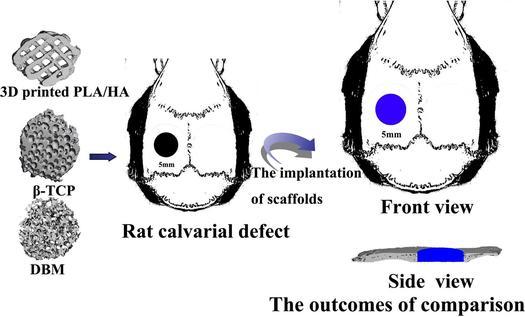
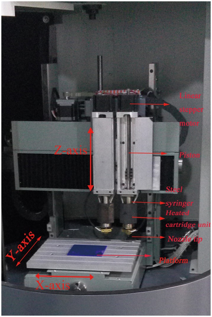
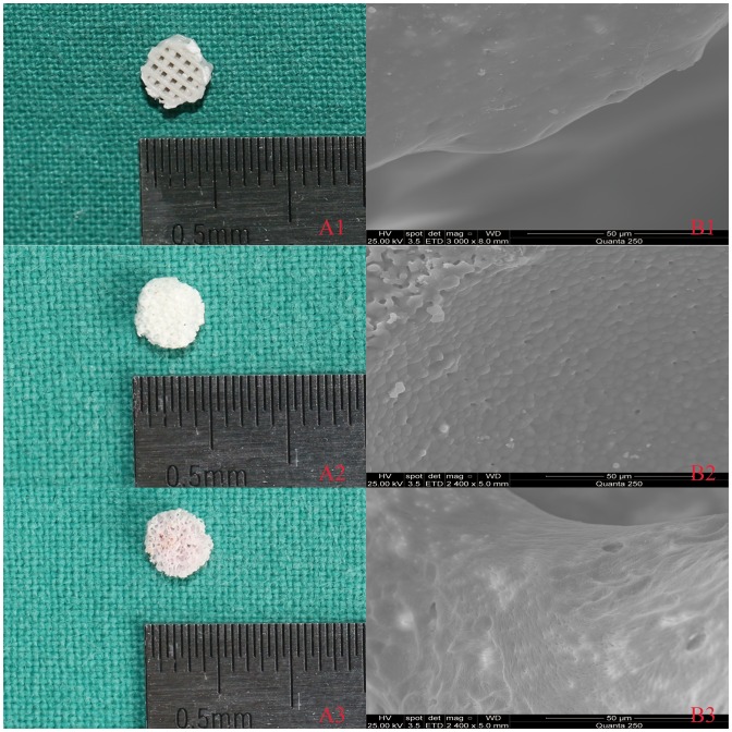
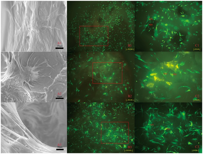
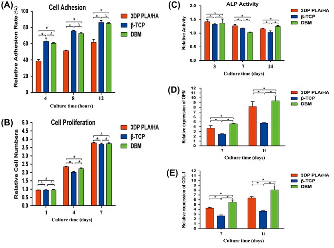
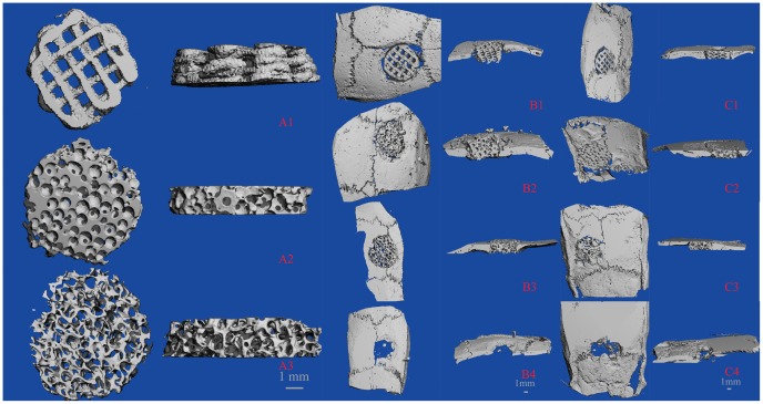
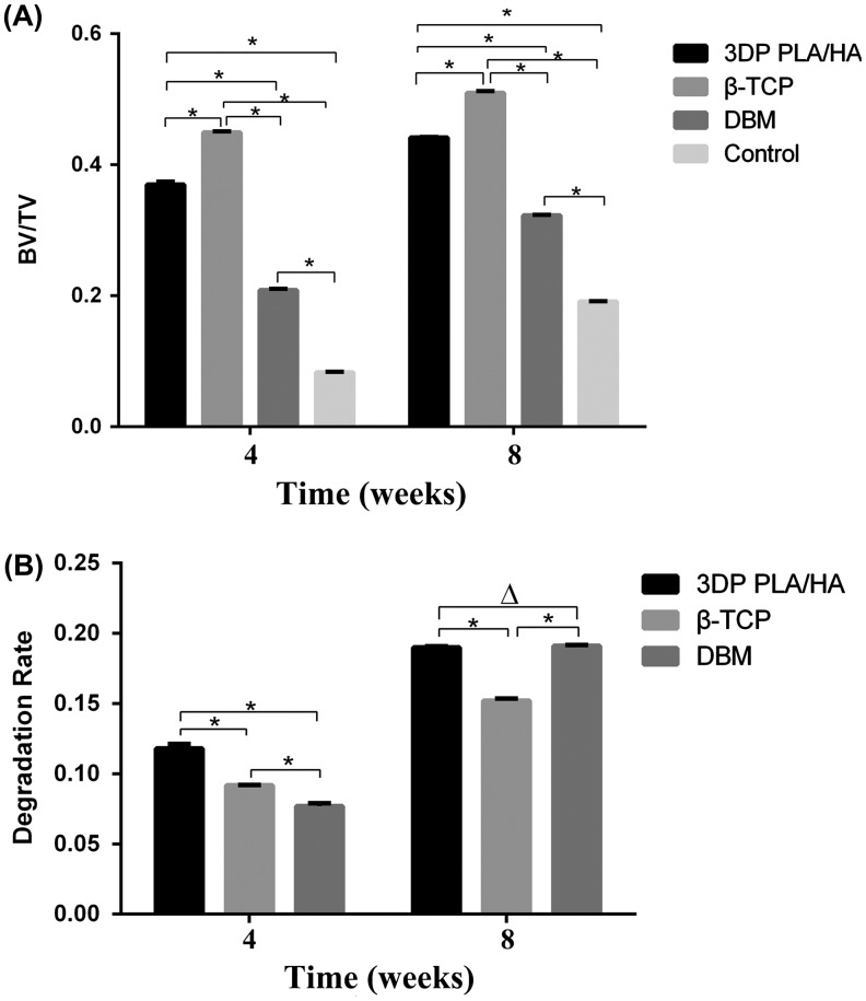
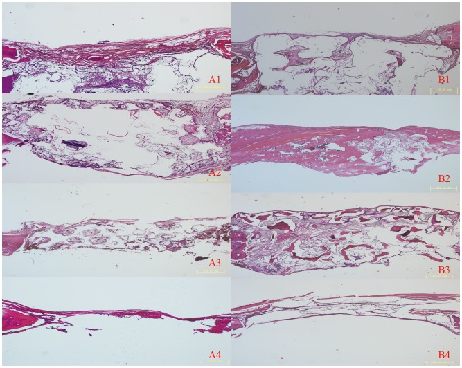
We have explored the applicability of printed scaffold by comparing osteogenic ability and biodegradation property of three resorbable biomaterials. A polylactic acid/hydroxyapatite (PLA/HA) composite with a pore size of 500 μm and 60% porosity was fabricated by three-dimensional printing. Three-dimensional printed PLA/HA, β-tricalcium phosphate (β-TCP) and partially demineralized bone matrix (DBM) seeded with bone marrow stromal cells (BMSCs) were evaluated by cell adhesion, proliferation, alkaline phosphatase activity and osteogenic gene expression of osteopontin (OPN) and collagen type I (COL-1). Moreover, the biocompatibility, bone repairing capacity and degradation in three different bone substitute materials were estimated using a critical-size rat calvarial defect model in vivo. The defects were evaluated by micro-computed tomography and histological analysis at four and eight weeks after surgery, respectively. The results showed that each of the studied scaffolds had its own specific merits and drawbacks. Three-dimensional printed PLA/HA scaffolds possessed good biocompatibility and stimulated BMSC cell proliferation and differentiation to osteogenic cells. The outcomes in vivo revealed that 3D printed PLA/HA scaffolds had good osteogenic capability and biodegradation activity with no difference in inflammation reaction. Therefore, 3D printed PLA/HA scaffolds have potential applications in bone tissue engineering and may be used as graft substitutes in reconstructive surgery.
Overview
The claims and statistics of this paper haven’t been checked yet.
Run the check — free, no account needed. It reads the open-access text and checks the sources it cites.
Integrity signals
Registry
Cites
Statistics
Figures
Verification
Claims the paper makes
The claims and statistics of this paper haven’t been checked yet.
Run the check — free, no account needed. It reads the open-access text and checks the sources it cites.
Claims other papers make about this one
Each is checked against this paper’s own text: does it say what the citing paper says it does?
In the citing paper
“Some studies evaluated PLA scaffolds made using 3D-printing technology, finding that it results in less inflammation, making it a promising material for repairing bone damage [139,140].”
From Synthesis and Biological Application of Polylactic Acid (2020) · cites this paper as [140]
Self-check
Percentages, sums and reported differences re-computed from the paper’s own numbers.
The self-check needs the paper’s full text.
Citations
Cites
Analysis of Relative Gene Expression Data Using Real-Time Quantitative PCR and the 2−ΔΔCT Method
Livak, K. J., & Schmittgen, T. D. (2001) · Methods · doi:10.1006/meth.2001.1262 · Cited by 180,860
Biodegradable and bioactive porous polymer/inorganic composite scaffolds for bone tissue engineering
Rezwan, K., Chen, Q. Z., Blaker, J. J., & Boccaccini, A. R. (2006) · Biomaterials · doi:10.1016/j.biomaterials.2006.01.039 · Cited by 3,840
Mutation of the mouse klotho gene leads to a syndrome resembling ageing
Kuro-o, M., Matsumura, Y., Aizawa, H., Kawaguchi, H., Suga, T., Utsugi, T., Ohyama, Y., Kurabayashi, M., Kaname, T., Kume, E., et al. (1997) · Nature · doi:10.1038/36285 · Cited by 3,837
Poly(lactic acid) modifications
Rasal, R. M., Janorkar, A. V., & Hirt, D. E. (2010) · Progress in Polymer Science · doi:10.1016/j.progpolymsci.2009.12.003 · Cited by 2,204
Bone regeneration: current concepts and future directions
Dimitriou, R. I., Jones, E., McGonagle, D., & Giannoudis, P. V. (2011) · BMC Medicine · doi:10.1186/1741-7015-9-66 · Cited by 2,024
Properties of osteoconductive biomaterials: calcium phosphates.
Legeros, R. (2002) · Clinical Orthopaedics and Related Research · doi:10.1097/00003086-200202000-00009 · Cited by 2,003
Navarro, M., Michiardi, A., Castaño, Ó., & Planell, J. A. (2008) · Journal of The Royal Society Interface · doi:10.1098/rsif.2008.0151 · Cited by 1,521
Naldini, L., Blömer, U., Gage, F. H., Trono, D., & Verma, I. M. (1996) · Proceedings of the National Academy of Sciences · doi:10.1073/pnas.93.21.11382 · Cited by 1,499
Athanasiou, K. A., Niederauer, G. G., & Agrawal, C. M. (1996) · Biomaterials · doi:10.1016/0142-9612(96)85754-1 · Cited by 1,497
Bioceramics of calcium orthophosphates
Dorozhkin, S. V. (2009) · Biomaterials · doi:10.1016/j.biomaterials.2009.11.050 · Cited by 1,346
Research works printed but not linked: 10
- John HD, 2004, Int J Oral Maxillofac Implants, 19, 199
- Aghaloo TL, 2007, Int J Oral Maxillofac Implants., 22, 49
- Bandyopadhyay A, 2001, MRS Bulletin., 25, 273
- Rasal RM, 2009, Prog Polym Sci., 89, 252
- Suganuma J, 2001, J Appl Biomater, 165, 2091
- Esposito M, 2008, Cochrane Database Syst Rev
- Jiang W, 2006, Polym Eng Sci., 12, 1821
- Kim K, 2001, Biomacromolecules, 25, 402
- van Lenthe GH, 2001, Biomaterials, 25, 402
- 10.2147/CCIDE.S13115 · DOI
Cited by
Sorted by citations
Items 201–205 of 205
Ray, S. S., Banerjee, R., Bandyopadhyay, J., & Orasugh, J. T. (2025) · Carl Hanser Verlag GmbH & Co. KG eBooks · doi:10.1007/978-1-56990-327-8_4
Biomaterials for bone regeneration
Hazarika, K., Vyas, A., Ghosh, S. B., Gupta, T., Sain, M. M., & Bandyopadhyay‐Ghosh, S. (2026) · Elsevier eBooks · doi:10.1016/b978-0-443-26739-0.00001-6
A review of FDM methods, microstructure, and structure–property relationships in polymer composites
Bharti, R., Mursaleen, M., & Dey, A. (2026) · Advanced Composite Materials · doi:10.1080/09243046.2026.2713975
Chen, J., Wang, Q., Zheng, X., Gu, X., Pei, L., Dai, J., Li, R., Wang, X., & Wang, S. (2026) · Polymer Engineering and Science · doi:10.1002/pen.70767
Composite biomaterials for 3D printing in biomedical applications
Debnath, A., Shafoyat, M. U., Tanha, N. J., Haque, S. R., Mannoor, K., Ali, S. M., & Asif, M. A. R. (2026) · Academia Materials Science · doi:10.20935/acadmatsci8450
Metadata
- Authors
- Zhang, H., Mao, X., Du, Z., Jiang, W., Han, X., Zhao, D., Han, D., & Li, Q.
- Affiliations
- Shanghai Jiao Tong University (CN)
- Corresponding author
- Han, Dong
- Venue
- Science and Technology of Advanced Materials
- Publisher
- Informa UK Limited
- Published
- 2016-01-01
- Volume
- 17
- Issue
- 1
- Pages
- 136-148
- ISSN
- 1468-6996 (ISSN-L)
- 1878-5514
- PMC
- PMC5101962
- OpenAlex
- W2330522579
- Cited by
- 208
- References
- 61
- Citations by year
- 2026: 11 · 2025: 18 · 2024: 14 · 2023: 24 · 2022: 31 · 2021: 32 · 2020: 27 · 2019: 23 · 2018: 13 · 2017: 14 · 2016: 1
- FWCI
- 8.18
- Type
- journal article
- Language
- en
- Open access
- gold
- doi.org
- Locations
- Science and Technology of Advanced Materials (publishedVersion, cc-by)
- PubMed Central (publishedVersion, cc-by)
- PubMed (publishedVersion)
- DOAJ (DOAJ: Directory of Open Access Journals) (submittedVersion, cc-by-sa)
- Funders
- National Natural Science Foundation of China (81272132)
- Topics
- Physical Sciences › Engineering › Biomedical Engineering › Bone Tissue Engineering Materials
- Physical Sciences › Engineering › Biomedical Engineering › 3D Printing in Biomedical Research
- Physical Sciences › Engineering › Industrial and Manufacturing Engineering › Additive Manufacturing and 3D Printing Technologies
- Keywords
- 3D-printed scaffolds, macroporous scaffolds, bone tissue engineering, critical-size calvarial defect, mesenchymal stem cells, osteogenic differentiation, biodegradation, beta-tricalcium phosphate, demineralized bone matrix, bone substitutes, osteopontin, type I collagen, biocompatibility
About these checks
Registry, retraction, Cites and statistics results are computed, not judged, with one exception: a printed reference that no index lists is read once by a model before it counts as a citation error. Claims are extracted by a model and kept only when their quote appears verbatim in the text; each cited source is then checked, with a model’s help, for whether it says what the claim relies on. Those verdicts can be wrong — read the claim’s quote and the cited source before relying on one.
Statistics and arithmetic are recomputed, not judged.
Confidence bands are shown only for fields calibrated on our annotated test set. None is calibrated yet, so no band is shown.
What counts as a claim ›How we measure confidence ›How we check this ›
Are you an author, or do you see a problem with this page? Request a review
