

The DOI matches its Crossref record; 24 of the 24 references it cites were found in a registry or an index.
Mitophagy is required for mitochondrial biogenesis and myogenic differentiation of C2C12 myoblasts
Open accessAbstract
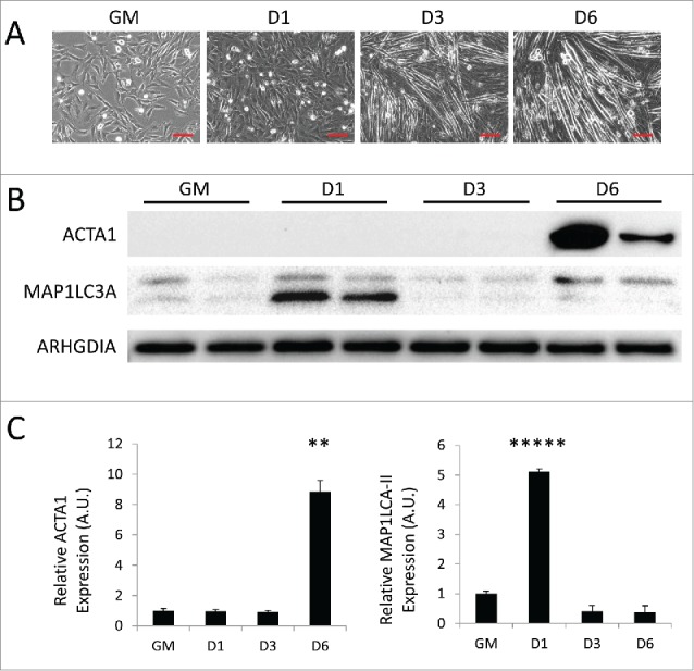
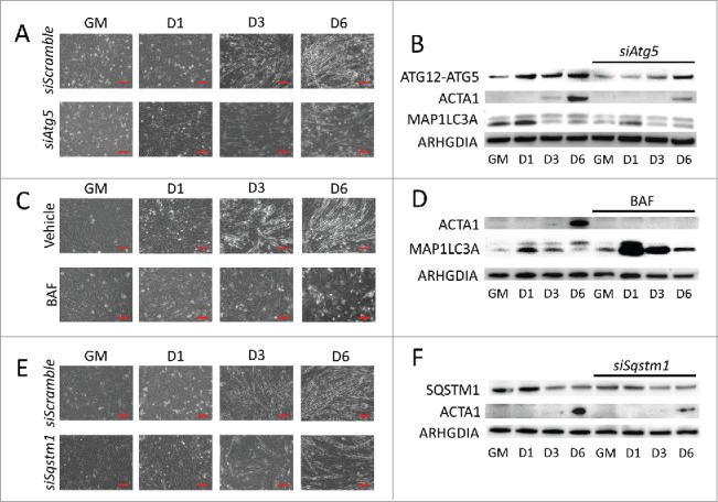
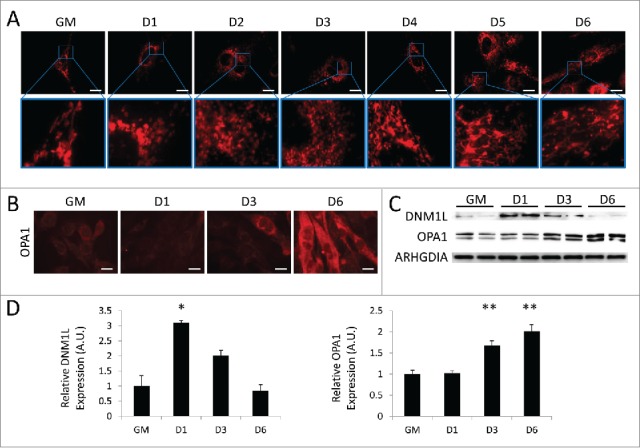
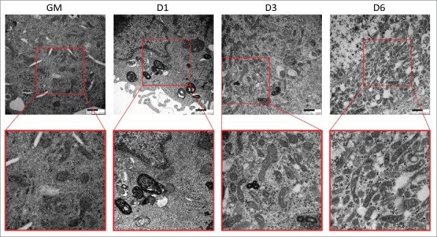
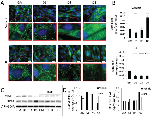
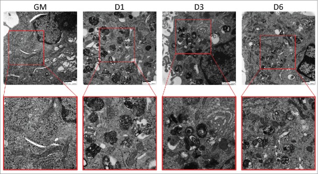
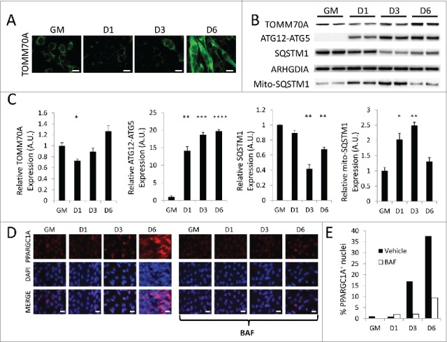
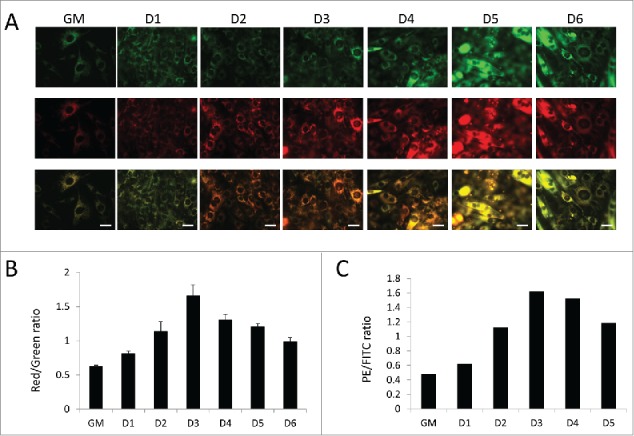
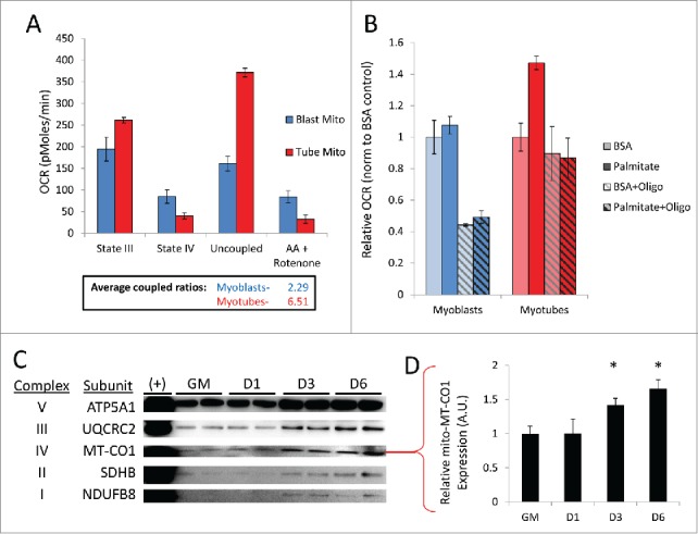
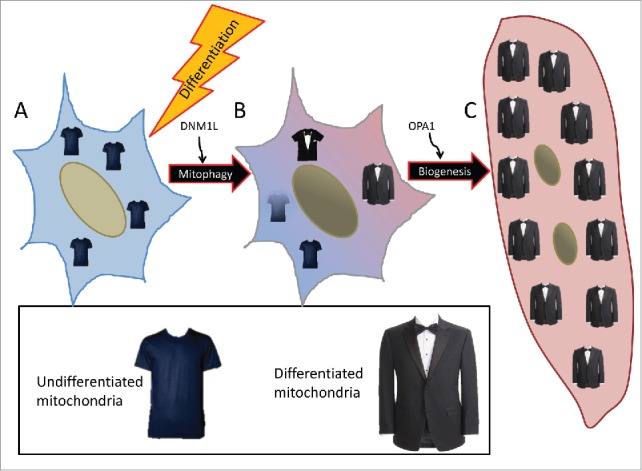
Myogenesis is a crucial process governing skeletal muscle development and homeostasis. Differentiation of primitive myoblasts into mature myotubes requires a metabolic switch to support the increased energetic demand of contractile muscle. Skeletal myoblasts specifically shift from a highly glycolytic state to relying predominantly on oxidative phosphorylation (OXPHOS) upon differentiation. We have found that this phenomenon requires dramatic remodeling of the mitochondrial network involving both mitochondrial clearance and biogenesis. During early myogenic differentiation, autophagy is robustly upregulated and this coincides with DNM1L/DRP1 (dynamin 1-like)-mediated fragmentation and subsequent removal of mitochondria via SQSTM1 (sequestosome 1)-mediated mitophagy. Mitochondria are then repopulated via PPARGC1A/PGC-1α (peroxisome proliferator-activated receptor gamma, coactivator 1 alpha)-mediated biogenesis. Mitochondrial fusion protein OPA1 (optic atrophy 1 [autosomal dominant]) is then briskly upregulated, resulting in the reformation of mitochondrial networks. The final product is a myotube replete with new mitochondria. Respirometry reveals that the constituents of these newly established mitochondrial networks are better primed for OXPHOS and are more tightly coupled than those in myoblasts. Additionally, we have found that suppressing autophagy with various inhibitors during differentiation interferes with myogenic differentiation. Together these data highlight the integral role of autophagy and mitophagy in myogenic differentiation.
Overview
The claims and statistics of this paper haven’t been checked yet.
Run the check — free, no account needed. It reads the open-access text and checks the sources it cites.
Integrity signals
Registry
Cites
Statistics
Figures
Verification
Claims the paper makes
The claims and statistics of this paper haven’t been checked yet.
Run the check — free, no account needed. It reads the open-access text and checks the sources it cites.
Claims other papers make about this one
Each is checked against this paper’s own text: does it say what the citing paper says it does?
The cited source says what the claim attributes to it.
In the citing paper
“autophagy is critical for mitochondrial remodeling and function in skeletal muscle cells in vitro (Sin et al., 2016)”
From Skeletal Muscle Ribosome and Mitochondrial Biogenesis in Response to Different Exercise Training Modalities (2021) · cites this paper as Sin et al., 2016
Self-check
Percentages, sums and reported differences re-computed from the paper’s own numbers.
The self-check needs the paper’s full text.
Citations
Cites
Autophagy in mammalian development and differentiation
Mizushima, N., & Levine, B. (2010) · Nature Cell Biology · doi:10.1038/ncb0910-823 · Cited by 1,555
Defective Hepatic Autophagy in Obesity Promotes ER Stress and Causes Insulin Resistance
Yang, L., Li, P., Fu, S., Calay, E. S., & Hotamisligil, G. S. (2010) · Cell Metabolism · doi:10.1016/j.cmet.2010.04.005 · Cited by 1,274
Notch-Mediated Restoration of Regenerative Potential to Aged Muscle
Conboy, I. M., Conboy, M. J., Smythe, G. M., & Rando, T. A. (2003) · Science · doi:10.1126/science.1087573 · Cited by 1,121
Folmes, C. D. L., Nelson, T. J., Martinez‐Fernandez, A., Arrell, D. K., Lindor, J. Z., Dzeja, P. P., Ikeda, Y., Pérez-Terzic, C. M., & Terzic, A. (2011) · Cell Metabolism · doi:10.1016/j.cmet.2011.06.011 · Cited by 980
AGE‐RELATED CHANGES IN THE STRUCTURE AND FUNCTION OF SKELETAL MUSCLES
Faulkner, J. A., Larkin, L. M., Claflin, D. R., & Brooks, S. V. (2007) · Clinical and Experimental Pharmacology and Physiology · doi:10.1111/j.1440-1681.2007.04752.x · Cited by 802
Endogenous Drp1 Mediates Mitochondrial Autophagy and Protects the Heart Against Energy Stress
Ikeda, Y., Shirakabe, A., Maejima, Y., Zhai, P., Sciarretta, S., Toli, J., Nomura, M., Mihara, K., Egashira, K., Ohishi, M., et al. (2015) · Circulation Research · doi:10.1161/circresaha.116.303356 · Cited by 571
Autophagy and aging: keeping that old broom working
Cuervo, A. M. (2008) · Trends in Genetics · doi:10.1016/j.tig.2008.10.002 · Cited by 527
In vivo satellite cell activation via Myf5 and MyoD in regenerating mouse skeletal muscle
Cooper, R. N., Tajbakhsh, S., Mouly, V., Cossu, G., BUCKINGHAM, M. E., & Butler-Browne, G. S. (1999) · Journal of Cell Science · doi:10.1242/jcs.112.17.2895 · Cited by 457
Burattini, S., Ferri, P., Battistelli, M., Curci, R., Luchetti, F., & Falcieri, E. (2004) · PubMed · doi:10.4081/891 · Cited by 353
Shefer, G., Rauner, G., Yablonka‐Reuveni, Z., & Benayahu, D. (2010) · PLoS ONE · doi:10.1371/journal.pone.0013307 · Cited by 220
Research works printed but not linked: 2
- Dedieu S, 2002, Int J Dev Biol, 46, 235
- Burattini S, 2004, Eur J Histochem, 48, 223
Cited by
Sorted by citations
Molecular mechanisms and physiological functions of mitophagy
Onishi, M., Yamano, K., Sato, M., Matsuda, N., & Okamoto, K. (2021) · The EMBO Journal · doi:10.15252/embj.2020104705 · Cited by 1,469
The mitophagy pathway and its implications in human diseases
Wang, S., Long, H., Hou, L., Feng, B., Ma, Z., Wu, Y., Zeng, Y., Cai, J., Zhang, D., & Zhao, G. (2023) · Signal Transduction and Targeted Therapy · doi:10.1038/s41392-023-01503-7 · Cited by 991
Mitophagy in human health, ageing and disease
Picca, A., Faitg, J., Auwerx, J., Ferrucci, L., & D’Amico, D. (2023) · Nature Metabolism · doi:10.1038/s42255-023-00930-8 · Cited by 635
Autophagy: The spotlight for cellular stress responses
Ravanan, P., Florance, I., & Talwar, P. (2017) · Life Sciences · doi:10.1016/j.lfs.2017.08.029 · Cited by 595
The Role of Autophagy in the Heart
Sciarretta, S., Maejima, Y., Zablocki, D., & Sadoshima, J. (2018) · Annual Review of Physiology · doi:10.1146/annurev-physiol-021317-121427 · Cited by 547
Metadata
- Authors
- Sin, J., Andres, A. M., Taylor, D. J. R., Weston, T., Hiraumi, Y., Stotland, A., Kim, B. J., Huang, C., Doran, K. S., & Gottlieb, R. A.
- Affiliations
- Cedars-Sinai Medical Center (US)
- San Diego State University (US)
- Corresponding author
- Gottlieb, Roberta A.
- ORCID
- Venue
- Autophagy
- Publisher
- Informa UK Limited
- Published
- 2016-02-01
- Volume
- 12
- Issue
- 2
- Pages
- 369-380
- ISSN
- 1554-8627 (ISSN-L)
- 1554-8635
- PMC
- PMC4836019
- OpenAlex
- W2280853449
- Cited by
- 385
- References
- 24
- Citations by year
- 2026: 27 · 2025: 27 · 2024: 50 · 2023: 39 · 2022: 38 · 2021: 46 · 2020: 48 · 2019: 45 · 2018: 30 · 2017: 25 · 2016: 8
- FWCI
- 16.75
- Type
- journal article
- Language
- en
- Open access
- hybrid
- doi.org
- Licence
- Locations
- Autophagy (publishedVersion, cc-by-nc)
- PubMed Central (publishedVersion, cc-by-nc)
- PubMed (publishedVersion)
- Funders
- National Heart, Lung, and Blood Institute (P01HL112730, T32HL116273)
- Topics
- Life Sciences › Biochemistry, Genetics and Molecular Biology › Cell Biology › Autophagy in Disease and Therapy
- Life Sciences › Biochemistry, Genetics and Molecular Biology › Molecular Biology › Muscle Physiology and Disorders
- Life Sciences › Biochemistry, Genetics and Molecular Biology › Molecular Biology › Mitochondrial Function and Pathology
- Keywords
- mitophagy, mitochondrial biogenesis, myogenesis, C2C12 myoblasts, Drp1, mitochondrial fragmentation, p62, PGC-1α, OPA1, mitochondrial fusion, oxidative phosphorylation, mitochondrial network remodeling, myotube formation, skeletal muscle development
- MeSH terms
- Animals, Organelle Biogenesis, Cell Differentiation, Cell Line, Mitochondria, Models, Biological, Oxidative Phosphorylation, Muscle Fibers, Skeletal, Macrolides, Muscle Development, Myoblasts, Mice, Mitophagy
About these checks
Registry, retraction, Cites and statistics results are computed, not judged, with one exception: a printed reference that no index lists is read once by a model before it counts as a citation error. Claims are extracted by a model and kept only when their quote appears verbatim in the text; each cited source is then checked, with a model’s help, for whether it says what the claim relies on. Those verdicts can be wrong — read the claim’s quote and the cited source before relying on one.
Statistics and arithmetic are recomputed, not judged.
Confidence bands are shown only for fields calibrated on our annotated test set. None is calibrated yet, so no band is shown.
What counts as a claim ›How we measure confidence ›How we check this ›
Are you an author, or do you see a problem with this page? Request a review
