

The DOI matches its Crossref record.
Induction of the differentiation of porcine bone marrow mesenchymal stem cells into premature hepatocyte-like cells in an indirect coculture system with primary hepatocytes
Open accessAbstract
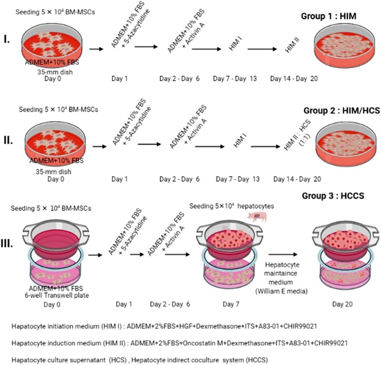
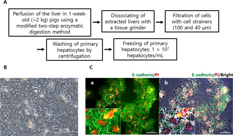
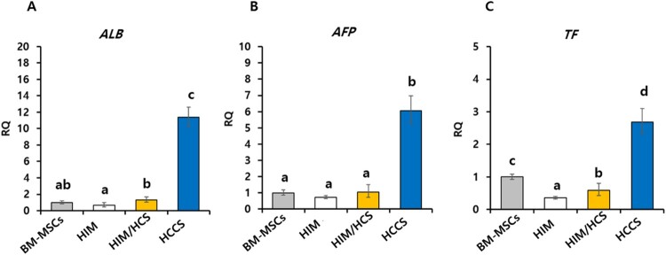
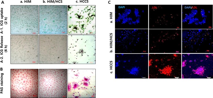
Liver transplantation is currently the only option for patients with end-stage liver disease. Thus, other alternate therapeutic strategies are needed. Bone marrow mesenchymal stem cells (BM-MSCs) are nonhematopoietic cells present in the bone marrow stroma that serve as precursors cells for various other cells. In this study, we evaluated the differentiation of porcine BM-MSCs into hepatocyte-like cells using three types of culture systems: hepatic induction medium (HIM), HIM/primary hepatocyte culture supernatant (HCS; 1:1 ratio), and a hepatocyte coculture system (HCCS; primary hepatocytes in the upper chamber, and BM-MSCs in the lower chamber). Primary hepatocytes were isolated from anesthetized healthy 1-month-old pigs by enzymatic digestion. Hepatic-specific marker expression (albumin [ALB], transferrin [TF], α-fetoprotein [AFP]), glycogen storage, low-density lipoprotein, and indocyanine green uptake were evaluated. Upregulation of hepatic-specific markers (ALB, TF, and AFP) was observed by real-time polymerase chain reaction in the HCCS group. Periodic acid-Schiff staining revealed enhanced glycogen storage in hepatocyte-like cells from the HCCS group compared with that from the HIM/HCS group. Furthermore, hepatocyte like-cells in the HCCS group showed improved LDL and ICG uptake than those in the other groups. Overall, our current study revealed that indirect coculture of primary hepatocytes and BM-MSCs enhanced the differentiation efficacy of BM-MSCs into hepatocyte-like cells by unknown useful soluble factors, including paracrine factors.
Overview
The claims and statistics of this paper haven’t been checked yet.
Run the check — free, no account needed. It reads the open-access text and checks the sources it cites.
Integrity signals
Registry
Cites
Statistics
Figures
Verification
Claims the paper makes
The claims and statistics of this paper haven’t been checked yet.
Run the check — free, no account needed. It reads the open-access text and checks the sources it cites.
Claims other papers make about this one
Each is checked against this paper’s own text: does it say what the citing paper says it does?
The cited source says what the claim attributes to it.
In the citing paper
“In an indirect co-culture system, the PHs enhance the hepatic-differentiation efficacy of porcine BM-MSCs through the exchange of soluble factors [357].”
From Mechanisms, efficacy, and future perspectives of cellular-based therapies for liver fibrosis/cirrhosis: focusing on mesenchymal stromal cells (2025) · cites this paper as [357]
Self-check
Percentages, sums and reported differences re-computed from the paper’s own numbers.
The self-check needs the paper’s full text.
Citations
Cites
No references could be resolved yet.
Research works printed but not linked: 42
- 10.1016/j.jhep.2017.06.009 · DOI
- 10.1387/ijdb.052072hb · DOI · Paper page
- 10.1016/j.diff.2019.01.005 · DOI
- 10.1002/jcp.21200 · DOI · Paper page
- 10.1634/stemcells.2007-0197 · DOI
- 10.1016/j.cell.2012.02.031 · DOI
- 10.1007/s00204-016-1901-x · DOI
- 10.1016/j.jhep.2017.04.004 · DOI
- 10.1038/nbt1163 · DOI · Paper page
- 10.1002/jcp.21481 · DOI
- 10.1096/fasebj.3.2.2914628 · DOI
- 10.1093/emboj/18.8.2127 · DOI
- 10.1016/j.yexcr.2003.10.011 · DOI
- 10.1016/j.archoralbio.2012.08.010 · DOI
- 10.1007/s00441-011-1249-8 · DOI
- 10.1073/pnas.96.13.7265 · DOI
- 10.1016/S0092-8674(01)00328-2 · DOI
- 10.1056/NEJM199312163292508 · DOI
- 10.1002/hep.20469 · DOI · Paper page
- 10.1146/annurev.cellbio.21.012704.131525 · DOI
- 10.1016/j.celrep.2016.03.071 · DOI
- 10.1038/nature07056 · DOI
- 10.1002/jcp.1136 · DOI
- 10.1126/science.1110542 · DOI
- 10.1089/ten.tec.2010.0149 · DOI
- 10.1155/2017/7567958 · DOI
- 10.1016/j.yexcr.2013.10.005 · DOI
- 10.1073/pnas.1220637110 · DOI
- 10.1002/stem.2575 · DOI · Paper page
- 10.1136/esmoopen-2016-000042 · DOI
- 10.1089/ten.2006.12.2965 · DOI
- 10.1155/2013/762196 · DOI
- 10.1038/373699a0 · DOI
- 10.1016/S0091-679X(08)61797-5 · DOI
- 10.1111/j.1872-034X.2007.00162.x · DOI
- 10.1038/nprot.2007.18 · DOI
- Sundback CA, 2000, Gastroenterology, 118, 438
- 10.1111/j.1349-7006.2005.00103.x · DOI
- 10.1016/j.lfs.2016.04.026 · DOI
- 10.1042/BSR20150025 · DOI
- Yu YB, 2018, Mol Med Rep, 18, 2009
- 10.1111/j.1478-3231.2009.02054.x · DOI
Cited by
Sorted by citations
Pan, X., Gao, T., & Wang, B. (2025) · Cell & Bioscience · doi:10.1186/s13578-025-01520-9 · Cited by 2
“In an indirect co-culture system, the PHs enhance the hepatic-differentia-tion efficacy of porcine BM-MSCs through the exchange of soluble factors [357].”
Metadata
- Authors
- Ullah, I., Seo, K., Wi, H., Kim, Y., Lee, S., & Ock, S. A.
- Affiliations
- Quaid-i-Azam University (PK)
- Rural Development Administration (KR)
- Animal Biotechnology Division, National Institute of Animal Science, Rural Development Administration, Wanju-gun, Republic of Korea
- Corresponding author
- Ock, Sun A
- Venue
- Animal Cells and Systems
- Publisher
- Informa UK Limited
- Published
- 2020-09-02
- Volume
- 24
- Issue
- 5
- Pages
- 289-298
- ISSN
- 1976-8354 (ISSN-L)
- 2151-2485
- PMC
- PMC7646558
- OpenAlex
- W3092516100
- Cited by
- 4
- References
- 0
- Citations by year
- 2025: 1 · 2023: 1 · 2022: 1 · 2021: 1
- FWCI
- 0.39
- Type
- journal article
- Language
- en
- Open access
- gold
- doi.org
- Locations
- Animal Cells and Systems (publishedVersion, cc-by)
- PubMed Central (publishedVersion, cc-by)
- PubMed (publishedVersion)
- DOAJ (DOAJ: Directory of Open Access Journals) (submittedVersion)
- Topics
- Life Sciences › Biochemistry, Genetics and Molecular Biology › Cell Biology › Mesenchymal stem cell research
- Health Sciences › Medicine › Hepatology › Liver physiology and pathology
- Life Sciences › Biochemistry, Genetics and Molecular Biology › Molecular Biology › Pluripotent Stem Cells Research
- Keywords
- bone marrow mesenchymal stem cells, hepatocyte-like cells, stem cell differentiation, primary hepatocytes, pigs, paracrine factors, glycogen storage, liver regeneration
About these checks
Registry, retraction, Cites and statistics results are computed, not judged, with one exception: a printed reference that no index lists is read once by a model before it counts as a citation error. Claims are extracted by a model and kept only when their quote appears verbatim in the text; each cited source is then checked, with a model’s help, for whether it says what the claim relies on. Those verdicts can be wrong — read the claim’s quote and the cited source before relying on one.
Statistics and arithmetic are recomputed, not judged.
Confidence bands are shown only for fields calibrated on our annotated test set. None is calibrated yet, so no band is shown.
What counts as a claim ›How we measure confidence ›How we check this ›
Are you an author, or do you see a problem with this page? Request a review
How to cite
Cite CoChat for the checks on this page, and the paper for its own findings.
- This page
- CoChat. (2026). Citation check: Induction of the differentiation of porcine bone marrow mesenchymal stem cells into premature hepatocyte-like cells in an indirect coculture system with primary hepatocytes. https://app.cochat.ai/papers/10.1080/19768354.2020.1823473
- The paper
- Ullah, I., Seo, K., Wi, H., Kim, Y., Lee, S., & Ock, S. A. (2020). Induction of the differentiation of porcine bone marrow mesenchymal stem cells into premature hepatocyte-like cells in an indirect coculture system with primary hepatocytes. Animal Cells and Systems. https://doi.org/10.1080/19768354.2020.1823473
