

The DOI matches its Crossref record; 4 of the 4 references it cites were found in a registry or an index.
Immortalization of primary marmoset skin fibroblasts by CRISPR-Cas9-mediated gene targeting
Overview
The claims and statistics of this paper haven’t been checked yet.
Run the check — free, no account needed. It reads the open-access text and checks the sources it cites.
Integrity signals
Registry
Cites
Statistics
Figures
Verification
Claims the paper makes
The claims and statistics of this paper haven’t been checked yet.
Run the check — free, no account needed. It reads the open-access text and checks the sources it cites.
Claims other papers make about this one
Each is checked against this paper’s own text: does it say what the citing paper says it does?
In the citing paper
“Certainly, the study demonstrates that CRISPR-Cas9-mediated targeting of the p53 gene or CDKN2A locus proves to be an effective method for immortalizing primary marmoset skin fibroblasts.”
From Cell Immortality: In Vitro Effective Techniques to Achieve and Investigate Its Applications and Challenges (2024) · cites this paper as [159]
In the citing paper
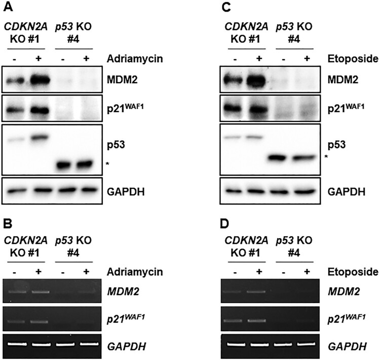
“The research reveals that, similar to Cdkn2a-deficient mouse cells, CDKN2A-deficient marmoset cells express wild-type p53 proteins and respond normally to genotoxic stresses such as adriamycin and etoposide.”
From Cell Immortality: In Vitro Effective Techniques to Achieve and Investigate Its Applications and Challenges (2024) · cites this paper as [159]
In the citing paper
“These findings collectively underscore that Cas9-mediated gene targeting of the p53 gene or CDKN2A locus is a potent tool for establishing immortalized marmoset cell lines with specific genetic modifications [159].”
From Cell Immortality: In Vitro Effective Techniques to Achieve and Investigate Its Applications and Challenges (2024) · cites this paper as [159]
Self-check
Percentages, sums and reported differences re-computed from the paper’s own numbers.
The self-check needs the paper’s full text.
Citations
Cites
Loss of p16Ink4a with retention of p19Arf predisposes mice to tumorigenesis
Sharpless, N. E., Bardeesy, N., Lee, K.-H., Carrasco, D., Castrillon, D. H., Aguirre, A. J., Wu, E. A., Horner, J. W., & DePinho, R. A. (2001) · Nature · doi:10.1038/35092592 · Cited by 848
Germline mutations in the p16INK4a binding domain of CDK4 in familial melanoma
Zuo, L., Weger, J., Yang, Q., Goldstein, A. M., Tucker, M. A., Walker, G. J., Hayward, N., & Dracopoli, N. C. (1996) · Nature Genetics · doi:10.1038/ng0196-97 · Cited by 818 · cited 2×
“melanoma and abolishes the ability of CDK4 protein to bind p16 protein (Wolfel et al. 1995; Zuo et al. 1996).”
BackgroundThe INK4A/ARF locus and its two gene products.
Sharpless, N. E., & DePinho, R. A. (1999) · Current Opinion in Genetics & Development · doi:10.1016/s0959-437x(99)80004-5 · Cited by 533
Genome-wide target specificities of CRISPR-Cas9 nucleases revealed by multiplex Digenome-seq
Kim, D., Kim, S., Kim, S., Park, J., & Kim, J.-S. (2016) · Genome Research · doi:10.1101/gr.199588.115 · Cited by 208
Research works printed but not linked: 29
- 10.1038/sj.onc.1209046 · DOI
- 10.1038/cdd.2017.180 · DOI
- 10.1038/79594 · DOI
- 10.1126/science.279.5349.349 · DOI · Paper page
- 10.1038/nbt.2507 · DOI · Paper page
- 10.1126/science.1231143 · DOI · Paper page
- Dumenco L, 1995, Hepatology, 22, 1279
- Gao Q, 1996, Cancer Res, 56, 3129
- 10.1038/s41421-018-0020-7 · DOI
- 10.1080/19768354.2020.1752306 · DOI
- 10.1101/gad.5.12b.2375 · DOI · Paper page
- Harvey M, 1993, Oncogene, 8, 2457
- 10.1016/0014-4827(65)90211-9 · DOI · Paper page
- 10.1016/j.stem.2014.01.003 · DOI · Paper page
- 10.1016/S0092-8674(00)80452-3 · DOI · Paper page
- 10.1080/19768354.2020.1847731 · DOI
- 10.1111/dgd.12109 · DOI
- 10.1126/science.1232033 · DOI · Paper page
- 10.1371/journal.pone.0127268 · DOI
- 10.1016/j.bbrc.2020.12.072 · DOI
- 10.1016/j.tice.2022.101848 · DOI
- 10.1371/journal.pone.0204580 · DOI
- 10.1016/0092-8674(95)90214-7 · DOI · Paper page
- Seluanov A, 2010, J Vis Exp, 44, e2033
- 10.1016/S0092-8674(00)81079-X · DOI · Paper page
- 10.1093/hmg/10.7.677 · DOI
- 10.1074/jbc.M113.477570 · DOI
- 10.1038/nbt.3117 · DOI · Paper page
- 10.1126/science.7652577 · DOI
Cited by
A Beginner’s Guide to Cell Culture: Practical Advice for Preventing Needless Problems
Weiskirchen, S., Schröder, S. K., Buhl, E. M., & Weiskirchen, R. (2023) · Cells · doi:10.3390/cells12050682 · Cited by 148
Chalak, M., Hesaraki, M., Mirbahari, S. N., Yeganeh, M., Abdi, S., Rajabi, S., & Hemmatzadeh, F. (2024) · Life · doi:10.3390/life14030417 · Cited by 66
Metadata
- Authors
- Jeong, Y.-J., Cho, J., Kwak, J., Sung, Y. H., & Kang, B.-C.
- Affiliations
- Convergence Medicine Research Center, Asan Institute for Life Sciences, Asan Medical Center, Seoul, Republic of Korea
- Graduate School of Translational Medicine, Seoul National University College of Medicine, Seoul, Republic of Korea
- ORCID
- Sung, Young Hoon
- Venue
- Animal Cells and Systems
- Publisher
- Informa UK Limited
- Published
- 2022-11-02
- Volume
- 26
- Issue
- 6
- Pages
- 266-274
- ISSN
- 1976-8354
- 2151-2485
- PMC
- PMC9809370
- Cited by
- 11
- References
- 4
- Language
- en
- Funders
- National Research Foundation of Korea
- Ministry of Science, ICT, and Future Planning (2021IP0050)
- Asan Institute for Life Sciences, Asan Medical Center
About these checks
Registry, retraction, Cites and statistics results are computed, not judged, with one exception: a printed reference that no index lists is read once by a model before it counts as a citation error. Claims are extracted by a model and kept only when their quote appears verbatim in the text; each cited source is then checked, with a model’s help, for whether it says what the claim relies on. Those verdicts can be wrong — read the claim’s quote and the cited source before relying on one.
Statistics and arithmetic are recomputed, not judged.
Confidence bands are shown only for fields calibrated on our annotated test set. None is calibrated yet, so no band is shown.
What counts as a claim ›How we measure confidence ›How we check this ›
Are you an author, or do you see a problem with this page? Request a review
