

The DOI matches its Crossref record; 44 of the 48 references it cites were found in a registry or an index.
New Component of the Vacuolar Class C-Vps Complex Couples Nucleotide Exchange on the Ypt7 Gtpase to Snare-Dependent Docking and Fusion
Open accessAbstract
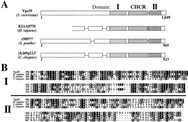
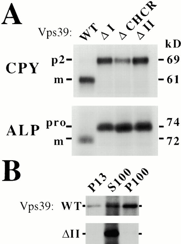
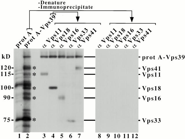
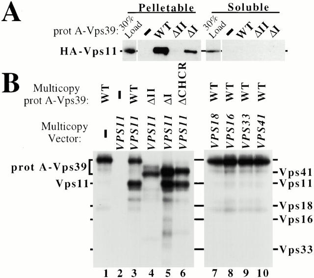
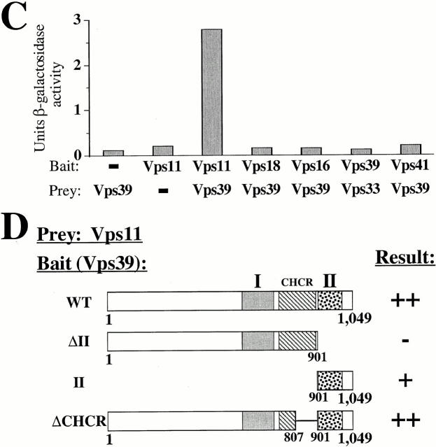
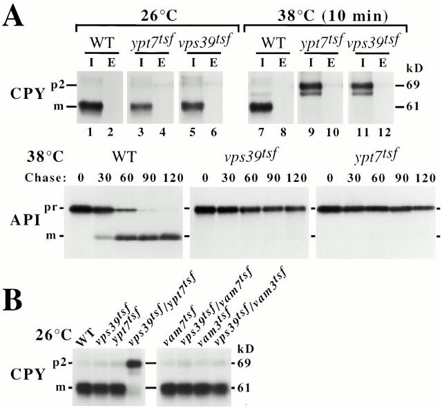
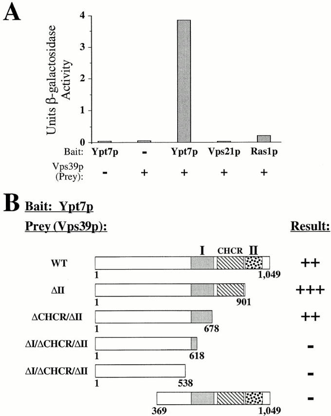
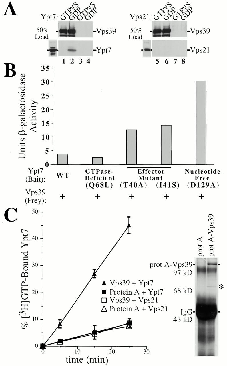
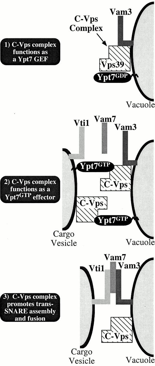
The class C subset of vacuolar protein sorting (Vps) proteins (Vps11, Vps18, Vps16 and Vps33) assembles into a vacuole/prevacuole-associated complex. Here we demonstrate that the class C-Vps complex contains two additional proteins, Vps39 and Vps41. The COOH-terminal 148 amino acids of Vps39 direct its association with the class C-Vps complex by binding to Vps11. A previous study has shown that a large protein complex containing Vps39 and Vps41 functions as a downstream effector of the active, GTP-bound form of Ypt7, a rab GTPase required for the fusion of vesicular intermediates with the vacuole (Price, A., D. Seals, W. Wickner, and C. Ungermann. 2000. J. Cell Biol. 148:1231–1238). Here we present data that indicate that this complex also functions to stimulate nucleotide exchange on Ypt7. We show that Vps39 directly binds the GDP-bound and nucleotide-free forms of Ypt7 and that purified Vps39 stimulates nucleotide exchange on Ypt7. We propose that the class C-Vps complex both promotes Vps39-dependent nucleotide exchange on Ypt7 and, based on the work of Price et al., acts as a Ypt7 effector that tethers transport vesicles to the vacuole. Thus, the class C-Vps complex directs multiple reactions during the docking and fusion of vesicles with the vacuole, each of which contributes to the overall specificity and efficiency of this transport process.
Overview
The claims and statistics of this paper haven’t been checked yet.
Run the check — free, no account needed. It reads the open-access text and checks the sources it cites.
Integrity signals
Registry
Cites
Statistics
Figures
Verification
Claims the paper makes
The claims and statistics of this paper haven’t been checked yet.
Run the check — free, no account needed. It reads the open-access text and checks the sources it cites.
Claims other papers make about this one
Each is checked against this paper’s own text: does it say what the citing paper says it does?
In the citing paper
“Fusion of the autophagosome and lysosome is mediated by the homotypic fusion and protein sorting (HOPS) complex, whose subunits include VPS11, VPS16, VPS18, VPS33A, VPS39, and VPS41 [45,46].”
From Autophagy and ALS: mechanistic insights and therapeutic implications (2022) · cites this paper as [46]
Self-check
Percentages, sums and reported differences re-computed from the paper’s own numbers.
The self-check needs the paper’s full text.
Citations
Cites
Robinson, J. S., Klionsky, D. J., Banta, L. M., & Emr, S. D. (1988) · Molecular and Cellular Biology · doi:10.1128/mcb.8.11.4936 · Cited by 981
Self-citationThe Exocyst is a multiprotein complex required for exocytosis in Saccharomyces cerevisiae.
Terbush, D. R., Maurice, T. C., Roth, D., & Novick, P. (1996) · The EMBO Journal · doi:10.1002/j.1460-2075.1996.tb01039.x · Cited by 843
Raymond, C. K., Howald-Stevenson, I., Vater, C. A., & Stevens, T. H. (1992) · Molecular Biology of the Cell · doi:10.1091/mbc.3.12.1389 · Cited by 840
Inhibition of NIH 3T3 Cell Proliferation by a Mutant ras Protein with Preferential Affinity for GDP
Feig, L. A., & Cooper, G. M. (1988) · Molecular and Cellular Biology · doi:10.1128/mcb.8.8.3235-3243.1988 · Cited by 799
The fungal vacuole: composition, function, and biogenesis
Klionsky, D. J., Herman, P. K., & Emr, S. D. (1990) · Microbiological Reviews · doi:10.1128/mr.54.3.266-292.1990 · Cited by 781
Self-citationThe exocyst is an effector for Sec4p, targeting secretory vesicles to sites of exocytosis
Guo, W., Roth, D., Walch-Solimena, C., & Novick, P. (1999) · The EMBO Journal · doi:10.1093/emboj/18.4.1071 · Cited by 627
Horiuchi, H., Lippé, R., McBride, H. M., Rubino, M., Woodman, P. G., Stenmark, H., Rybin, V., Wilm, M. S., Ashman, K., Mann, M., et al. (1997) · Cell · doi:10.1016/s0092-8674(00)80380-3 · Cited by 595
Biological and biochemical properties of human rasH genes mutated at codon 61
Der, C. J., Finkel, T., & Cooper, G. M. (1986) · Cell · doi:10.1016/0092-8674(86)90495-2 · Cited by 536
McBride, H. M., Rybin, V., Murphy, C., Giner, A., Teasdale, R., & Zerial, M. (1999) · Cell · doi:10.1016/s0092-8674(00)81966-2 · Cited by 520
Seals, D. F., Eitzen, G., Margolis, N., Wickner, W. T., & Price, A. (2000) · Proceedings of the National Academy of Sciences · doi:10.1073/pnas.97.17.9402 · Cited by 477
Cited by
Sorted by citations
Items 301–400 of 421
The phosphoinositide-associated protein Rush hour regulates endosomal trafficking inDrosophila
Gailite, I., Egger‐Adam, D., & Wodarz, A. (2011) · Molecular Biology of the Cell · doi:10.1091/mbc.e11-02-0154 · Cited by 16
Multiple functions of the vacuolar sorting protein Ccz1p in Saccharomyces cerevisiae
Hoffman‐Sommer, M., Migdalski, A., Rytka, J., & Kucharczyk, R. (2005) · Biochemical and Biophysical Research Communications · doi:10.1016/j.bbrc.2005.01.107 · Cited by 15
Lin, Y., Wei, D., He, X., Huo, L., Wang, J., Zhang, X., Wu, Y., Zhang, R., Gao, Y., & Kang, T. (2024) · Journal of Extracellular Vesicles · doi:10.1002/jev2.12494 · Cited by 14
Zhang, C., Calderin, J. D., Hurst, L. R., Gokbayrak, Z. D., Hrabak, M. R., Balutowski, A., Rivera‐Kohr, D. A., Kazmirchuk, T. D. D., Brett, C. L., & Fratti, R. A. (2024) · Journal of Biological Chemistry · doi:10.1016/j.jbc.2024.107808 · Cited by 14
Wagner, M. C., Molnar, E. E., Molitoris, B. A., & Goebl, M. G. (2006) · Antimicrobial Agents and Chemotherapy · doi:10.1128/aac.50.2.587-595.2006 · Cited by 14
Terawaki, S., Vasilyev, F. F., Moriwaki, T., & Otomo, T. (2023) · Scientific Reports · doi:10.1038/s41598-023-45418-3 · Cited by 13
Yang, H.-J., & Neiman, A. M. (2010) · Molecular Biology of the Cell · doi:10.1091/mbc.e09-10-0842 · Cited by 13
Fell, G. L., Munson, A. M., Croston, M. A., & Rosenwald, A. (2011) · G3 Genes Genomes Genetics · doi:10.1534/g3.111.000166 · Cited by 13
Jackson, C. L. (2014) · Cellular Logistics · doi:10.4161/21592780.2014.943616 · Cited by 11
Fan, C.-L., & Liu, T. (2021) · Journal of Fungi · doi:10.3390/jof7060418 · Cited by 11
UVRAG reveals its second nature
Peplowska, K., Cabrera, M., & Ungermann, C. (2008) · Nature Cell Biology · doi:10.1038/ncb0708-759 · Cited by 11
Distinct roles of the two VPS33 proteins in the endolysosomal system in Caenorhabditis elegans
Gengyo‐Ando, K., Kage‐Nakadai, E., Yoshina, S., Otori, M., Kagawa‐Nagamura, Y., Nakai, J., & Mitani, S. (2016) · Traffic · doi:10.1111/tra.12430 · Cited by 11
Is Autophagy a Friend or Foe in SARS-CoV-2 Infection?
Khan, A., Ling, J., & Li, J. (2024) · Viruses · doi:10.3390/v16091491 · Cited by 10
Guo, Y., & Linstedt, A. D. (2013) · Cellular Logistics · doi:10.4161/cl.27687 · Cited by 10
Vam6 reduces iNKT cell function in tumor via modulating AMPK/mTOR pathways
Bai, S., Wu, Q., Zhu, S., Zhang, Y., Chen, X., Su, M., Pan, J., Li, S., Yue, T., Xu, L., et al. (2023) · Frontiers in Immunology · doi:10.3389/fimmu.2022.1051045 · Cited by 10
Efficient fusion requires a membrane anchor on the vacuolar Qa-SNARE
Wickner, W. T., Lopes, K. L., Song, H., Rizo, J., & Orr, A. S. (2023) · Molecular Biology of the Cell · doi:10.1091/mbc.e23-02-0052 · Cited by 10
Ma, Y., Sugiura, R., Zhang, L., Zhou, X., Takeuchi, M., He, Y., & Kuno, T. (2010) · Molecular Genetics and Genomics · doi:10.1007/s00438-010-0550-7 · Cited by 10
Toh, W. H., & Gleeson, P. A. (2016) · Frontiers in Cell and Developmental Biology · doi:10.3389/fcell.2016.00012 · Cited by 10
VPS41 recruits biosynthetic LAMP-positive vesicles through interaction with Arl8b
Sanzà, P., van der Beek, J., Draper, D., de Heus, C., Veenendaal, T., ten Brink, C., Farı́as, G. G., Liv, N., & Klumperman, J. (2025) · The Journal of Cell Biology · doi:10.1083/jcb.202405002 · Cited by 9
Class IA Phosphatidylinositol 3-Kinase p110α Regulates Phagosome Maturation
Thi, E. P., Lambertz, U., & Reiner, N. E. (2012) · PLoS ONE · doi:10.1371/journal.pone.0043668 · Cited by 9
Rab5-independent activation and function of yeast Rab7-like protein, Ypt7p, in the AP-3 pathway
Shimamura, H., Nagano, M., Nakajima, K., Toshima, J. Y., & Toshima, J. (2019) · PLoS ONE · doi:10.1371/journal.pone.0210223 · Cited by 9
Trafficking to the lysosome: HOPS paves the way
van der Beek, J., & Klumperman, J. (2025) · Current Opinion in Cell Biology · doi:10.1016/j.ceb.2025.102515 · Cited by 8
PI3P regulates multiple stages of membrane fusion
Orr, A. S., & Wickner, W. T. (2023) · Molecular Biology of the Cell · doi:10.1091/mbc.e22-10-0486 · Cited by 8
Li, H., Gong, W., Sun, W., Yao, Y., & Han, Y. (2023) · Journal of Cellular Biochemistry · doi:10.1002/jcb.30396 · Cited by 7
Identification of New Interactions between Endolysosomal Tethering Factors
Simon‐Vecsei, Z., Sőth, Á., Lőrincz, P., Rubics, A., Tálas, A., Kulcsár, P. I., & Juhász, G. (2021) · Journal of Molecular Biology · doi:10.1016/j.jmb.2021.166965 · Cited by 7
The autophagosome: current understanding of formation and maturation
Lane, J. D., & Mannack, L. (2015) · Research and Reports in Biochemistry · doi:10.2147/rrbc.s57405 · Cited by 8
A splice variant of RILP induces lysosomal clustering independent of dynein recruitment
Marsman, M., Jordens, I., Rocha, N., Kuijl, C. P., Janssen, L., & Neefjes, J. (2006) · Biochemical and Biophysical Research Communications · doi:10.1016/j.bbrc.2006.03.178 · Cited by 7
Kim, B. Y., & Akazawa, C. (2007) · Biochemical and Biophysical Research Communications · doi:10.1016/j.bbrc.2007.08.046 · Cited by 7
Mesén-Ramírez, P., Fuchs, G., Burmester, J., Farias, G. B., Alape-Flores, A.-M., Singla, S., Alder, A., Cubillán-Marín, J., Castro-Peña, C., Lemcke, S., et al. (2025) · PLoS Pathogens · doi:10.1371/journal.ppat.1013053 · Cited by 6
Sec18 binds the tethering/SM complex HOPS to engage the Qc-SNARE for membrane fusion
Orr, A. S., & Wickner, W. T. (2024) · Molecular Biology of the Cell · doi:10.1091/mbc.e24-02-0060 · Cited by 6
Multifaceted Housekeeping Functions of Autophagy
Chinchwadkar, S., Padmanabhan, S., Mishra, P., Singh, S., Suresh, S., Vats, S., Barve, G., Ammanathan, V., & Manjithaya, R. R. (2017) · Journal of the Indian Institute of Science · doi:10.1007/s41745-016-0015-z · Cited by 6
Hoffman‐Sommer, M., Kucharczyk, R., Piekarska, I., Kozłowska, E., & Rytka, J. (2009) · European Journal of Cell Biology · doi:10.1016/j.ejcb.2009.07.003 · Cited by 6
van der Welle, R. E. N., Jobling, R., Burns, C. M., Sanzà, P., ten Brink, C., Fasano, A. A., Chen, L., Zwartkruis, F. J. T., Zwakenberg, S., Griffin, E., et al. (2019) · bioRxiv (Cold Spring Harbor Laboratory) · doi:10.1101/2019.12.18.867333 · Cited by 6
Sőth, Á., Molnár, M., Lőrincz, P., Simon‐Vecsei, Z., & Juhász, G. (2024) · Scientific Reports · doi:10.1038/s41598-024-59775-0 · Cited by 5
Vps41 functions as a molecular ruler for HOPS tethering complex-mediated membrane fusion
König, C., Shvarev, D., Gao, J., Haar, E., Susan, N., Auffarth, K., Langemeyer, L., Moeller, A., & Ungermann, C. (2025) · Journal of Cell Science · doi:10.1242/jcs.263788 · Cited by 5
Vps41, a protein involved in lysosomal trafficking, interacts with caspase-8.
Wang, L., Pan, X., He, L., Zhang, R., Chen, W., Zhang, J., Lu, M., & Hua, Z.-C. (2012) · Acta Biochimica Polonica · doi:10.18388/abp.2013_1948 · Cited by 5
Protein Transport to the Yeast Vacuole
Graham, T. R., & Nothwehr, S. F. (2002) · Elsevier eBooks · doi:10.1016/b978-012200731-6.50016-1 · Cited by 5
Endocytic Trafficking and Human Diseases
Puertollano, R. (2008) · Springer eBooks · doi:10.1007/978-0-387-39951-5_10 · Cited by 5
Endolysosomal transport at the crossroads of cellular signaling
Füllbrunn, N., Ungermann, C., & Langemeyer, L. (2026) · Trends in Cell Biology · doi:10.1016/j.tcb.2025.12.002 · Cited by 6
MARCKS Effector Domain, a reversible lipid ligand, illuminates late stages of membrane fusion
Orr, A. S., & Wickner, W. T. (2023) · Molecular Biology of the Cell · doi:10.1091/mbc.e23-06-0228 · Cited by 4
Glueck, N. K., O’Brien, K. M., Seguin, D. C., & Starai, V. J. (2021) · Traffic · doi:10.1111/tra.12807 · Cited by 4
Computational Model Explains High Activity and Rapid Cycling of Rho GTPases within Protein Complexes
Goryachev, A. B., & Pokhilko, A. V. (2005) · PLoS Computational Biology · doi:10.1371/journal.pcbi.0020172.eor · Cited by 4
Naik, U., & Harrison, R. E. (2013) · Colloquium Series on Building Blocks of the Cell Cell Structure and Function · doi:10.4199/c00081ed1v01y201304bbc004 · Cited by 3
The inner workings of intracellular heterotypic and homotypic membrane fusion mechanisms
Cruz, M. D., & Kim, K. (2019) · Journal of Biosciences · doi:10.1007/s12038-019-9913-3 · Cited by 4
Sphingolipids regulate the tethering stage of vacuole fusion by affecting membrane fluidityPreprint
Zhang, C., Hurst, L. R., Gokbayrak, Z. D., Calderin, J. D., Hrabak, M. R., Balutowski, A., Rivera‐Kohr, D. A., Kazmirchuk, T. D. D., Brett, C. L., & Fratti, R. A. (2020) · bioRxiv (Cold Spring Harbor Laboratory) · doi:10.1101/2020.02.17.953331 · Cited by 4
Calvo, I. A., Sharma, S., Paulo, J. A., Gulka, A. O. D., Boeszoermenyi, A., Zhang, J., Lombana, J. M., Palmieri, C. M., Laviolette, L. A., Arthanari, H., et al. (2021) · iScience · doi:10.1016/j.isci.2021.103338 · Cited by 3
Fusion of tethered membranes can be driven by Sec18/NSF and Sec17/αSNAP without HOPS
Song, H., & Wickner, W. T. (2021) · eLife · doi:10.7554/elife.73240 · Cited by 3
E3 Ligases Regulate Organelle Inheritance in Yeast
Obara, K., Nishimura, K., & Kamura, T. (2024) · Cells · doi:10.3390/cells13040292 · Cited by 3
Hastened Fusion-Dependent Endosomal Escape Improves Activity of Delivered Enzyme Cargo
Vázquez-Maldonado, Á. L., Chen, T., Rodriguez, D. I., Zoltek, M., & Schepartz, A. (2025) · ACS Central Science · doi:10.1021/acscentsci.5c00012 · Cited by 3
Hirota, Y., Fujimoto, K., & Tanak, Y. (2013) · InTech eBooks · doi:10.5772/54578 · Cited by 3
The Roles of ESCRT Proteins in Healthy Cells and in Disease
Ilievska, J., Naomi, E., Sarah, J. M., & Paul, R. (2012) · InTech eBooks · doi:10.5772/33537 · Cited by 2
Rab7a: the master regulator of vesicular trafficking
BasuRay, S. (2014) · Biomedical Reviews · doi:10.14748/bmr.v25.1049 · Cited by 2
The Antimicrobial Functions of Macrophages
Flannagan, R. S. (2014) · Springer eBooks · doi:10.1007/978-1-4939-1311-4_6 · Cited by 2
Functional dissection of alternative secretory pathways in the yeast S. cerevisiae
Bastos, R. (2008) · Cited by 2
Tethering proteins in membrane traffic
Chu, S., & Guo, W. (2004) · Topics in current genetics · doi:10.1007/b98495 · Cited by 2
Lupashin, V. V., & Sztul, E. S. (2009) · Springer eBooks · doi:10.1007/978-0-387-93877-6_13 · Cited by 2
Niu, K., Fang, Y., Deng, Y., Wang, Z., Xie, S., Zhu, J., Song, B., Wu, W., Jing, Z., & Peng, C. (2024) · bioRxiv (Cold Spring Harbor Laboratory) · doi:10.1101/2024.06.13.598619 · Cited by 2
Fraga, O. T., Silva, L. A. C., Silva, J. C. F., Bevitori, R., Silva, F. D. A., Pereira, W. A., Reis, P. A. B., & Fontes, E. P. B. (2024) · Planta · doi:10.1007/s00425-024-04538-4 · Cited by 1
Regulation of vacuole fusion in stomata by dephosphorylation of the HOPS subunit VPS39
Pullen, A., Billings, G., Hodgens, C., White, G., Akpa, B. S., & Rojas‐Pierce, M. (2025) · The Plant Journal · doi:10.1111/tpj.70640 · Cited by 1
Multiple overlapping SNARE complexes drive endosome maturation in Drosophila nephrocytes
Hargitai, D., Molnár, M., Rubics, A., Bodor, I., Baukál, D., Nagy, A., Balogh, V., Simon‐Vecsei, Z., Juhász, G., & Lőrincz, P. (2026) · Nature Communications · doi:10.1038/s41467-026-77479-z · Cited by 1
Rag GTPases in TORC1 Activation and Nutrient Signaling
(2010) · The Enzymes · doi:10.1016/s1874-6047(10)27005-1 · Cited by 1
Hu, X., Hu, X., Song, F., & Zheng, Z. (2006) · DNA sequence · doi:10.1080/10425170600700212 · Cited by 1
Rab Proteins and the Organization of Organelle Membrane Domains
Wieffer, M., McShane, M. P., & Zerial, M. (2014) · Springer eBooks · doi:10.1007/978-3-319-07761-1_2 · Cited by 1
AUTOPHAGOSOME MATURATION, ENDOCYTOSIS AND NEURODEGENERATIVE DISEASE
Yamamoto, A., & Simonsen, A. (2012) · WORLD SCIENTIFIC eBooks · doi:10.1142/9789814350457_0002 · Cited by 1
CCDC115 inhibits autophagy‐mediated degradation of YAP to promote cell proliferation
Feng, H., Liu, X., Zhou, C., Gu, Q., Li, Y., Chen, J., Teng, J., & Zheng, P. (2023) · FEBS Letters · doi:10.1002/1873-3468.14575 · Cited by 1
Autophagy-Independent Tumor Suppression
He, S., & Liang, C. (2013) · Elsevier eBooks · doi:10.1016/b978-0-12-405877-4.00025-1 · Cited by 1
Götte, M. (2003) · Genetics and Molecular Biology · doi:10.1590/s1415-47572003000100015 · Cited by 1
Molecular Interactions of Neuronal Ceroid Lipofuscinosis Protein CLN3
Uusi-Rauva, K. (2012) · Työväentutkimus Vuosikirja · Cited by 1
Pearce, D. A. (2004) · doi:10.1093/acprof:oso/9780198508786.003.0010 · Cited by 1
Functions of Rab GTPases in organelle biogenesis
Burd, C. G., & Collins, R. (2004) · Topics in current genetics · doi:10.1007/b97781 · Cited by 1
Binding and Uptake of Single and Dual-opsonized Target Particles by Macrophages
Naik, U. (2020) · TSpace (University of Toronto) · Cited by 1
Structure of the endosomal CORVET tethering complexPreprint
Shvarev, D., König, C., Susan, N., Langemeyer, L., Walter, S., Perz, A., Fröhlich, F., Ungermann, C., & Moeller, A. (2023) · bioRxiv (Cold Spring Harbor Laboratory) · doi:10.1101/2023.10.13.562242 · Cited by 1
Sasidharan, N. (2011) · eDiss (Georg-August-Universität Göttingen) · doi:10.53846/goediss-3162 · Cited by 1
McCray, B. A. (2010) · ScholarlyCommons (University of Pennsylvania)
Liu, X. (2011) · D-Scholarship@Pitt (University of Pittsburgh)
Regulation of endocytosis and secretion by Rab GTPase activating proteins
Haas, A. K. (2008) · Elektronische Hochschulschriften der LMU München (Ludwig-Maximilians-Universität München) · doi:10.5282/edoc.8660
The HOPS complex and Vps33 in Dictyostelium discoideum
Caffarelli, N. (2017) · Texas ScholarWorks (Texas Digital Library) · doi:10.15781/t2z60ch7g
Phagocytosis and signaling in the innate immune system
Gonzalez, E. A. (2012) · University Libraries (University of Maryland)
Chotard, L. (2011) · eScholarship@McGill (McGill) · doi:10.82308/51120
Endosomal Maturation Is Controlled By A Rab7 Feedback Loop Concomitant With Vesicular Interactions
Berg-Rolness, H. S. (2012) · NORA - Norwegian Open Research Archives
Cell-cycle-dependent trafficking in the endocytic pathway
Bergeland, T. (2008) · NORA - Norwegian Open Research Archives
Roles of the HOPS complex and Sec1/Munc18 proteins in membrane fusion
Lobingier, B. T. (2013) · ResearchWorks at the University of Washington (University of Washington)
Membrane protein degradation is intrinsic to vacuole fusion
Richard, J. (2013) · Spectrum Research Repository (Concordia University)
Studies of Intracellular Transport and Anticancer Drug Action by Functional Genomics in Yeast
Gustavsson, M. (2008) · KTH Publication Database DiVA (KTH Royal Institute of Technology)
The Na + /H + exchanger Nhx1 of Saccharomyces cerevisiae is
Khodami-Pour, A. (2009)
The role of autophagy in clearance of protein aggregates
Isakson, P. (2011) · NORA - Norwegian Open Research Archives
The Mechanism of Tubular Recycling Endosome Biogenesis
Xie, S. (2016) · DigitalCommons@UNMC
Structural and functional characterization of interactions of myotubularin-related proteins
Gegenschatz‐Schmid, K. (2012) · Repository for Publications and Research Data (ETH Zurich) · doi:10.3929/ethz-a-007313479
The Exocyst Subunit Sec6 Interacts with Assembled Exocytic Snare Complexes: A Dissertation
Dubuke, M. L. (2015) · eScholarship@UMassChan · doi:10.13028/m2b01m
Characterization of VPS33B and VPS16B in Megakaryocyte and Platelet α-granule Biogenesis
Chen, C. (2016) · TSpace (University of Toronto)
Identifying Potential Binding Partners of VPS16B and VPS33B in Mammalian Cells
Chen, S. Z. (2013) · TSpace (University of Toronto)
A Global Mapping of Protein Complexes in S. cerevisiae
Vlasblom, J. (2013) · TSpace (University of Toronto)
The Molecular Mechanisms Underlying Fusion Events at the Lysosome Membrane
Karim, M. A. (2017) · Spectrum Research Repository (Concordia University)
Memeli hücrelerinde otofajinin moleküler mekanizması
Şahin, Y., & Yildiz, D. A. (2017) · Mehmet Akif Ersoy Üniversitesi Sağlık Bilimleri Enstitüsü Dergisi · doi:10.24998/maeusabed.355164
Role of Rab20 in phagosome maturation and mycobacterial killing
Pei, G. (2013) · Digitale Bibliothek Braunschweig (Verbundzentrale Göttingen (VZG)) · doi:10.24355/dbbs.084-201311211006-0
Tasrip, N. A., Mokhtar, N. F. K., Hanapi, U. K., Ali, M. E., Cheah, Y. K., Mustafa, S., & Desa, M. N. M. (2017) · Universiti Putra Malaysia Institutional Repository (Universiti Putra Malaysia)
Kulkarni, A. (2016) · MOJ Proteomics & Bioinformatics
Identification and Characterization of the Interaction between VPS33B and SNAREs
Puhacz, M. (2011) · TSpace (University of Toronto)
Metadata
- Authors
- Wurmser, A. E., Sato, T. K., & Emr, S. D.
- Affiliations
- Howard Hughes Medical Institute (US)
- Venue
- The Journal of Cell Biology
- Publisher
- Rockefeller University Press
- Published
- 2000-10-30
- Volume
- 151
- Issue
- 3
- Pages
- 551-562
- ISSN
- 0021-9525 (ISSN-L)
- 1540-8140
- PMC
- PMC2185595
- OpenAlex
- W2132005005
- Cited by
- 425
- References
- 48
- Citations by year
- 2026: 4 · 2025: 8 · 2024: 10 · 2023: 12 · 2022: 7 · 2021: 12 · 2020: 10 · 2019: 13 · 2018: 8 · 2017: 16 · 2016: 16 · 2015: 15 · 2014: 23 · 2013: 17 · 2012: 28
- FWCI
- 8.09
- Type
- journal article
- Language
- en
- Open access
- green
- www.ncbi.nlm.nih.gov
- Licence
- cc-by-nc-sa
- Locations
- The Journal of Cell Biology (publishedVersion)
- PubMed Central (publishedVersion, cc-by-nc-sa)
- PubMed (publishedVersion)
- Funders
- National Institutes of Health (5p01ca058689-10)
- National Cancer Institute (CA 58689)
- Topics
- Life Sciences › Biochemistry, Genetics and Molecular Biology › Cell Biology › Cellular transport and secretion
- Life Sciences › Biochemistry, Genetics and Molecular Biology › Molecular Biology › Fungal and yeast genetics research
- Life Sciences › Biochemistry, Genetics and Molecular Biology › Cell Biology › Autophagy in Disease and Therapy
- Keywords
- VPS39, VPS41, Ypt7, Rab GTPases, nucleotide exchange, vacuole fusion, vesicle docking, SNARE proteins, vacuolar protein sorting, yeast vacuole, protein complex formation
- MeSH terms
- Amino Acid Sequence, Biological Transport, Carrier Proteins, Fungal Proteins, Guanosine Triphosphate, Membrane Fusion, Membrane Proteins, Models, Biological, Molecular Sequence Data, Mutation, Nuclear Proteins, Protein Binding, Saccharomyces cerevisiae, Substrate Specificity, Vacuoles, Sequence Alignment, RNA-Binding Proteins, Conserved Sequence, Protein Structure, Tertiary, Genes, Essential, rab GTP-Binding Proteins, Two-Hybrid System Techniques, Saccharomyces cerevisiae Proteins, Vesicular Transport Proteins, Adaptor Proteins, Vesicular Transport, Macromolecular Substances, SNARE Proteins
About these checks
Registry, retraction, Cites and statistics results are computed, not judged, with one exception: a printed reference that no index lists is read once by a model before it counts as a citation error. Claims are extracted by a model and kept only when their quote appears verbatim in the text; each cited source is then checked, with a model’s help, for whether it says what the claim relies on. Those verdicts can be wrong — read the claim’s quote and the cited source before relying on one.
Statistics and arithmetic are recomputed, not judged.
Confidence bands are shown only for fields calibrated on our annotated test set. None is calibrated yet, so no band is shown.
What counts as a claim ›How we measure confidence ›How we check this ›
Are you an author, or do you see a problem with this page? Request a review
