

The DOI matches its Crossref record; 72 of the 72 references it cites were found in a registry or an index; none of the 18 sources its claims cite is checked for support yet.
Deficiency in glutamine but not glucose induces MYC-dependent apoptosis in human cells
Open accessAbstract
The idea that conversion of glucose to ATP is an attractive target for cancer therapy has been supported in part by the observation that glucose deprivation induces apoptosis in rodent cells transduced with the proto-oncogene MYC, but not in the parental line. Here, we found that depletion of glucose killed normal human cells irrespective of induced MYC activity and by a mechanism different from apoptosis. However, depletion of glutamine, another major nutrient consumed by cancer cells, induced apoptosis depending on MYC activity. This apoptosis was preceded by depletion of the Krebs cycle intermediates, was prevented by two Krebs cycle substrates, but was unrelated to ATP synthesis or several other reported consequences of glutamine starvation. Our results suggest that the fate of normal human cells should be considered in evaluating nutrient deprivation as a strategy for cancer therapy, and that understanding how glutamine metabolism is linked to cell viability might provide new approaches for treatment of cancer.
Overview
AI snapshot
Written from the paper’s full text.
TL;DR
In human cells, glucose deprivation killed normal fibroblasts independently of MYC and not by apoptosis, whereas glutamine deprivation induced MYC-dependent apoptosis. The glutamine-dependent death was linked to depletion of Krebs cycle intermediates and was prevented by pyruvate or oxaloacetate, but not by ATP, glutathione, DNA damage, protein synthesis, or mTOR mechanisms.
Key findings
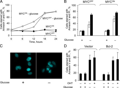
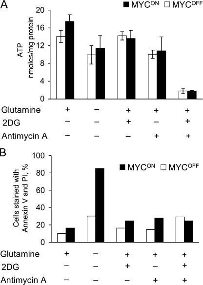
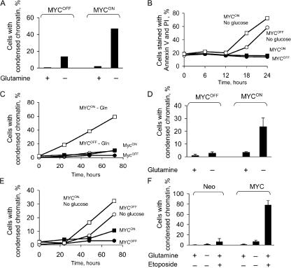
- Glucose depletion killed IER-MYC human fibroblasts irrespective of ERTM-MYC activity; 0.5 mM glucose (2% of normal) killed more slowly but also independently of MYC. Death was not apoptosis, as shown by absence of chromatin condensation and failure of Bcl-2 to prevent death.
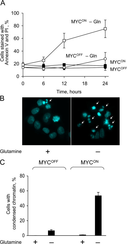
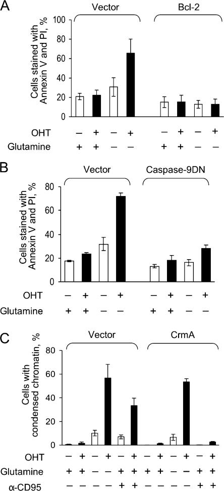
- Glutamine depletion killed IER-MYC cells faster when ERTM-MYC was active; IER control cells remained viable without glutamine regardless of OHT. Death was apoptosis via the intrinsic pathway: Bcl-2 and caspase-9 dominant-negative prevented death, while crmA had no effect.
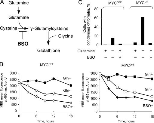
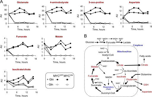
- Apoptosis after glutamine depletion was not caused by ATP deficiency, glutathione deficiency, DNA damage, protein synthesis deficiency, mTOR inhibition, or cell cycle arrest. Glutamine deprivation depleted Krebs cycle intermediates except citrate and isocitrate; pyruvate or oxaloacetate completely prevented apoptosis but did not restore proliferation, and the NADH/NAD ratio remained unchanged.
- Nutrient requirements varied by cell type: HFF-ER-MYC reacted like IER-MYC; HA1E cells underwent apoptosis after glucose depletion and reacted to glutamine depletion like IER-MYC; RPE-hTERT cells survived without glutamine irrespective of ectopic MYC and died without glucose independently of MYC by a non-apoptotic mechanism.
Methods
- Normal human fibroblasts IMR90 were retrovirally transduced with ERTM-MYC (IER-MYC) or ERTM alone (IER); HFF, HA1E, and RPE-hTERT cells were also tested. ERTM-MYC was activated with 200 nM 4-hydroxytamoxifen (OHT), with ethanol as vehicle.
- Nutrient depletion used glucose-free or glutamine-free DMEM; standard concentrations were 10 mM glucose and 2 mM glutamine. Viability and apoptosis were assessed by annexin V/PI, Hoechst 33342 nuclear morphology, Trypan Blue, and immunoblotting/immunofluorescence.
- Metabolite and viability analyses included ATP by luciferin-luciferase, reduced glutathione by flow cytometry, NADH/NAD by enzymatic cycling, lactate by LDH reaction, and capillary electrophoresis coupled to mass spectrometry (CE-MS) for metabolite profiling. Results are presented as averages of independent experiments with SDs where indicated.
Limitations
- Stated: how abnormal MYC synergizes with glutamine depletion to induce apoptosis remains to be determined; the authors cannot exclude a combination of tested mechanisms or an unknown glutamine function. Mitochondrial NADH/NAD+ and FADH/FAD+ ratios were not measured.
- Stated: the effect of nutrient depletion varied across cell types, so conclusions may be difficult to extrapolate from one or a few experimental systems.
- Evident: the study relies on ectopically expressed inducible ERTM-MYC in cultured human cell lines rather than endogenous MYC or in vivo tumor models; several supporting observations are described as unpublished or are based on two independent experiments.
Model-written summary. Check it against the paper before relying on it.
Integrity signals
Registry
Cites
Statistics
Figures
Verification
Claims the paper makes
28 Own result · 13 Not checked yet · 11 Own interpretation · 6 Own method
A result the paper reports from its own data — it cites no other source for it, so there is none to check. The quote shows it is in the paper.
In the paper
“depletion of glucose killed normal human cells irrespective of induced MYC activity and by a mechanism different from apoptosis.”
- Also stated in: § Glucose deprivation kills normal human fibroblasts not by apoptosis and largely independently of the MYC activity · plain
“depletion of glucose killed IER-MYC cells irrespective of ERTM-MYC activity (Fig. 1, A and B).”
§ Deficiency in glutamine but not glucose induces MYC-dependent apoptosis in human cells > Abstract
plaindirect- Asserted: passes — The authors put it forward as true.
- Checkable: passes — It can be checked against evidence.
- Load-bearing: passes — The paper’s conclusions depend on it.
- Based: passes — The paper gives a reason why it holds.
- Also stated in: § Glucose deprivation kills normal human fibroblasts not by apoptosis and largely independently of the MYC activity · plain
A result the paper reports from its own data — it cites no other source for it, so there is none to check. The quote shows it is in the paper.
In the paper
“by a mechanism different from apoptosis.”
- Also stated in: § Glucose deprivation kills normal human fibroblasts not by apoptosis and largely independently of the MYC activity · hedged
“cells in our system died by a process other than apoptosis, as indicated by the absence of characteristic chromatin condensation (Fig. 1 C) and the failure of Bcl-2, an inhibitor of apoptosis, to prevent cell death (Fig. 1 D).”
§ Deficiency in glutamine but not glucose induces MYC-dependent apoptosis in human cells > Abstract
plaindirect- Asserted: passes — The authors put it forward as true.
- Checkable: passes — It can be checked against evidence.
- Load-bearing: passes — The paper’s conclusions depend on it.
- Based: passes — The paper gives a reason why it holds.
- Also stated in: § Glucose deprivation kills normal human fibroblasts not by apoptosis and largely independently of the MYC activity · hedged
A result the paper reports from its own data — it cites no other source for it, so there is none to check. The quote shows it is in the paper.
In the paper
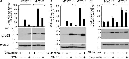
“glutamine depletion even prevented the increase in p53 concentration caused by activation of ERTM-MYC”
§ Deficiency in glutamine but not glucose induces MYC-dependent apoptosis in human cells > Discussion > How does ectopic activity of MYC synergize with glutamine depletion to induce apoptosis?
plaindirect- Asserted: passes — The authors put it forward as true.
- Checkable: passes — It can be checked against evidence.
- Load-bearing: passes — The paper’s conclusions depend on it.
- Based: passes — The paper gives a reason why it holds.
A result the paper reports from its own data — it cites no other source for it, so there is none to check. The quote shows it is in the paper.
In the paper
“Apoptosis was induced through the intrinsic pathway because Bcl-2 and a caspase-9 dominant-negative mutant (C9DN) prevented death (Fig. 3, A and B), whereas crmA, a viral inhibitor of the extrinsic pathway (Enari et al., 1995), had no effect (Fig. 3 D).”
§ Deficiency in glutamine but not glucose induces MYC-dependent apoptosis in human cells > Results > Glutamine deprivation induces apoptosis selectively in cells with acutely activated ER-MYC
plaindirect- Asserted: passes — The authors put it forward as true.
- Checkable: passes — It can be checked against evidence.
- Load-bearing: passes — The paper’s conclusions depend on it.
- Based: passes — The paper gives a reason why it holds.
A result the paper reports from its own data — it cites no other source for it, so there is none to check. The quote shows it is in the paper.
In the paper
“the antioxidants that we tested, tiron and N-acetyl-cysteine, failed to rescue the depleted cells from apoptosis (unpublished data)”
§ Deficiency in glutamine but not glucose induces MYC-dependent apoptosis in human cells > Discussion > How does glutamine depletion kill?
plain- Asserted: passes — The authors put it forward as true.
- Checkable: passes — It can be checked against evidence.
- Load-bearing: passes — The paper’s conclusions depend on it.
- Based: passes — The paper gives a reason why it holds.
A result the paper reports from its own data — it cites no other source for it, so there is none to check. The quote shows it is in the paper.
In the paper
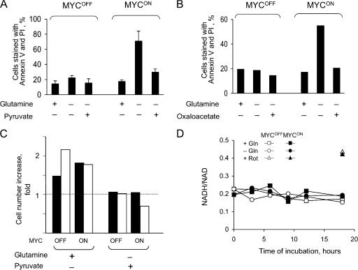
“oxaloacetate, which is a membrane-permeable Krebs cycle intermediate, or pyruvate, which can enter the cycle by converting into acetyl-CoA or oxaloacetate, completely prevented apoptosis in the absence of glutamine (Fig. 8, A and B).”
§ Deficiency in glutamine but not glucose induces MYC-dependent apoptosis in human cells > Results > Apoptosis caused by glutamine depletion is prevented by substrates of the Krebs cycle
plaindirect- Asserted: passes — The authors put it forward as true.
- Checkable: passes — It can be checked against evidence.
- Load-bearing: passes — The paper’s conclusions depend on it.
- Based: passes — The paper gives a reason why it holds.
- Rests on an assumption: Oxaloacetate is a membrane-permeable Krebs cycle intermediate and pyruvate can enter the Krebs cycle by converting into acetyl-CoA or oxaloacetate.
A result the paper reports from its own data — it cites no other source for it, so there is none to check. The quote shows it is in the paper.
In the paper
“BSO failed to induce apoptosis in IER-MYC cells (Fig. 6 C), suggesting that glutathione deficiency alone could not be the cause of apoptosis induced by glutamine depletion.”
§ Deficiency in glutamine but not glucose induces MYC-dependent apoptosis in human cells > Results > Apoptosis is not caused by glutathione deficiency
hedged- Asserted: passes — The authors put it forward as true.
- Checkable: passes — It can be checked against evidence.
- Load-bearing: passes — The paper’s conclusions depend on it.
- Based: passes — The paper gives a reason why it holds.
A result the paper reports from its own data — it cites no other source for it, so there is none to check. The quote shows it is in the paper.
In the paper
“IER-MYC cells ceased to proliferate irrespective of ERTM-MYC activity if either any of the essential amino acids or glutamine were absent (unpublished data). Therefore, deregulation of cell cycle arrest by itself could not explain cell death caused by glutamine depletion.”
§ Deficiency in glutamine but not glucose induces MYC-dependent apoptosis in human cells > Results > A cell cycle arrest is not the cause of apoptosis
hedged- Asserted: passes — The authors put it forward as true.
- Checkable: passes — It can be checked against evidence.
- Load-bearing: passes — The paper’s conclusions depend on it.
- Based: passes — The paper gives a reason why it holds.
A result the paper reports from its own data — it cites no other source for it, so there is none to check. The quote shows it is in the paper.
In the paper
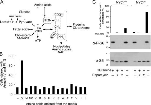
“omitting any of the essential amino acids from the tissue culture media failed to kill cells, at least over the time sufficient to kill by omitting glutamine (Fig. 4 B). Therefore, protein synthesis was not the cause of apoptosis induced by glutamine depletion.”
§ Deficiency in glutamine but not glucose induces MYC-dependent apoptosis in human cells > Results > A deficiency in protein synthesis or inhibition of mTOR did not cause apoptosis induced by glutamine depletion
plain- Asserted: passes — The authors put it forward as true.
- Checkable: passes — It can be checked against evidence.
- Load-bearing: passes — The paper’s conclusions depend on it.
- Based: passes — The paper gives a reason why it holds.
A result the paper reports from its own data — it cites no other source for it, so there is none to check. The quote shows it is in the paper.
In the paper
“Fig. S1 C demonstrates that the MYC moiety of ER-MYC makes cells sensitive to glutamine depletion”
§ Deficiency in glutamine but not glucose induces MYC-dependent apoptosis in human cells > Materials and methods > Online supplemental material
boosteddirect- Asserted: passes — The authors put it forward as true.
- Checkable: passes — It can be checked against evidence.
- Load-bearing: passes — The paper’s conclusions depend on it.
- Based: passes — The paper gives a reason why it holds.
A result the paper reports from its own data — it cites no other source for it, so there is none to check. The quote shows it is in the paper.
In the paper
“The finding that the effect of glutamine depletion on cell viability was different both quantitatively and qualitatively from DON”
§ Deficiency in glutamine but not glucose induces MYC-dependent apoptosis in human cells > Discussion > Relevance of glutamine metabolism to cancer therapy
plain- Asserted: passes — The authors put it forward as true.
- Checkable: passes — It can be checked against evidence.
- Load-bearing: passes — The paper’s conclusions depend on it.
- Based: passes — The paper gives a reason why it holds.
A result the paper reports from its own data — it cites no other source for it, so there is none to check. The quote shows it is in the paper.
In the paper
“glutamine deprivation caused rapid depletion of glutamate, 4-aminobutyrate, 5-oxoproline, and aspartate, which are products of glutamine metabolism (Fig. 9 and Table S1).”
§ Deficiency in glutamine but not glucose induces MYC-dependent apoptosis in human cells > Results > Apoptosis caused by glutamine depletion is prevented by substrates of the Krebs cycle
plaindirect- Asserted: passes — The authors put it forward as true.
- Checkable: passes — It can be checked against evidence.
- Load-bearing: passes — The paper’s conclusions depend on it.
- Based: passes — The paper gives a reason why it holds.
A result the paper reports from its own data — it cites no other source for it, so there is none to check. The quote shows it is in the paper.
In the paper
“we found that glutamine deprivation was much more potent than DON in inducing apoptosis”
§ Deficiency in glutamine but not glucose induces MYC-dependent apoptosis in human cells > Discussion > Relevance of glutamine metabolism to cancer therapy
plaindirect- Asserted: passes — The authors put it forward as true.
- Checkable: passes — It can be checked against evidence.
- Load-bearing: passes — The paper’s conclusions depend on it.
- Based: passes — The paper gives a reason why it holds.
A result the paper reports from its own data — it cites no other source for it, so there is none to check. The quote shows it is in the paper.
In the paper
“Modification of nutrient requirements by acute activation of ectopically expressed MYC vary with cell type”
§ Deficiency in glutamine but not glucose induces MYC-dependent apoptosis in human cells > Results > Modification of nutrient requirements by acute activation of ectopically expressed MYC vary with cell type
plaincorrelational- Asserted: passes — The authors put it forward as true.
- Checkable: passes — It can be checked against evidence.
- Load-bearing: passes — The paper’s conclusions depend on it.
- Based: passes — The paper gives a reason why it holds.
A result the paper reports from its own data — it cites no other source for it, so there is none to check. The quote shows it is in the paper.
In the paper
“we detected no increase in the concentration of ROS or DNA damage either after activation of ERTM-MYC or depleting glutathione”
§ Deficiency in glutamine but not glucose induces MYC-dependent apoptosis in human cells > Discussion > How does ectopic activity of MYC synergize with glutamine depletion to induce apoptosis?
plain- Asserted: passes — The authors put it forward as true.
- Checkable: passes — It can be checked against evidence.
- Load-bearing: passes — The paper’s conclusions depend on it.
- Based: passes — The paper gives a reason why it holds.
A result the paper reports from its own data — it cites no other source for it, so there is none to check. The quote shows it is in the paper.
In the paper
“and more selective for cells with ectopically activated MYC”
§ Deficiency in glutamine but not glucose induces MYC-dependent apoptosis in human cells > Discussion > Relevance of glutamine metabolism to cancer therapy
plaincorrelational- Asserted: passes — The authors put it forward as true.
- Checkable: passes — It can be checked against evidence.
- Load-bearing: passes — The paper’s conclusions depend on it.
- Based: passes — The paper gives a reason why it holds.
A result the paper reports from its own data — it cites no other source for it, so there is none to check. The quote shows it is in the paper.
In the paper
“In contrast to DON, glutamine depletion was not followed by an increase in p53 concentration”
§ Deficiency in glutamine but not glucose induces MYC-dependent apoptosis in human cells > Discussion > Relevance of glutamine metabolism to cancer therapy
plain- Asserted: passes — The authors put it forward as true.
- Checkable: passes — It can be checked against evidence.
- Load-bearing: passes — The paper’s conclusions depend on it.
- Based: passes — The paper gives a reason why it holds.
A result the paper reports from its own data — it cites no other source for it, so there is none to check. The quote shows it is in the paper.
In the paper
“neither pyruvate nor oxaloacetate restored cell proliferation (Fig. 8 C; and unpublished data), which suggested that the rescue was not due to replenishing depleted glutamine but rather due to specific effect of these metabolites on cell viability.”
§ Deficiency in glutamine but not glucose induces MYC-dependent apoptosis in human cells > Results > Apoptosis caused by glutamine depletion is prevented by substrates of the Krebs cycle
plain- Asserted: passes — The authors put it forward as true.
- Checkable: passes — It can be checked against evidence.
- Load-bearing: passes — The paper’s conclusions depend on it.
- Based: passes — The paper gives a reason why it holds.
A result the paper reports from its own data — it cites no other source for it, so there is none to check. The quote shows it is in the paper.
In the paper
“depletion of glutamine, another major nutrient consumed by cancer cells, induced apoptosis depending on MYC activity.”
- Also stated in: § Glutamine deprivation induces apoptosis selectively in cells with acutely activated ER-MYC · plain
“depletion of glutamine killed IER-MYC cells faster if ERTM-MYC was active (Fig. 2 A).”
- Also stated in: § Glutamine deprivation induces apoptosis selectively in cells with acutely activated ER-MYC · hedged
“The IER-MYC cells died by apoptosis, as indicated by the characteristic nuclear morphology of the dead cells (Fig. 2, B and C).”
§ Deficiency in glutamine but not glucose induces MYC-dependent apoptosis in human cells > Abstract
plaindirect- Asserted: passes — The authors put it forward as true.
- Checkable: passes — It can be checked against evidence.
- Load-bearing: passes — The paper’s conclusions depend on it.
- Based: passes — The paper gives a reason why it holds.
- Also stated in: § Glutamine deprivation induces apoptosis selectively in cells with acutely activated ER-MYC · plain
A result the paper reports from its own data — it cites no other source for it, so there is none to check. The quote shows it is in the paper.
In the paper
“a much larger decrease induced by a combination of 2-deoxyglucose, an inhibitor of glycolysis, and antimycin A, an inhibitor of mitochondrial respiration, failed to kill IER-MYC cells as efficiently as did glutamine depletion (Fig. 5, A and B). Therefore, apoptosis caused by glutamine depletion could not be mediated by ATP deficiency alone.”
§ Deficiency in glutamine but not glucose induces MYC-dependent apoptosis in human cells > Results > Apoptosis induced by glutamine depletion is not caused by ATP deficiency
hedged- Asserted: passes — The authors put it forward as true.
- Checkable: passes — It can be checked against evidence.
- Load-bearing: passes — The paper’s conclusions depend on it.
- Based: passes — The paper gives a reason why it holds.
A result the paper reports from its own data — it cites no other source for it, so there is none to check. The quote shows it is in the paper.
In the paper
“we detected no ROS accumulation after glutamine depletion”
§ Deficiency in glutamine but not glucose induces MYC-dependent apoptosis in human cells > Discussion > How does glutamine depletion kill?
plain- Asserted: passes — The authors put it forward as true.
- Checkable: passes — It can be checked against evidence.
- Load-bearing: passes — The paper’s conclusions depend on it.
- Based: passes — The paper gives a reason why it holds.
A result the paper reports from its own data — it cites no other source for it, so there is none to check. The quote shows it is in the paper.
In the paper
“All of the detected Krebs cycle intermediates, with the notable exception of citrate and isocitrate (Fig. 9), were depleted as well.”
§ Deficiency in glutamine but not glucose induces MYC-dependent apoptosis in human cells > Results > Apoptosis caused by glutamine depletion is prevented by substrates of the Krebs cycle
plaindirect- Asserted: passes — The authors put it forward as true.
- Checkable: passes — It can be checked against evidence.
- Load-bearing: passes — The paper’s conclusions depend on it.
- Based: passes — The paper gives a reason why it holds.
A conclusion the paper draws from its own results — it cites no other source for it, so there is none to check. The quote shows it is in the paper.
In the paper
“which indicated that these treatments activated different pathways”
§ Deficiency in glutamine but not glucose induces MYC-dependent apoptosis in human cells > Discussion > Relevance of glutamine metabolism to cancer therapy
hedgeddirect- Asserted: passes — The authors put it forward as true.
- Checkable: passes — It can be checked against evidence.
- Load-bearing: passes — The paper’s conclusions depend on it.
- Based: passes — The paper gives a reason why it holds.
None of the sources cited for this claim have been checked yet.
In the paper
“glutamine can be converted via glutamate into 2-oxoglutarate, a Krebs cycle intermediate (Kovacevic, 1972; Baggetto, 1992)”
§ Deficiency in glutamine but not glucose induces MYC-dependent apoptosis in human cells > Results > Apoptosis caused by glutamine depletion is prevented by substrates of the Krebs cycle
What the sources say
- Not checked yetcites Kovacevic, 1972
- Not checked yetcites Baggetto, 1992
plaindirect- Asserted: passes — The authors put it forward as true.
- Checkable: passes — It can be checked against evidence.
- Load-bearing: passes — The paper’s conclusions depend on it.
- Based: passes — The paper gives a reason why it holds.
What the paper did — it cites no source for this, so there is none to check. The quote shows it is in the paper.
In the paper
“The changes in ATP and glutathione concentrations detected by CE-MS reflected those obtained by biochemical methods (Fig. S4 and Table S1, available at http://www.jcb.org/cgi/content/full/jcb.200703099/DC1), indicating that the approach was reliable in our hands in detecting relative changes in metabolite concentrations.”
§ Deficiency in glutamine but not glucose induces MYC-dependent apoptosis in human cells > Results > Apoptosis caused by glutamine depletion is prevented by substrates of the Krebs cycle
hedged- Asserted: passes — The authors put it forward as true.
- Checkable: passes — It can be checked against evidence.
- Load-bearing: passes — The paper’s conclusions depend on it.
- Based: passes — The paper gives a reason why it holds.
In the paper
“The first part of our study reiterated the previously raised concern (Zu and Guppy, 2004) that the knowledge about the relative requirement of normal and transformed cells for glucose is still insufficient to make a reliable judgment as to whether agents that mimic glucose deprivation can be effective in cancer therapy.”
§ Deficiency in glutamine but not glucose induces MYC-dependent apoptosis in human cells > Discussion
What the sources say
- Not checked yetcites Zu and Guppy, 2004
plain- Asserted: passes — The authors put it forward as true.
- Checkable: passes — It can be checked against evidence.
- Load-bearing: passes — The paper’s conclusions depend on it.
- Based: passes — The paper gives a reason why it holds.
A conclusion the paper draws from its own results — it cites no other source for it, so there is none to check. The quote shows it is in the paper.
In the paper
“which implied that in our experimental system glutamine metabolism links MYC activity and increase in p53 concentration”
§ Deficiency in glutamine but not glucose induces MYC-dependent apoptosis in human cells > Discussion > How does ectopic activity of MYC synergize with glutamine depletion to induce apoptosis?
hedgedcorrelational- Asserted: passes — The authors put it forward as true.
- Checkable: passes — It can be checked against evidence.
- Load-bearing: passes — The paper’s conclusions depend on it.
- Based: passes — The paper gives a reason why it holds.
A conclusion the paper draws from its own results — it cites no other source for it, so there is none to check. The quote shows it is in the paper.
In the paper
“Because pyruvate and oxaloacetate contain no nitrogen but rescue from apoptosis induced by glutamine deficiency, the cause of apoptosis is likely to be associated with the fate of the glutamine carbon chain.”
§ Deficiency in glutamine but not glucose induces MYC-dependent apoptosis in human cells > Discussion > How does glutamine depletion kill?
hedgedcorrelational- Asserted: passes — The authors put it forward as true.
- Checkable: passes — It can be checked against evidence.
- Load-bearing: passes — The paper’s conclusions depend on it.
- Based: passes — The paper gives a reason why it holds.
What the paper did — it cites no source for this, so there is none to check. The quote shows it is in the paper.
In the paper
“This system allows studying immediate effects of MYC by minimizing the artifacts associated with clonal selection of cells that because of additional genetic changes avoided apoptosis induced by ectopic expression of MYC.”
§ Deficiency in glutamine but not glucose induces MYC-dependent apoptosis in human cells > Results
plaindirect- Asserted: passes — The authors put it forward as true.
- Checkable: passes — It can be checked against evidence.
- Load-bearing: passes — The paper’s conclusions depend on it.
- Based: passes — The paper gives a reason why it holds.
A conclusion the paper draws from its own results — it cites no other source for it, so there is none to check. The quote shows it is in the paper.
In the paper
“and that glutamine deprivation may not need p53 to kill”
§ Deficiency in glutamine but not glucose induces MYC-dependent apoptosis in human cells > Discussion > Relevance of glutamine metabolism to cancer therapy
hedgedconditional- Asserted: passes — The authors put it forward as true.
- Checkable: passes — It can be checked against evidence.
- Load-bearing: passes — The paper’s conclusions depend on it.
- Based: passes — The paper gives a reason why it holds.
A conclusion the paper draws from its own results — it cites no other source for it, so there is none to check. The quote shows it is in the paper.
In the paper
“These results indicated that glutamine depletion indeed inhibited the Krebs cycle progression.”
§ Deficiency in glutamine but not glucose induces MYC-dependent apoptosis in human cells > Results > Apoptosis caused by glutamine depletion is prevented by substrates of the Krebs cycle
hedgeddirect- Asserted: passes — The authors put it forward as true.
- Checkable: passes — It can be checked against evidence.
- Load-bearing: passes — The paper’s conclusions depend on it.
- Based: passes — The paper gives a reason why it holds.
None of the sources cited for this claim have been checked yet.
In the paper
“Some cancer cells can even survive in vitro without glucose at all if supplied with glutamine and nucleosides (Reitzer et al., 1979; Wice et al., 1981; Linker et al., 1985).”
§ Deficiency in glutamine but not glucose induces MYC-dependent apoptosis in human cells > Introduction
What the sources say
- Not checked yetcites Reitzer et al., 1979
- Not checked yetcites Wice et al., 1981
- Not checked yetcites Linker et al., 1985
plainconditional- Asserted: passes — The authors put it forward as true.
- Checkable: passes — It can be checked against evidence.
- Load-bearing: passes — The paper’s conclusions depend on it.
- Based: passes — The paper gives a reason why it holds.
A conclusion the paper draws from its own results — it cites no other source for it, so there is none to check. The quote shows it is in the paper.
In the paper
“Therefore, the findings that the Krebs cycle is deficient and the substrates of the cycle rescue from glutamine depletion suggested that a deficiency in glutamine causes apoptosis by affecting the Krebs cycle.”
§ Deficiency in glutamine but not glucose induces MYC-dependent apoptosis in human cells > Results > Apoptosis caused by glutamine depletion is prevented by substrates of the Krebs cycle
hedgeddirect- Asserted: passes — The authors put it forward as true.
- Checkable: passes — It can be checked against evidence.
- Load-bearing: passes — The paper’s conclusions depend on it.
- Based: passes — The paper gives a reason why it holds.
- It may state two separate claims as one. 11
- Probably not a substantial claim. 15
A result the paper reports from its own data — it cites no other source for it, so there is none to check. The quote shows it is in the paper.
In the paper
“glutamine depletion not only failed to increase the p53 concentration, but even reversed the increase induced by activation of ERTM-MYC (Fig. 7).”
- Also stated in: § Glutamine depletion does not cause DNA damage · plain
“Phosphorylation of H2AX, as detected by immunofluorescence, was induced by etoposide and by activation of ERTM-MYC, but not by glutamine depletion (Fig. S2, available at http://www.jcb.org/cgi/content/full/jcb.200703099/DC1).”
- Also stated in: § Glutamine depletion does not cause DNA damage · hedged
“Hence, depletion of glutamine was unlikely to cause apoptosis by inducing DNA damage.”
§ Deficiency in glutamine but not glucose induces MYC-dependent apoptosis in human cells > Results > Glutamine depletion does not cause DNA damage
plain- Asserted: passes — The authors put it forward as true.
- Checkable: passes — It can be checked against evidence.
- Load-bearing: passes — The paper’s conclusions depend on it.
- Based: passes — The paper gives a reason why it holds.
It may state two separate claims as one.
- Also stated in: § Glutamine depletion does not cause DNA damage · plain
A result the paper reports from its own data — it cites no other source for it, so there is none to check. The quote shows it is in the paper.
In the paper
“the NADH/NAD ratio remained unchanged (Fig. 8 D) and lactate was still produced (Fig. S3, available at http://www.jcb.org/cgi/content/full/jcb.200703099/DC1) after glutamine depletion, even though at a lower rate, indicating that pyruvate was unlikely to rescue from glutamine depletion by being a substrate of LDH.”
§ Deficiency in glutamine but not glucose induces MYC-dependent apoptosis in human cells > Results > Apoptosis caused by glutamine depletion is prevented by substrates of the Krebs cycle
plain- Asserted: passes — The authors put it forward as true.
- Checkable: passes — It can be checked against evidence.
- Load-bearing: passes — The paper’s conclusions depend on it.
- Based: passes — The paper gives a reason why it holds.
It may state two separate claims as one.
A result the paper reports from its own data — it cites no other source for it, so there is none to check. The quote shows it is in the paper.
In the paper
“rapamycin caused S6 dephosphorylation but had no effect on cell viability over the time sufficient to kill cells by glutamine depletion (Fig. 4 C). Hence, glutamine depletion caused apoptosis by affecting pathways that did not require mTOR activity.”
§ Deficiency in glutamine but not glucose induces MYC-dependent apoptosis in human cells > Results > A deficiency in protein synthesis or inhibition of mTOR did not cause apoptosis induced by glutamine depletion
plain- Asserted: passes — The authors put it forward as true.
- Checkable: passes — It can be checked against evidence.
- Load-bearing: passes — The paper’s conclusions depend on it.
- Based: passes — The paper gives a reason why it holds.
It may state two separate claims as one.
A result the paper reports from its own data — it cites no other source for it, so there is none to check. The quote shows it is in the paper.
In the paper
“in our hands depletion of essential amino acids caused cell cycle arrest but not apoptosis”
§ Deficiency in glutamine but not glucose induces MYC-dependent apoptosis in human cells > Discussion > How does ectopic activity of MYC synergize with glutamine depletion to induce apoptosis?
plaindirect- Asserted: passes — The authors put it forward as true.
- Checkable: passes — It can be checked against evidence.
- Load-bearing: unclear — It is unclear whether the paper’s conclusions depend on it.
- Based: passes — The paper gives a reason why it holds.
It may state two separate claims as one.
A result the paper reports from its own data — it cites no other source for it, so there is none to check. The quote shows it is in the paper.
In the paper
“Fig. S3 documents that lactate production remains largely unchanged after depletion of glutamine and is independent of ER-MYC activity”
§ Deficiency in glutamine but not glucose induces MYC-dependent apoptosis in human cells > Materials and methods > Online supplemental material
plain- Asserted: passes — The authors put it forward as true.
- Checkable: passes — It can be checked against evidence.
- Load-bearing: unclear — It is unclear whether the paper’s conclusions depend on it.
- Based: passes — The paper gives a reason why it holds.
It may state two separate claims as one.
A result the paper reports from its own data — it cites no other source for it, so there is none to check. The quote shows it is in the paper.
In the paper
“glutamine depletion caused cell cycle arrest irrespective of the ERTM-MYC activity (unpublished data)”
§ Deficiency in glutamine but not glucose induces MYC-dependent apoptosis in human cells > Discussion > How does ectopic activity of MYC synergize with glutamine depletion to induce apoptosis?
plaindirect- Asserted: passes — The authors put it forward as true.
- Checkable: passes — It can be checked against evidence.
- Load-bearing: fails — The paper’s conclusions may not depend on it.
- Based: passes — The paper gives a reason why it holds.
Probably not a substantial claim. The paper’s conclusions may not depend on it.
In the paper
“we used IER-MYC that expressed C9DN (IER-MYC–C9DN), which prevents cell disassembly but not the preceding steps of apoptosis (Fearnhead et al., 1998), thus avoiding artifacts associated with cell decomposition.”
§ Deficiency in glutamine but not glucose induces MYC-dependent apoptosis in human cells > Results > Apoptosis induced by glutamine depletion is not caused by ATP deficiency
What the sources say
- Not checked yetcites Fearnhead et al., 1998
plaindirect- Asserted: passes — The authors put it forward as true.
- Checkable: passes — It can be checked against evidence.
- Load-bearing: passes — The paper’s conclusions depend on it.
- Based: passes — The paper gives a reason why it holds.
It may state two separate claims as one.
What the paper did — it cites no source for this, so there is none to check. The quote shows it is in the paper.
In the paper
“Identification was based on retention time and exact mass measurements after alignment of both axes with reference compounds, and confirmed by spiked standards.”
§ Deficiency in glutamine but not glucose induces MYC-dependent apoptosis in human cells > Materials and methods > CE-MS analysis
plain- Asserted: passes — The authors put it forward as true.
- Checkable: passes — It can be checked against evidence.
- Load-bearing: passes — The paper’s conclusions depend on it.
- Based: passes — The paper gives a reason why it holds.
Probably not a substantial claim.
In the paper
“metabolism of human cells is changed by oncogenes in various ways (Ramanathan et al., 2005)”
§ Deficiency in glutamine but not glucose induces MYC-dependent apoptosis in human cells > Results > Modification of nutrient requirements by acute activation of ectopically expressed MYC vary with cell type
What the sources say
- Not checked yetcites Ramanathan et al., 2005
plaindirect- Asserted: passes — The authors put it forward as true.
- Checkable: passes — It can be checked against evidence.
- Load-bearing: fails — The paper’s conclusions may not depend on it.
- Based: passes — The paper gives a reason why it holds.
Probably not a substantial claim. The paper’s conclusions may not depend on it.
None of the sources cited for this claim have been checked yet.
In the paper
“previous reports suggested that depletion of glutamine kills because of stress caused by deficiency in proteins, ATP, glutathione, or nucleotides (for review see Ahluwalia et al., 1990; Fuchs and Bode, 2006).”
§ Deficiency in glutamine but not glucose induces MYC-dependent apoptosis in human cells > Results > Glutamine deprivation induces apoptosis selectively in cells with acutely activated ER-MYC
What the sources say
- Not checked yetcites Ahluwalia et al., 1990
- Not checked yetcites Fuchs and Bode, 2006
hedgeddirect- Asserted: fails — The authors may not put it forward as true.
- Checkable: passes — It can be checked against evidence.
- Load-bearing: passes — The paper’s conclusions depend on it.
- Based: passes — The paper gives a reason why it holds.
Probably not a substantial claim. The authors may not put it forward as true.
None of the sources cited for this claim have been checked yet.
In the paper
“Adding OHT to the tissue culture medium caused translocation of ERTM-MYC to the nucleus (Fig. S1 A, available at http://www.jcb.org/cgi/content/full/jcb.200703099/DC1) and increased sensitivity of the cells to DNA damage (Fig. S1 B), which confirmed that the system functioned as expected (Eilers et al., 1989; Littlewood et al., 1995).”
§ Deficiency in glutamine but not glucose induces MYC-dependent apoptosis in human cells > Results
What the sources say
- Not checked yetcites Eilers et al., 1989
- Not checked yetcites Littlewood et al., 1995
boosteddirect- Asserted: passes — The authors put it forward as true.
- Checkable: passes — It can be checked against evidence.
- Load-bearing: passes — The paper’s conclusions depend on it.
- Based: passes — The paper gives a reason why it holds.
It may state two separate claims as one.
In the paper
“Pyruvate might affect cell viability by being a substrate of lactate dehydrogenase (LDH), which oxidizes NADH to NAD (Zielke et al., 1980).”
§ Deficiency in glutamine but not glucose induces MYC-dependent apoptosis in human cells > Results > Apoptosis caused by glutamine depletion is prevented by substrates of the Krebs cycle
What the sources say
- Not checked yetcites Zielke et al., 1980
hedgedconditional- Asserted: passes — The authors put it forward as true.
- Checkable: passes — It can be checked against evidence.
- Load-bearing: fails — The paper’s conclusions may not depend on it.
- Based: passes — The paper gives a reason why it holds.
Probably not a substantial claim. The paper’s conclusions may not depend on it.
In the paper
“Treatments that induce glutamine deficiency or interfere with its metabolism as a substrate for nucleotide synthesis were highly successful in animals, but turned out to be unacceptably toxic in people and were largely abandoned (Ahluwalia et al., 1990).”
§ Deficiency in glutamine but not glucose induces MYC-dependent apoptosis in human cells > Discussion > Relevance of glutamine metabolism to cancer therapy
What the sources say
- Not checked yetcites Ahluwalia et al., 1990
plaindirect- Asserted: passes — The authors put it forward as true.
- Checkable: passes — It can be checked against evidence.
- Load-bearing: passes — The paper’s conclusions depend on it.
- Based: passes — The paper gives a reason why it holds.
It may state two separate claims as one.
A conclusion the paper draws from its own results — it cites no other source for it, so there is none to check. The quote shows it is in the paper.
In the paper
“Overall, our study is consistent with the view that metabolic changes associated with carcinogenesis, and the energy metabolism in particular, might be much more complex than Warburg postulated”
§ Deficiency in glutamine but not glucose induces MYC-dependent apoptosis in human cells > Discussion > Relevance of glutamine metabolism to cancer therapy
hedgedcorrelational- Asserted: passes — The authors put it forward as true.
- Checkable: fails — It may be too vague to check against evidence.
- Load-bearing: fails — The paper’s conclusions may not depend on it.
- Based: passes — The paper gives a reason why it holds.
Probably not a substantial claim. It may be too vague to check against evidence. The paper’s conclusions may not depend on it.
A conclusion the paper draws from its own results — it cites no other source for it, so there is none to check. The quote shows it is in the paper.
In the paper
“our observations indicated that glutamine in our system is either the main source of Krebs cycle intermediates, as in some proliferating normal and cancer cells (Reitzer et al., 1979; Moreadith and Lehninger, 1984; Zielke et al., 1984; Kovacevic et al., 1991; Baggetto, 1992; Petch and Butler, 1994), or regulates the Krebs cycle activity.”
§ Deficiency in glutamine but not glucose induces MYC-dependent apoptosis in human cells > Discussion > How does glutamine depletion kill?
hedgedconditional- Asserted: passes — The authors put it forward as true.
- Checkable: passes — It can be checked against evidence.
- Load-bearing: passes — The paper’s conclusions depend on it.
- Based: passes — The paper gives a reason why it holds.
It may state two separate claims as one.
A conclusion the paper draws from its own results — it cites no other source for it, so there is none to check. The quote shows it is in the paper.
In the paper
“suggests that the potential of glutamine metabolism as a target for cancer therapy may need revisiting”
§ Deficiency in glutamine but not glucose induces MYC-dependent apoptosis in human cells > Discussion > Relevance of glutamine metabolism to cancer therapy
hedged- Asserted: passes — The authors put it forward as true.
- Checkable: fails — It may be too vague to check against evidence.
- Load-bearing: passes — The paper’s conclusions depend on it.
- Based: passes — The paper gives a reason why it holds.
Probably not a substantial claim. It may be too vague to check against evidence.
In the paper
“the NADH/NAD ratio determines rates of many processes, from glycolysis to histone modifications (for review see Ladurner, 2006)”
§ Deficiency in glutamine but not glucose induces MYC-dependent apoptosis in human cells > Results > Apoptosis caused by glutamine depletion is prevented by substrates of the Krebs cycle
What the sources say
- Not checked yetcites Ladurner, 2006
plaindirect- Asserted: passes — The authors put it forward as true.
- Checkable: passes — It can be checked against evidence.
- Load-bearing: fails — The paper’s conclusions may not depend on it.
- Based: passes — The paper gives a reason why it holds.
Probably not a substantial claim. The paper’s conclusions may not depend on it.
In the paper
“capillary electrophoresis coupled to mass spectrometry (CE-MS), an approach that estimates concentration of multiple metabolites in one sample (Soga et al., 2003).”
§ Deficiency in glutamine but not glucose induces MYC-dependent apoptosis in human cells > Results > Apoptosis caused by glutamine depletion is prevented by substrates of the Krebs cycle
What the sources say
- Not checked yetcites Soga et al., 2003
plain- Asserted: passes — The authors put it forward as true.
- Checkable: passes — It can be checked against evidence.
- Load-bearing: passes — The paper’s conclusions depend on it.
- Based: passes — The paper gives a reason why it holds.
Probably not a substantial claim.
What the paper did — it cites no source for this, so there is none to check. The quote shows it is in the paper.
In the paper
“All changes in brightness or contrast, if any done, were applied to the entire image.”
§ Deficiency in glutamine but not glucose induces MYC-dependent apoptosis in human cells > Materials and methods > Immunofluorescence
plain- Asserted: passes — The authors put it forward as true.
- Checkable: passes — It can be checked against evidence.
- Load-bearing: fails — The paper’s conclusions may not depend on it.
- Based: passes — The paper gives a reason why it holds.
Probably not a substantial claim. The paper’s conclusions may not depend on it.
What the paper did — it cites no source for this, so there is none to check. The quote shows it is in the paper.
In the paper
“To further decrease the chances of clonal selection, we used retroviral transduction rather than transient transfection to generate populations of normal human fibroblasts IMR90 that expressed ERTM-MYC (IER-MYC) or ERTM (IER); the latter served to exclude effects of OHT unrelated to MYC activity.”
§ Deficiency in glutamine but not glucose induces MYC-dependent apoptosis in human cells > Results
plaindirect- Asserted: passes — The authors put it forward as true.
- Checkable: passes — It can be checked against evidence.
- Load-bearing: passes — The paper’s conclusions depend on it.
- Based: passes — The paper gives a reason why it holds.
Probably not a substantial claim.
A conclusion the paper draws from its own results — it cites no other source for it, so there is none to check. The quote shows it is in the paper.
In the paper
“our results demonstrated that dependence of cells on glucose and the effects of its depletion vary among cell types and can depend on the combination of oncogenes expressed in these cells.”
§ Deficiency in glutamine but not glucose induces MYC-dependent apoptosis in human cells > Discussion
boostedcorrelational- Asserted: passes — The authors put it forward as true.
- Checkable: passes — It can be checked against evidence.
- Load-bearing: passes — The paper’s conclusions depend on it.
- Based: passes — The paper gives a reason why it holds.
It may state two separate claims as one.
None of the sources cited for this claim have been checked yet.
In the paper
“the Krebs cycle is not only a source of NADH for oxidative phosphorylation, but also a “hub” of numerous metabolic pathways (http://www.tcd.ie/Biochemistry/IUBMB-Nicholson/minimaps.html)”
§ Deficiency in glutamine but not glucose induces MYC-dependent apoptosis in human cells > Results > Apoptosis caused by glutamine depletion is prevented by substrates of the Krebs cycle
plain- Asserted: passes — The authors put it forward as true.
- Checkable: passes — It can be checked against evidence.
- Load-bearing: passes — The paper’s conclusions depend on it.
- Based: passes — The paper gives a reason why it holds.
Probably not a substantial claim.
A conclusion the paper draws from its own results — it cites no other source for it, so there is none to check. The quote shows it is in the paper.
In the paper
“Our results suggest that the fate of normal human cells should be considered in evaluating nutrient deprivation as a strategy for cancer therapy”
§ Deficiency in glutamine but not glucose induces MYC-dependent apoptosis in human cells > Abstract
hedged- Asserted: passes — The authors put it forward as true.
- Checkable: fails — It may be too vague to check against evidence.
- Load-bearing: passes — The paper’s conclusions depend on it.
- Based: passes — The paper gives a reason why it holds.
Probably not a substantial claim. It may be too vague to check against evidence.
None of the sources cited for this claim have been checked yet.
In the paper
“depletion of glucose induced apoptosis in rodent cells that were transduced with MYC, an oncogene that is abnormally expressed in many human cancers (Nesbit et al., 1999), but not in the parental cell line (Shim et al., 1998).”
§ Deficiency in glutamine but not glucose induces MYC-dependent apoptosis in human cells > Introduction
What the sources say
- Not checked yetcites Shim et al., 1998
- Not checked yetcites Nesbit et al., 1999
plaindirect- Asserted: passes — The authors put it forward as true.
- Checkable: passes — It can be checked against evidence.
- Load-bearing: passes — The paper’s conclusions depend on it.
- Based: passes — The paper gives a reason why it holds.
Probably not a substantial claim. It may state two separate claims as one.
What the paper did — it cites no source for this, so there is none to check. The quote shows it is in the paper.
In the paper
“Fig. S1, A and B document that ER-MYC is activated as expected”
§ Deficiency in glutamine but not glucose induces MYC-dependent apoptosis in human cells > Materials and methods > Online supplemental material
plain- Asserted: passes — The authors put it forward as true.
- Checkable: passes — It can be checked against evidence.
- Load-bearing: passes — The paper’s conclusions depend on it.
- Based: passes — The paper gives a reason why it holds.
Probably not a substantial claim.
The paper relies on this without support.
Metabolism can vary among species.
“Considering that metabolism can vary among species, and that human rather than animal tumors are of primary interest, we used human cells.”
The paper relies on this without support.
Glutamine is made by cells, making the requirement for exogenous glutamine appear unnecessary.
“However, glutamine is made by cells, which makes the requirement for exogenous glutamine appear unnecessary.”
The paper relies on this without support.
Oxaloacetate is a membrane-permeable Krebs cycle intermediate and pyruvate can enter the Krebs cycle by converting into acetyl-CoA or oxaloacetate.
“oxaloacetate, which is a membrane-permeable Krebs cycle intermediate, or pyruvate, which can enter the cycle by converting into acetyl-CoA or oxaloacetate, completely prevented apoptosis in the absence of glutamine (Fig. 8, A and B).”
The paper relies on this without support.
Why glutamine is required for cell viability is not entirely clear.
“Surprisingly, why glutamine is required for cell viability, or, in other words, why depletion of glutamine would kill cells, is not entirely clear.”
The paper relies on this without support.
p53 is mutated in many cancers.
“which is an advantage as this protein is mutated in many cancers”
Claims other papers make about this one
Each is checked against this paper’s own text: does it say what the citing paper says it does?
The source backs part of the claim, not all of it — or backs it for a different population, condition or comparison.
In the citing paper
“Furthermore, depending on the tissue of origin, cancer cells use various routes of glutamine metabolism. Some have been shown to require an exogenous glutamine supply for malignant proliferation [71,72], whereas others do not [73] and can synthesize it de novo [74].”
From HIF-1-Independent Mechanisms Regulating Metabolic Adaptation in Hypoxic Cancer Cells (2021) · cites this paper as [71]
Experimental — accuracy not yet measuredThe source backs part of the claim, not all of it — or backs it for a different population, condition or comparison.
In the citing paper
“Cancer cells often consume glutamine, and those dependent on Myc signaling are highly sensitive to glutamine withdrawal (Yuneva et al., 2007).”
From Therapeutic targeting of hypoxia and hypoxia-inducible factors in cancer (2016) · cites this paper as Yuneva et al., 2007
Experimental — accuracy not yet measuredIn the citing paper
“Similarly, checkpoint kinase 1 (CHK1) inhibitors625–627 and glutaminase (GLS) inhibitors628–634 are also considered potential candidates for treating Myc-overexpressing cancers.”
From Recent advances in targeting the “undruggable” proteins: from drug discovery to clinical trials (2023) · cites this paper as [628]
In the citing paper
“Providing cells with αKG, oxaloacetate, or pyruvate is sufficient to rescue cancer cell proliferation in conditions of glutamine starvation, confirming that glutamine supports proliferation by replenishing depleted TCA cycle intermediates, a process termed anaplerosis (Altman et al., 2016; Weinberg et al., 2010; Yuneva et al., 2007).”
From Targeting Metabolism for Cancer Therapy (2017) · cites this paper as Yuneva et al., 2007
In the citing paper
“In certain contexts, MYC-transformed cancers become glutamine dependent and undergo apoptosis in the absence of glutamine (Yuneva et al., 2007).”
From Targeting Metabolism for Cancer Therapy (2017) · cites this paper as Yuneva et al., 2007
In the citing paper
“In c-myc-transformed cells in particular, glutamine deprivation leads to the collapse of the TCA cycle and triggers cell death, which can be rescued by the addition of oxaloacetate or a membrane-permeable form of α-ketoglutarate (Wise et al., 2008; Yuneva et al., 2007).”
From The emerging hallmarks of cancer metabolism (2016) · cites this paper as Yuneva et al., 2007
Self-check
Percentages, sums and reported differences re-computed from the paper’s own numbers.
Each contradiction between two passages listed here was confirmed by an independent check.
Rated major: it would touch a result — a value a result rests on, reported and recomputed p-values on opposite sides of the significance level, two passages that conflict with no sign which is right, or a cited work retracted before this paper was published.
In the paper
§ Deficiency in glutamine but not glucose induces MYC-dependent apoptosis in human cells > Discussion > How does ectopic activity of MYC synergize with glutamine depletion to induce apoptosis?
“However, we detected no increase in the concentration of ROS or DNA damage either after activation of ERTM-MYC or depleting glutathione.”
§ Deficiency in glutamine but not glucose induces MYC-dependent apoptosis in human cells > Results > Glutamine depletion does not cause DNA damage
“Phosphorylation of H2AX, as detected by immunofluorescence, was induced by etoposide and by activation of ERTM-MYC, but not by glutamine depletion (Fig. S2, available at http://www.jcb.org/cgi/content/full/jcb.200703099/DC1).”
Rated minor: it would not touch a result — a value no result rests on, a p-value that stays on the same side of the significance level, or a conflict that is clerical, resolved elsewhere in the paper, or outside its own findings.
In the paper
§ Deficiency in glutamine but not glucose induces MYC-dependent apoptosis in human cells > Results > Glutamine deprivation induces apoptosis selectively in cells with acutely activated ER-MYC
“whereas crmA, a viral inhibitor of the extrinsic pathway (Enari et al., 1995), had no effect (Fig. 3 D).”
§ Deficiency in glutamine but not glucose induces MYC-dependent apoptosis in human cells > Results > Glutamine deprivation induces apoptosis selectively in cells with acutely activated ER-MYC
“(C) IER-MYC transduced with CrmA or vector alone were treated as in A except anti-Fas (α-CD95) antibody was added as indicated.”
Rated minor: it would not touch a result — a value no result rests on, a p-value that stays on the same side of the significance level, or a conflict that is clerical, resolved elsewhere in the paper, or outside its own findings.
In the paper
§ Deficiency in glutamine but not glucose induces MYC-dependent apoptosis in human cells > Results > Apoptosis is not caused by glutathione deficiency
“(C) IER-MYC cells were treated as in A except the cells were collected at 24 h of incubation in the indicated media to determine the rate of apoptosis.”
§ Deficiency in glutamine but not glucose induces MYC-dependent apoptosis in human cells > Results > Apoptosis is not caused by glutathione deficiency
“(A) Biosynthesis of glutathione. (B) IER-MYC–C9DN were incubated with OHT (MYCON) or ethanol (MYCOFF) for 24 h, and then transferred to the medium depleted of glutamine or supplemented with 100 μM BSO.”
Citations
Cites
(1975) · Journal of AOAC INTERNATIONAL · doi:10.1093/jaoac/58.5.1085 · Cited by 20,078
Warburg, O. (1956) · Science · doi:10.1126/science.123.3191.309 · Cited by 13,425 · cited 1×
“Glucose metabolism has drawn much of the attention (for review see Garber, 2006) from the time when Otto Warburg found that some cancer cells consume more glucose than normal cells and proposed that a failure to make ATP by oxidative phosphorylation is the cause of cancer (Warburg, 1956).”
BackgroundDNA double-stranded breaks induce histone H2AX phosphorylation on serine 139
Rogakou, E. P., Pilch, D. R., Orr, A. H., Ivanova, V. S., & Bonner, W. M. (1998) · Journal of Biological Chemistry · doi:10.1074/jbc.273.10.5858 · Cited by 5,438 · cited 4×
“S2 visualizes localization of phosphorylated histone H2AX in cells deprived of glutamine or treated with etoposide.”
BackgroundInfluentialCillà, S. D., Farruggio, S., Vujosevic, S., Raina, G., Filippini, D. A., Gatti, V., Clemente, N., Mary, D., Vezzola, D., Casini, G., et al. (2017) · Cellular Physiology and Biochemistry · doi:10.1159/000479441 · Cited by 5,154
The Pathophysiology of Mitochondrial Cell Death
Green, D. R., & Kroemer, G. (2004) · Science · doi:10.1126/science.1099320 · Cited by 3,487 · cited 1×
“For example, citrate is used for the synthesis of fatty acids whose defi ciency might change permeability of mitochondrial membranes and affect their regulation by the Bcl-2 proteins (Green and Kroemer, 2004).”
BackgroundSuccinate links TCA cycle dysfunction to oncogenesis by inhibiting HIF-α prolyl hydroxylase
Selak, M. A., Armour, S. M., MacKenzie, E. D., Boulahbel, H., Watson, D. G., Mansfield, K. D., Pan, Y., Simon, M. C., Thompson, C. B., & Gottlieb, E. (2005) · Cancer Cell · doi:10.1016/j.ccr.2004.11.022 · Cited by 2,108 · cited 1×
“Some of the Krebs cycle intermediates, such as fumarate, succinate, and 2-oxoglutarate, can function as messenger and cofactor molecules directly regulating activity of enzymes and thereby contributing to regulation of gene transcription and/or tumorigenesis (Selak et al., 2005; Chen et al., 2006).”
BackgroundRas, PI(3)K and mTOR signalling controls tumour cell growth
Shaw, R. J., & Cantley, L. C. (2006) · Nature · doi:10.1038/nature04869 · Cited by 2,019 · cited 1×
“The differences between cancer and normal cells in consumption and metabolism of basic nutrients have been considered a promising cancer therapy target for several decades (for review see Thompson et al., 2005; Shaw and Cantley, 2006).”
BackgroundGriffith, O. W., & Meister, A. (1979) · Journal of Biological Chemistry · doi:10.1016/s0021-9258(18)35980-5 · Cited by 1,683
Translational control in stress and apoptosis
Holčı́k, M., & Sonenberg, N. (2005) · Nature Reviews Molecular Cell Biology · doi:10.1038/nrm1618 · Cited by 1,409 · cited 1×
“…review see Baggetto, 1992; Curi et al., 2005), abnormal expression of MYC accelerates cell proliferation and changes metabolism of mitochondria (Morrish et al., 2003; Li et al., 2005), and even a transient decrease in ATP concentration can cause apoptosis (Izyumov et al., 2004; Shaw et al., 2004).”
BackgroundmTOR and cancer: insights into a complex relationship
Sabatini, D. M. (2006) · Nature Reviews Cancer · doi:10.1038/nrc1974 · Cited by 1,322 · cited 1×
“The availability of nitrogen sources, and glutamine in particular, regulates the activity of mTOR, a kinase that controls protein synthesis, cell proliferation, and other processes that require nitrogen (for review see Sabatini, 2006).”
Background
Research works printed but not linked: 6
- 1969, Cancer Res., 29, 669
- 1969, Cancer Res., 29, 1195
- 1985, Eur. J. Cell Biol., 36, 176
- 2004, Anticancer Res., 24, 1121
- 2003, Cancer Res., 63, 7330
- 1984, Fed. Proc., 43, 121
Cited by
Sorted by citations
Understanding the Warburg Effect: The Metabolic Requirements of Cell Proliferation
Vander Heiden, M. G., Cantley, L. C., & Thompson, C. B. (2009) · Science · doi:10.1126/science.1160809 · Cited by 16,197
The emerging hallmarks of cancer metabolism
Pavlova, N. N., & Thompson, C. B. (2016) · Cell Metabolism · doi:10.1016/j.cmet.2015.12.006 · Cited by 5,926
The Biology of Cancer: Metabolic Reprogramming Fuels Cell Growth and Proliferation
DeBerardinis, R. J., Lum, J. J., Hatzivassiliou, G., & Thompson, C. B. (2008) · Cell Metabolism · doi:10.1016/j.cmet.2007.10.002 · Cited by 4,088
“Impressively, glutamine deprivation from fibroblast cultures essentially eliminates pools of the TCA cycle intermediates fumarate and malate (Yuneva et al., 2007).”
BackgroundInfluentialDang, C. V. (2012) · Cell · doi:10.1016/j.cell.2012.03.003 · Cited by 3,390
“Likewise, glutamine withdrawal triggers apoptosis of Myc-overexpressing cells (Yuneva et al., 2007).”
BackgroundOtto Warburg's contributions to current concepts of cancer metabolism
Koppenol, W. H., Bounds, P. L., & Dang, C. V. (2011) · Nature Reviews Cancer · doi:10.1038/nrc3038 · Cited by 3,427
“Thus, overexpression of MYC in cancer cells renders them sensitive to glutamine withdrawa”
Background
Metadata
- Authors
- Yuneva, M., Zamboni, N., Oefner, P., Sachidanandam, R., & Lazebnik, Y.
- Affiliations
- University of California, San Francisco (US)
- ETH Zurich (CH)
- University of Regensburg (DE)
- Cold Spring Harbor Laboratory (US)
- Corresponding author
- Lazebnik, Yuri
- Venue
- The Journal of Cell Biology
- Publisher
- Rockefeller University Press
- Published
- 2007-07-02
- Volume
- 178
- Issue
- 1
- Pages
- 93-105
- ISSN
- 0021-9525 (ISSN-L)
- 1540-8140
- PMC
- PMC2064426
- OpenAlex
- W2112861594
- Cited by
- 690
- References
- 72
- Citations by year
- 2026: 16 · 2025: 27 · 2024: 26 · 2023: 46 · 2022: 36 · 2021: 36 · 2020: 32 · 2019: 40 · 2018: 39 · 2017: 56 · 2016: 49 · 2015: 47 · 2014: 50 · 2013: 46 · 2012: 42
- FWCI
- 7.28
- Type
- journal article
- Language
- en
- Open access
- green
- www.ncbi.nlm.nih.gov
- Licence
- cc-by-nc-sa
- Locations
- The Journal of Cell Biology (publishedVersion)
- PubMed Central (publishedVersion, cc-by-nc-sa)
- PubMed (publishedVersion)
- Cold Spring Harbor Laboratory Institutional Repository (Cold Spring Harbor Laboratory) (acceptedVersion)
- Funders
- National Cancer Institute (CA-13106-31, P01CA013106)
- Topics
- Life Sciences › Biochemistry, Genetics and Molecular Biology › Cancer Research › Cancer, Hypoxia, and Metabolism
- Life Sciences › Biochemistry, Genetics and Molecular Biology › Biochemistry › Amino Acid Enzymes and Metabolism
- Life Sciences › Biochemistry, Genetics and Molecular Biology › Molecular Biology › PI3K/AKT/mTOR signaling in cancer
- Keywords
- glutamine deprivation, glucose deprivation, c-Myc, apoptosis, Krebs cycle intermediates, nutrient deprivation, cancer therapy, glutamine metabolism, cell viability, glucose metabolism
- MeSH terms
- Proto-Oncogene Mas, Cell Line, Cells, Cultured, Fibroblasts, Gene Expression Regulation, Glucose, Glutamine, Humans, Lung, Male, Retroviridae, Skin, Transduction, Genetic, Proto-Oncogene Proteins c-myc, Apoptosis, Cell Culture Techniques
About these checks
Registry, retraction, Cites and statistics results are computed, not judged, with one exception: a printed reference that no index lists is read once by a model before it counts as a citation error. Claims are extracted by a model and kept only when their quote appears verbatim in the text; each cited source is then checked, with a model’s help, for whether it says what the claim relies on. Those verdicts can be wrong — read the claim’s quote and the cited source before relying on one.
A language model drew the claims from the paper on 2026-10-06, under version 3 of the claim definition (model deepseek-v4.1-flash, prompt extract-v3).
A second model labelled each claim on 2026-10-06 (model jev-1.13-20260917, definition revision gold-guideline-v0.2).
Statistics and arithmetic are recomputed, not judged.
Confidence bands are shown only for fields calibrated on our annotated test set. None is calibrated yet, so no band is shown.
What counts as a claim ›How we measure confidence ›How we check this ›
Are you an author, or do you see a problem with this page? Request a review
