

Co-operation between different targeting pathways during integration of a membrane protein.
Open accessThe DOI matches its Crossref record; 48 of the 48 references it cites were found in a registry or an index.
Abstract
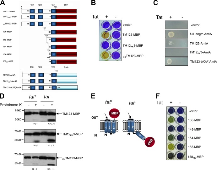
Membrane protein assembly is a fundamental process in all cells. The membrane-bound Rieske iron-sulfur protein is an essential component of the cytochrome bc1 and cytochrome b6f complexes, and it is exported across the energy-coupling membranes of bacteria and plants in a folded conformation by the twin arginine protein transport pathway (Tat) transport pathway. Although the Rieske protein in most organisms is a monotopic membrane protein, in actinobacteria, it is a polytopic protein with three transmembrane domains. In this work, we show that the Rieske protein of Streptomyces coelicolor requires both the Sec and the Tat pathways for its assembly. Genetic and biochemical approaches revealed that the initial two transmembrane domains were integrated into the membrane in a Sec-dependent manner, whereas integration of the third transmembrane domain, and thus the correct orientation of the iron-sulfur domain, required the activity of the Tat translocase. This work reveals an unprecedented co-operation between the mechanistically distinct Sec and Tat systems in the assembly of a single integral membrane protein.
Overview
The claims and statistics of this paper haven’t been checked yet.
Free: it reads the open-access text and checks the first 5 claims that cite a source we can check. Create a free account to check every claim.
Integrity signals
Registry
Cites
Statistics
Figures



Verification
Claims the paper makes
The claims and statistics of this paper haven’t been checked yet.
Free: it reads the open-access text and checks the first 5 claims that cite a source we can check. Create a free account to check every claim.
Claims other papers make about this one
Each is checked against this paper’s own text: does it say what the citing paper says it does?
The cited source says what the claim attributes to it.
In the citing paper
“But there are more examples, like the SecYEG-Tat interaction (Keller et al., 2012) or the SecYEG-Bam interaction (Alvira et al., 2020), and the collaboration between different transport systems needs to be further explored.”
From The Dynamic SecYEG Translocon (2021) · cites this paper as Keller et al., 2012
In the citing paper
“The SecYEG translocon also cooperates with additional protein transport systems (Figure 1), like the YidC insertase (Scotti et al., 2000; Sachelaru et al., 2015, 2017; Dalbey et al., 2017; Petriman et al., 2018), the Tat transport machinery (Keller et al., 2012; Kudva et al., 2013; Tooke et al., 2017) and the Bam complex”
From The Dynamic SecYEG Translocon (2021) · cites this paper as Keller et al., 2012
Self-check
Percentages, sums and reported differences re-computed from the paper’s own numbers.
The self-check needs the paper’s full text.
Citations
Cites
Cleavage of Structural Proteins during the Assembly of the Head of Bacteriophage T4
LAEMMLI, U. K. (1970) · Nature · doi:10.1038/227680a0 · Cited by 249,961 · cited 6×
“For Western blot analysis, protein samples were separated by SDS-PAGE, electroblotted, and probed with primary antibodies raised against the E. coli proteins MBP fusion (purchased from New England Biolabs, Inc.), TatA (a polyclonal anti–rabbit anti-serum raised against the purified E. coli TatA protein; Sargent et al.…”
InfluentialTowbin, H., Staehelin, T., & Gordon, J. (1979) · Proceedings of the National Academy of Sciences · doi:10.1073/pnas.76.9.4350 · Cited by 53,385 · cited 1×
“Pellets were solubilized in equivalent volumes of 0.1 N NaOH, mixed with SDS loading buffer (Bio-Rad Laboratories), and analyzed via SDS-PAGE (Laemmli, 1970) and Western blotting (Towbin et al., 1979).”
InfluentialGuzman, L. M., Belin, D., Carson, M. J., & Beckwith, J. (1995) · Journal of Bacteriology · doi:10.1128/jb.177.14.4121-4130.1995 · Cited by 4,935
X-ray structure of a protein-conducting channel
Berg, B. V. D., Clemons, W. M., Collinson, I., Modis, Y., Hartmann, E., Harrison, S. C., & Rapoport, T. A. (2004) · Nature · doi:10.1038/nature02218 · Cited by 1,250 · cited 1×
“The vast majority of bacterial inner membrane proteins are assembled cotranslationally, with the hydrophobic -helices proposed to escape into the lipid bilayer through a lateral gate in the Sec translocon (Van den Berg et al., 2004).”
Amann, E., Ochs, B., & Abel, K.-J. (1988) · Gene · doi:10.1016/0378-1119(88)90440-4 · Cited by 1,061
Casadaban, M. J., & Cohen, S. N. (1979) · Proceedings of the National Academy of Sciences · doi:10.1073/pnas.76.9.4530 · Cited by 924
A common export pathway for proteins binding complex redox cofactors?
Berks, B. C. (1996) · Molecular Microbiology · doi:10.1046/j.1365-2958.1996.00114.x · Cited by 669 · cited 2×
“…are several differences between Sec and Tat signal peptides, including the presence of a conserved S-R-R-x-F-L-K twin arginine motif in Tat signals where the arginines are almost always invariant, and are essential for efficient transport by the Tat pathway (Berks, 1996; Stanley et al., 2000).”
Protein Translocation Across the Bacterial Cytoplasmic Membrane
Driessen, A. J. M., & Nouwen, N. (2008) · Annual Review of Biochemistry · doi:10.1146/annurev.biochem.77.061606.160747 · Cited by 614 · cited 2×
“JCB: Article”
Self-citationThe twin-arginine translocation (Tat) protein export pathway
Palmer, T., & Berks, B. C. (2012) · Nature Reviews Microbiology · doi:10.1038/nrmicro2814 · Cited by 534 · cited 2×
“1) shows that there is a highly conserved (Cline and Theg, 2007; for review see Palmer and Berks, 2012).”
Self-citationBartolomé, B., Jubete, Y., Martínez, E., & de la Cruz, F. (1991) · Gene · doi:10.1016/0378-1119(91)90541-i · Cited by 469 · cited 2×
“…pBAD24 producing a fusion protein comprising aa 1–86 of E. coli MltA, aa 1–185 of Sco2149 and aa 27–396 of E. coli MBP This study pSU18 CmR Bartolomé et al., 1991 pSUAmiA pSU18, amiA+ Ize et al., 2003 pTM123-AmiA pBAD24 producing aa 1–185 of Sco2149 fused to aa 33–289 of E. coli AmiA…”
Cited by
Cao, Z., Casabona, M. G., Kneuper, H., Chalmers, J. D., & Palmer, T. (2016) · Nature Microbiology · doi:10.1038/nmicrobiol.2016.183 · Cited by 264
Self-citationKudva, R., Denks, K., Kuhn, P., Vogt, A., Müller, M., & Koch, H.-G. (2013) · Research in Microbiology · doi:10.1016/j.resmic.2013.03.016 · Cited by 177
The Sec translocon mediated protein transport in prokaryotes and eukaryotes.
Denks, K., Vogt, A., Sachelaru, I., Petriman, N.-A., Kudva, R., & Koch, H.-G. (2014) · Molecular Membrane Biology · doi:10.3109/09687688.2014.907455 · Cited by 170
The Model [NiFe]-Hydrogenases of Escherichia coli
Sargent, F. (2016) · Advances in microbial physiology/Advances in Microbial Physiology · doi:10.1016/bs.ampbs.2016.02.008 · Cited by 125
The Tat system of Gram-positive bacteria
Goosens, V. J., Monteferrante, C. G., & van Dijl, J. M. (2013) · Biochimica et Biophysica Acta (BBA) - Molecular Cell Research · doi:10.1016/j.bbamcr.2013.10.008 · Cited by 112
Oswald, J., Njenga, R., Natriashvili, A., Sarmah, P., & Koch, H.-G. (2021) · Frontiers in Molecular Biosciences · doi:10.3389/fmolb.2021.664241 · Cited by 88
Ulhuq, F. R., Gomes, M. C., Duggan, G. M., Guo, M., Mendonca, C., Buchanan, G., Chalmers, J. D., Cao, Z., Kneuper, H., Murdoch, S., et al. (2020) · Proceedings of the National Academy of Sciences · doi:10.1073/pnas.2006110117 · Cited by 87
“Preparation of urea-washed membrane fractions was adapted from Keller et al. (66).”
Self-citationTargeting of proteins to the twin‐arginine translocation pathway
Palmer, T., & Stansfeld, P. J. (2020) · Molecular Microbiology · doi:10.1111/mmi.14461 · Cited by 81
“(b) Biogenesis pathway for polytopic Rieske proteins through Sec, to enable initial transmembrane helix insertion (adapted from Keller et al., 2012; Tooke et al., 2017) and subsequently, (c) through Tat to allow insertion of the final transmembrane helix and transport of the soluble domain (a) (b)…”
BackgroundMethodSelf-citationBacterial Signal Peptides- Navigating the Journey of Proteins
Kaushik, S., He, H., & Dalbey, R. (2022) · Frontiers in Physiology · doi:10.3389/fphys.2022.933153 · Cited by 87
Anné, J., Vrancken, K., Mellaert, L. V., van Impe, J. F. M., & Bernaerts, K. (2014) · Biochimica et Biophysica Acta (BBA) - Molecular Cell Research · doi:10.1016/j.bbamcr.2013.12.023 · Cited by 97
Jauss, B., Petriman, N.-A., Drepper, F., Franz, L., Sachelaru, I., Welte, T., Steinberg, R., Warscheid, B., & Koch, H.-G. (2019) · Journal of Biological Chemistry · doi:10.1074/jbc.ra119.010686 · Cited by 43
“We also identified TatA, the translocase of the twin-arginine– dependent protein translocation pathway (50), suggesting that the previously observed cooperation between the Sec and Tat pathways in Gram-positive bacteria (51) also exists in E.”
BackgroundSignal Peptide Hydrophobicity Modulates Interaction with the Twin-Arginine Translocase
Huang, Q., & Palmer, T. (2017) · mBio · doi:10.1128/mbio.00909-17 · Cited by 38
Self-citationPetrů, M., Wideman, J. G., Moore, K., Alcock, F., Palmer, T., & Doležal, P. (2018) · BMC Biology · doi:10.1186/s12915-018-0607-3 · Cited by 33
Self-citationProtein Export into and across the Atypical Diderm Cell Envelope of Mycobacteria
van Winden, V. J. C., Houben, E. N. G., & Braunstein, M. (2019) · Microbiology Spectrum · doi:10.1128/microbiolspec.gpp3-0043-2018 · Cited by 32
“The fi rst example of a dual Sec-and Tat-dependent membrane protein was the Streptomyces Rieske iron-sulfur binding protein, which is a subunit of the cytochrome bc 1 complex (85).”
BackgroundLife at the border: Adaptation of proteins to anisotropic membrane environment
Pogozheva, I. D., Mosberg, H. I., & Lomize, A. L. (2014) · Protein Science · doi:10.1002/pro.2508 · Cited by 29
“It was proposed that some membrane proteins, such as Rieske iron-sulfur protein, are initially inserted into membrane by the Sec-machinery and then use Tat-complex to translocate folded globular domain containing Fe-S cluster.(107)”
Tooke, F. J., Babot, M., Chandra, G., Buchanan, G., & Palmer, T. (2017) · eLife · doi:10.7554/elife.26577 · Cited by 27
“…Sco2149 membrane assembly Previous work has shown that the S. coelicolor Rieske protein, Sco2149, has three transmembrane domains that require the combined action of two distinct protein translocases, Sec and Tat, for complete assembly into the membrane (Keller et al., 2012; Hopkins et al., 2014).”
BackgroundMethodSelf-citationHuang, Q., Alcock, F., Kneuper, H., Deme, J. C., Rollauer, S. E., Lea, S. M., Berks, B. C., & Palmer, T. (2017) · Proceedings of the National Academy of Sciences · doi:10.1073/pnas.1615056114 · Cited by 26
“Using a strain lacking chromosomal tat genes and amiA/amiC (37), coproduction of plasmid-encoded AmiA (from the medium copy construct pSUAmiA) (36) alongside a separate plasmid, pTAT1d (producing TatABC from a compatible medium copy plasmid) (38), permits the strain to grow on LB medium containing 2% (wt/vol) SDS (SI…”
BackgroundMethodSelf-citationTwin-Arginine Protein Translocation
Goosens, V. J., & van Dijl, J. M. (2016) · Current topics in microbiology and immunology · doi:10.1007/82_2016_7 · Cited by 30
Genome wide identification and experimental validation of Pseudomonas aeruginosa Tat substrates
Gimenez, M. R., Chandra, G., Overvelt, P. V., Voulhoux, R., Bleves, S., & Ize, B. (2018) · Scientific Reports · doi:10.1038/s41598-018-30393-x · Cited by 26
Signal peptide etiquette during assembly of a complex respiratory enzyme
James, M., Coulthurst, S. J., Palmer, T., & Sargent, F. (2013) · Molecular Microbiology · doi:10.1111/mmi.12373 · Cited by 24
“…assays were performed using E. coli strains MCDSSAC (as MC4100, amiA Δ2–33, amiC Δ2–32; Ize et al., 2003) and the isogenic tat mutant MCDSSACDtatABC (Keller et al., 2012) harbouring either of pSU-ssTtrASt-AmiA, pSUssTtrAQQSt-AmiA, pSU-ssTtrBSt-AmiA, pSU-ssTtrBKQStAmiA or the pSU40 vector…”
BackgroundMethodSelf-citationMychack, A., Amrutha, R. N., Chung, C., Arevalo, K. C., Reddy, M., & Janakiraman, A. (2018) · Molecular Microbiology · doi:10.1111/mmi.14157 · Cited by 23
“By contrast, the Tat machinery can transport folded proteins across the cytoplasmic membrane and relies entirely on energy derived from the PMF (Keller et al., 2012; Alcock et al., 2016).”
BackgroundParthasarathy, S., Parapatla, H., Nandavaram, A., Palmer, T., & Siddavattam, D. (2016) · Journal of Biological Chemistry · doi:10.1074/jbc.m116.715110 · Cited by 20
“OPH Is a Lipoprotein—The Tat system is known to assemble some membrane-anchored proteins (20, 35, 36).”
BackgroundSelf-citationGoosens, V. J., Monteferrante, C. G., & van Dijl, J. M. (2014) · Journal of Biological Chemistry · doi:10.1074/jbc.m113.529677 · Cited by 18
Mohammadi, S., Mostafavi‐Pour, Z., Ghasemi, Y., Barazesh, M., Pour, S. K., Atapour, A., Mokarram, P., & Morowvat, M. H. (2018) · International Journal of Peptide Research and Therapeutics · doi:10.1007/s10989-018-9775-9 · Cited by 17
Öztürk, Y., Andrei, A., Blaby-Haas, C. E., Daum, N., Daldal, F., & Koch, H.-G. (2023) · mBio · doi:10.1128/mbio.03040-22 · Cited by 13
“signal sequences in CutO and CutG is consistent with the observed physical contact 419 between the SecYEG translocon and the Tat translocase (38, 47).”
BackgroundHopkins, A. P., Buchanan, G., & Palmer, T. (2013) · Journal of Bacteriology · doi:10.1128/jb.00776-13 · Cited by 16
“coelicolor Rieske protein is strictly dependent upon the Tat pathway (12).”
BackgroundMethodResultSelf-citationBorges, A. D. S., de Keyzer, J., Driessen, A. J. M., & Scheffers, D. (2015) · Journal of Bacteriology · doi:10.1128/jb.02556-14 · Cited by 15
“After 30 min, a 10% synthesis control was collected, and the remainder of the reaction mixture was treated with 6 M urea (final concentration) for 30 min on ice to assay membrane integration (41).”
MethodSelf-citationOertel, D., Schmitz, S., & Freudl, R. (2015) · PLoS ONE · doi:10.1371/journal.pone.0123413 · Cited by 15
“coelicolor, it has been shown that the Sec and the Tat translocases cooperate in the assembly process [32].”
BackgroundPassmore, I. J., Dow, J. M., Coll, F., Cuccui, J., Palmer, T., & Wren, B. W. (2020) · Journal of Bacteriology · doi:10.1128/jb.00541-19 · Cited by 13
“Recently, the polytopic Rieske protein of Streptomyces coelicolor was shown to be an unusual Tat substrate because it utilizes the Sec machinery for the insertion of its first two transmembrane helices and has an internal Tat signal sequence that forms the third transmembrane domain and that mediates export of the fol…”
BackgroundSelf-citationSingle-Molecule Studies of Bacterial Protein Translocation
Kedrov, A., Küsters, I., & Driessen, A. J. M. (2013) · Biochemistry · doi:10.1021/bi400913x · Cited by 13
Self-citationThe contribution of mRNA targeting to spatial protein localization in bacteria
Shang, W., Lichtenberg, E., Mlesnita, A.-M., Wilde, A., & Koch, H. (2024) · FEBS Journal · doi:10.1111/febs.17054 · Cited by 11
“The best-studied examples are the cooperation of SRP and SecA during the insertion of membrane proteins with large periplasmic loops [69,70], the formation of a SecYEG-YidC-containing holotranslocon [71 – 73] or the interaction of TatA with the SecYEG translocon [74,75].”
The Cellular Mechanisms that Ensure an Efficient Secretion in Streptomyces
Gullón, S., & Mellado, R. (2018) · Antibiotics · doi:10.3390/antibiotics7020033 · Cited by 8
“lividans genome [3], where the YidC protein acts in Streptomyces in a Sec-dependent manner [47].”
BackgroundSeveri, E., Batista, M., Lannoy, A., Stansfeld, P. J., & Palmer, T. (2023) · Microbiology · doi:10.1099/mic.0.001298 · Cited by 6
“coli cultures were prepared as described previously (43).”
MethodSelf-citationPalmer, T. (2016) · FEMS Microbiology Letters · doi:10.1093/femsle/fnw271 · Cited by 5
Self-citationProtein Export into and across the Atypical Diderm Cell Envelope of Mycobacteria
van Winden, V. J. C., Houben, E. N. G., & Braunstein, M. (2019) · ASM Press eBooks · doi:10.1128/9781683670131.ch68 · Cited by 5
Sec and Tat collaborate in a Rieske business
Jermy, A. J. (2012) · Nature Reviews Microbiology · doi:10.1038/nrmicro2912 · Cited by 4
Ulhuq, F. R., Gomes, M. C., Duggan, G., Guo, M., Mendonca, C., Buchanan, G., Chalmers, J. D., Cao, Z., Kneuper, H., Murdoch, S. L., et al. (2018) · bioRxiv (Cold Spring Harbor Laboratory) · doi:10.1101/443630 · Cited by 3
Self-citationKhademi, S. M. H., Sahl, C., Happonen, L., Försberg, Å., & Påhlman, L. I. (2023) · Virulence · doi:10.1080/21505594.2023.2284513 · Cited by 2
“Moreover, it has been demonstrated that atypical Tat substrates can utilize the Sec machinery for the initial insertion, followed by an internal Tat signal to direct the remaining peptide via the Tat system [43,44].”
BackgroundYang, Q., Shi, J., Xu, L., Zhao, G., Chen, Y., Cheng, L., Li, X., 孙志刚, & Huang, S. (2026) · MedComm · doi:10.1002/mco2.70798 · Cited by 2
“…crosstalk is the Rieske iron-sulfur protein from Actinobacteria: the preceding transmembrane helices of this protein are cotranslationally inserted into the membrane via the Sec pathway, while the final transmembrane helix shares the characteristics of a canonical Tat signal sequence [ 67–69 ].”
Cammack, R., & Balk, J. (2014) · Royal Society of Chemistry eBooks · doi:10.1039/9781849739979-00333 · Cited by 1
Sec and Tat share the workload
Short, B. (2012) · The Journal of Cell Biology · doi:10.1083/jcb.1992iti2
The bacterial membrane insertase YidC : in vivo studies of substrate binding and membrane insertion
Klenner, C. (2016) · University writing server of the University of Hohenheim (Universität Hohenheim)
Membrane and secretory protein production in Escherichia coli
Karyolaimos, A. (2021) · DiVA (Stockholm University)
Biochemische und strukturelle Analysen bakterieller Tat-Translokasen
Werner, M.-H. (2025) · Institutional Repository of Leibniz Universität Hannover (Leibniz Universität Hannover) · doi:10.15488/18346
Twin-arginine transport complex plays an essential role in Caulobacter cell shape and viability
Chong, T. N., Christensen, K., Whitfield, D. L., Panjalingam, M., Pendar, N., Myat, P. P., & Chen Joseph, C. (2026) · Molecular Biology of the Cell · doi:10.1091/mbc.e24-12-0578
“…translocation is the simplest explanation, we cannot rule out that PBP2 translocation may involve cooperation between the Tat and Sec pathways (Keller et al. , 2012; Tooke et al. , 2017) or a hitchhiking mechanism with another Tat substrate (Rodrigue et al. , 1999; Gallego-Parrilla et al. ,…”
Architecture de l'Holotranslocon SecYEG-DF-YajC-YidC
Botte, M. (2013) · theses.fr (ABES) · doi:10.70675/eaedea1ez4c8bz461az8084zdb8c282113df
Metadata
- Authors
- Keller, R., de Keyzer, J., Driessen, A. J. M., & Palmer, T.
- Affiliations
- University of Dundee (GB)
- Divison of Molecular Microbiology Groningen Biomolecular Sciences and Biotechnology Institute and Zernike Institute for Advanced Materials, University of Groningen, 9747 AG Groningen, Netherlands 2
- University of Groningen (NL)
- Corresponding author
- Keller, Rebecca
- Palmer, Tracy
- Venue
- Journal of Cell Biology
- Publisher
- Rockefeller University Press
- Published
- 2012-10-08
- Volume
- 199
- Issue
- 2
- Pages
- 303-315
- ISSN
- 0021-9525 (ISSN-L)
- 1540-8140
- PMC
- PMC3471235
- OpenAlex
- W2118228305
- Cited by
- 46
- References
- 48
- Citations by year
- 2026: 2 · 2024: 1 · 2023: 3 · 2022: 2 · 2021: 2 · 2020: 3 · 2019: 4 · 2018: 6 · 2017: 3 · 2016: 6 · 2015: 2 · 2014: 5 · 2013: 5 · 2012: 2
- FWCI
- 2.68
- Type
- journal article
- Language
- en
- Open access
- hybrid
- doi.org
- Licence
- other-oa
- Locations
- The Journal of Cell Biology (publishedVersion, other-oa)
- Europe PMC (PubMed Central) (publishedVersion, cc-by-nc-sa)
- PubMed Central (publishedVersion, cc-by-nc-sa)
- PubMed (publishedVersion)
- Discovery Research Portal (University of Dundee) (publishedVersion)
- University of Groningen research database (University of Groningen / Centre for Information Technology) (submittedVersion, cc-by-nc-sa)
- Funders
- Deutsche Forschungsgemeinschaft
- Medical Research Council
- Topics
- Life Sciences › Biochemistry, Genetics and Molecular Biology › Genetics › Bacterial Genetics and Biotechnology
- Life Sciences › Biochemistry, Genetics and Molecular Biology › Molecular Biology › Photosynthetic Processes and Mechanisms
- Life Sciences › Biochemistry, Genetics and Molecular Biology › Cell Biology › Cellular transport and secretion
- Keywords
- Rieske iron-sulfur protein, membrane protein assembly, twin-arginine translocation, Tat translocase, Streptomyces coelicolor, polytopic membrane proteins, cytochrome bc1 complex, cytochrome b6f complex, protein export
- MeSH terms
- Amino Acid Sequence, Bacterial Proteins, Cell Membrane, Escherichia coli, Membrane Proteins, Molecular Sequence Data, Electron Transport Complex III, Sequence Alignment, Protein Structure, Tertiary, Protein Transport, Membrane Transport Proteins, Escherichia coli Proteins, Streptomyces coelicolor
About these checks
Registry, retraction, Cites and statistics results are computed, not judged, with one exception: a printed reference that no index lists is read once by a model before it counts as a citation error. Claims are extracted by a model and kept only when their quote appears verbatim in the text; each cited source is then checked, with a model’s help, for whether it says what the claim relies on. Those verdicts can be wrong — read the claim’s quote and the cited source before relying on one.
Statistics and arithmetic are recomputed, not judged.
Confidence bands are shown only for fields calibrated on our annotated test set. None is calibrated yet, so no band is shown.
What counts as a claim ›How we measure confidence ›How we check this ›
Are you an author, or do you see a problem with this page? Request a review
Data sources
Bibliographic data: Semantic Scholar, OpenAlex, Crossref.
Contains information from the Semantic Scholar Academic Graph Datasets, made available under the ODC Attribution License (ODC-BY 1.0).
Semantic Scholar’s data is described in: Kinney, R., et al. (2023). The Semantic Scholar Open Data Platform. arXiv:2301.10140.
