

The DOI matches its Crossref record; 55 of the 55 references it cites were found in a registry or an index.
Structure of the F-tractin–F-actin complex
Open accessPublished version of a preprint
Abstract
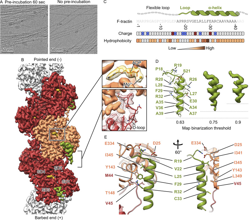
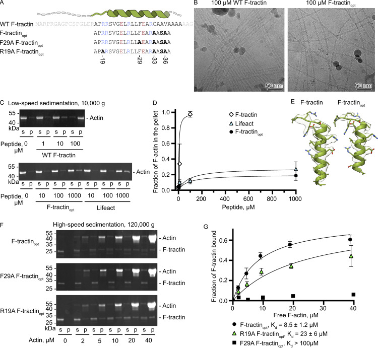
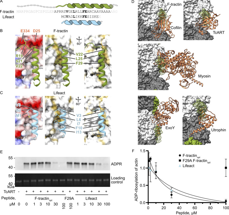
F-tractin is a peptide widely used to visualize the actin cytoskeleton in live eukaryotic cells but has been reported to impair cell migration and induce actin bundling at high expression levels. To elucidate these effects, we determined the cryo-EM structure of the F-tractin–F-actin complex, revealing that F-tractin consists of a flexible N-terminal region and an amphipathic C-terminal helix. The N-terminal part is dispensable for F-actin binding but responsible for the bundling effect. Based on these insights, we developed an optimized F-tractin, which eliminates the N-terminal region and minimizes bundling while retaining strong actin labeling. The C-terminal helix interacts with a hydrophobic pocket formed by two neighboring actin subunits, an interaction region shared by many actin-binding polypeptides, including the popular actin-binding probe Lifeact. Thus, rather than contrasting F-tractin and Lifeact, our data indicate that these peptides have analogous modes of interaction with F-actin. Our study dissects the structural elements of F-tractin and provides a foundation for developing future actin probes.
Overview
The claims and statistics of this paper haven’t been checked yet.
Run the check — free, no account needed. It reads the open-access text and checks the sources it cites.
Integrity signals
Registry
Cites
Statistics
Figures
Verification
Claims the paper makes
The claims and statistics of this paper haven’t been checked yet.
Run the check — free, no account needed. It reads the open-access text and checks the sources it cites.
Self-check
Percentages, sums and reported differences re-computed from the paper’s own numbers.
The self-check needs the paper’s full text.
Citations
Cites
UCSF Chimera—A visualization system for exploratory research and analysis
Pettersen, E. F., Goddard, T. D., Huang, C. C., Couch, G. S., Greenblatt, D. M., Meng, E. C., & Ferrin, T. E. (2004) · Journal of Computational Chemistry · doi:10.1002/jcc.20084 · Cited by 45,828
Highly accurate protein structure prediction with AlphaFold
Jumper, J., Evans, R., Pritzel, A., Green, T., Figurnov, M., Ronneberger, O., Tunyasuvunakool, K., Bates, R., Žídek, A., Potapenko, A., et al. (2021) · Nature · doi:10.1038/s41586-021-03819-2 · Cited by 43,764
Liebschner, D., Afonine, P. V., Baker, M. L., Bunkóczi, G., Chen, V. B., Croll, T. I., Hintze, B., Hung, L.-W., Jain, S., McCoy, A. J., et al. (2019) · Acta Crystallographica Section D Structural Biology · doi:10.1107/s2059798319011471 · Cited by 7,953
MolProbity: More and better reference data for improved all‐atom structure validation
Williams, C. J., Headd, J. J., Moriarty, N. W., Prisant, M. G., Videau, L. L., Deis, L. N., Verma, V., Keedy, D. A., Hintze, B. J., Chen, V. B., et al. (2018) · Protein Science · doi:10.1002/pro.3330 · Cited by 4,599
Lifeact: a versatile marker to visualize F-actin
Riedl, J., Crevenna, A. H., Kessenbrock, K., Yu, J. H., Neukirchen, D., Bista, M., Bradke, F., Jenne, D., Holak, T. A., Werb, Z., et al. (2008) · Nature Methods · doi:10.1038/nmeth.1220 · Cited by 2,189
Croll, T. I. (2018) · Acta Crystallographica Section D Structural Biology · doi:10.1107/s2059798318002425 · Cited by 1,985
Dominguez, R., & Holmes, K. C. (2011) · Annual Review of Biophysics · doi:10.1146/annurev-biophys-042910-155359 · Cited by 1,211
Fluorescent phallotoxin, a tool for the visualization of cellular actin.
Wulf, E., Deboben, A., Bautz, F. A., Faulstich, H., & Wieland, T. (1979) · Proceedings of the National Academy of Sciences · doi:10.1073/pnas.76.9.4498 · Cited by 789
Versatile fluorescent probes for actin filaments based on the actin‐binding domain of utrophin
Burkel, B. M., von Dassow, G., & Bement, W. M. (2007) · Cell Motility and the Cytoskeleton · doi:10.1002/cm.20226 · Cited by 472
Martin, B. R., Giepmans, B. N. G., Adams, S., & Tsien, R. Y. (2005) · Nature Biotechnology · doi:10.1038/nbt1136 · Cited by 424
Lifeact mice for studying F-actin dynamics
Riedl, J., Flynn, K. C., Raducanu, A., Gärtner, F. C., Beck, G., Bösl, M. R., Bradke, F., Maßberg, S., Aszódi, A., Sixt, M., et al. (2010) · Nature Methods · doi:10.1038/nmeth0310-168 · Cited by 333
Self-citationSTED Nanoscopy Reveals the Ubiquity of Subcortical Cytoskeleton Periodicity in Living Neurons
D’Este, E., Kamin, D., Göttfert, F., Hady, A. E., & Hell, S. W. (2015) · Cell Reports · doi:10.1016/j.celrep.2015.02.007 · Cited by 324
Yi, J., Wu, X., Crites, T. J., & Hammer, J. A. (2012) · Molecular Biology of the Cell · doi:10.1091/mbc.e11-08-0731 · Cited by 271
Sheahan, M. B., Staiger, C. J., Rose, R. J., & McCurdy, D. W. (2004) · PLANT PHYSIOLOGY · doi:10.1104/pp.104.049411 · Cited by 265
Monitoring Actin Cortex Thickness in Live Cells
Clark, A. G., Dierkes, K., & Paluch, E. K. (2013) · Biophysical Journal · doi:10.1016/j.bpj.2013.05.057 · Cited by 248
Actin visualization at a glance
Melak, M., Plessner, M., & Grosse, R. (2017) · Journal of Cell Science · doi:10.1242/jcs.189068 · Cited by 239
Cryo-EM structure of a human cytoplasmic actomyosin complex at near-atomic resolution
von der Ecken, J., Heissler, S. M., Pathan-Chhatbar, S., Manstein, D. J., & Raunser, S. (2016) · Nature · doi:10.1038/nature18295 · Cited by 233
Structural basis for cofilin binding and actin filament disassembly
Tanaka, K., Takeda, S., Mitsuoka, K., Oda, T., Kimura-Sakiyama, C., Maéda, Y., & Narita, A. (2018) · Nature Communications · doi:10.1038/s41467-018-04290-w · Cited by 208
Nuclear localization of enhanced green fluorescent protein homomultimers
Seibel, N. M., Eljouni, J., Nalaskowski, M. M., & Hampe, W. (2007) · Analytical Biochemistry · doi:10.1016/j.ab.2007.05.025 · Cited by 207
Wehland, J., Osborn, M., & Weber, K. K. (1977) · Proceedings of the National Academy of Sciences · doi:10.1073/pnas.74.12.5613 · Cited by 197
Structural transitions of F-actin upon ATP hydrolysis at near-atomic resolution revealed by cryo-EM
Merino, F., Pospich, S., Funk, J., Wagner, T., Küllmer, F., Arndt, H., Bieling, P., & Raunser, S. (2018) · Nature Structural & Molecular Biology · doi:10.1038/s41594-018-0074-0 · Cited by 191
Comparative analysis of tools for live cell imaging of actin network architecture
Belin, B. J., Goins, L. M., & Mullins, R. D. (2014) · BioArchitecture · doi:10.1080/19490992.2014.1047714 · Cited by 172
Johnson, H. W., & Schell, M. J. (2009) · Molecular Biology of the Cell · doi:10.1091/mbc.e09-01-0083 · Cited by 155
Structural basis of actin filament assembly and aging
Oosterheert, W., Klink, B. U., Belyy, A., Pospich, S., & Raunser, S. (2022) · Nature · doi:10.1038/s41586-022-05241-8 · Cited by 152
Self-citationF- and G-Actin Concentrations in Lamellipodia of Moving Cells
Koestler, S. A., Rottner, K., Lai, F. P. L., Block, J., Vinzenz, M., & Small, J. V. (2009) · PLoS ONE · doi:10.1371/journal.pone.0004810 · Cited by 144
Biodegradable Mg corrosion and osteoblast cell culture studies
Yun, Y., Dong, Z., Yang, D., Schulz, M. J., Shanov, V. N., Yarmolenko, S. N., Xu, Z., Kumta, P. N., & Sfeir, C. S. (2009) · Materials Science and Engineering C · doi:10.1016/j.msec.2009.02.008 · Cited by 136
Phalloidin enhances actin assembly by preventing monomer dissociation.
Coluccio, L. M., & Tilney, L. G. (1984) · The Journal of Cell Biology · doi:10.1083/jcb.99.2.529 · Cited by 133
Courtemanche, N., Pollard, T. D., & Qian, C. (2016) · Nature Cell Biology · doi:10.1038/ncb3351 · Cited by 132
Spracklen, A. J., Fagan, T. N., Lovander, K. E., & Tootle, T. L. (2014) · Developmental Biology · doi:10.1016/j.ydbio.2014.06.022 · Cited by 119
Flores, R., Janeiro, R., & Viegas, J. (2019) · Scientific Reports · doi:10.1038/s41598-019-45909-2 · Cited by 97
Structural insights into actin filament recognition by commonly used cellular actin markers
Kumari, A., Kesarwani, S., Javoor, M. G., Vinothkumar, K. R., & Sirajuddin, M. (2020) · The EMBO Journal · doi:10.15252/embj.2019104006 · Cited by 97
Engineering Af1521 improves ADP-ribose binding and identification of ADP-ribosylated proteins
Nowak, K., Rosenthal, F., Karlberg, T., Bütepage, M., Thorsell, A. G., Dreier, B., Grossmann, J., Sobek, J., Imhof, R., Lüscher, B., et al. (2020) · Nature Communications · doi:10.1038/s41467-020-18981-w · Cited by 83
Aizawa, H., Sameshima, M., & Yahara, I. (1997) · Cell Structure and Function · doi:10.1247/csf.22.335 · Cited by 75
Structure of the Lifeact–F-actin complex
Belyy, A., Merino, F., Sitsel, O., & Raunser, S. (2020) · PLoS Biology · doi:10.1371/journal.pbio.3000925 · Cited by 75
Self-citationSliogeryte, K., Thorpe, S. D., Wang, Z., Thompson, C. L., Gavara, N., & Knight, M. M. (2015) · Journal of Biomechanics · doi:10.1016/j.jbiomech.2015.12.034 · Cited by 63
Actin chromobody imaging reveals sub-organellar actin dynamics
Schiavon, C. R., Zhang, T., Zhao, B. Q., Moore, A. S., Wales, P., de Andrade, L. R., Wu, M., Sung, T.-C., Dayn, Y., Feng, J. W., et al. (2020) · Nature Methods · doi:10.1038/s41592-020-0926-5 · Cited by 63
Affimer proteins for F-actin: novel affinity reagents that label F-actin in live and fixed cells
Lopata, A., Hughes, R., Tiede, C., et al. (2018) · Scientific Reports · doi:10.1038/s41598-018-24953-4 · Cited by 49
Measuring F-actin properties in dendritic spines
Koskinen, M., & Hotulainen, P. (2014) · Frontiers in Neuroanatomy · doi:10.3389/fnana.2014.00074 · Cited by 48
The Position of the GFP Tag on Actin Affects the Filament Formation in Mammalian Cells
Nagasaki, A., Kijima, S. T., Yumoto, T., Imaizumi, M., Yamagishi, A., Kim, H., Nakamura, C., & Uyeda, T. Q. P. (2017) · Cell Structure and Function · doi:10.1247/csf.17016 · Cited by 42
Xu, R., & Du, S. (2021) · Frontiers in Cell and Developmental Biology · doi:10.3389/fcell.2021.746818 · Cited by 26
Mechanism of actin-dependent activation of nucleotidyl cyclase toxins from bacterial human pathogens
Belyy, A., Merino, F., Mechold, U., & Raunser, S. (2021) · Nature Communications · doi:10.1038/s41467-021-26889-2 · Cited by 25
Self-citationMechanism of threonine ADP-ribosylation of F-actin by a Tc toxin
Belyy, A., Lindemann, F., Roderer, D., Funk, J., Bardiaux, B., Protze, J., Bieling, P., Oschkinat, H., & Raunser, S. (2022) · Nature Communications · doi:10.1038/s41467-022-31836-w · Cited by 23
Self-citationSugizaki, A., Sato, K., Chiba, K., Saito, K., Kawagishi, M., Tomabechi, Y., Mehta, S. B., Ishii, H., Sakai, N., Shirouzu, M., et al. (2021) · Proceedings of the National Academy of Sciences · doi:10.1073/pnas.2019071118 · Cited by 22
Bubb, M., Knutson, J. R., Porter, D., & Korn, E. D. (1994) · Journal of Biological Chemistry · doi:10.1016/s0021-9258(18)47291-2 · Cited by 20
Ladner-Keay, C. L., Turner, R. J., & Edwards, R. A. (2018) · Methods in molecular biology · doi:10.1007/978-1-4939-8745-0_22 · Cited by 17
Designed Ankyrin Repeat Proteins as Actin Labels of Distinct Cytoskeletal Structures in Living Cells
Ivanova, J. R., Benk, A. S., Schaefer, J. V., Dreier, B., Hermann, L. O., Plückthun, A., Missirlis, D., & Spatz, J. P. (2024) · ACS Nano · doi:10.1021/acsnano.3c12265 · Cited by 17
Functional characterisation of filamentous actin probe expression in neuronal cells
Patel, S., Fok, S., Stefen, H., Tomanić, T., Parić, E., Herold, R., Brettle, M., Djordjevic, A., & Fath, T. (2017) · PLoS ONE · doi:10.1371/journal.pone.0187979 · Cited by 12
A novel Fiji/ImageJ plugin for the rapid analysis of blebbing cells
Vosatka, K. W., Lavenus, S. B., & Logue, J. S. (2022) · PLoS ONE · doi:10.1371/journal.pone.0267740 · Cited by 9
van Zwam, M. C., Dhar, A., Bosman, W., van Straaten, W., Weijers, S. A., Seta, E., Joosten, B., van Haren, J., Palani, S., & van den Dries, K. (2024) · PLoS Biology · doi:10.1371/journal.pbio.3002551 · Cited by 9
The Shot CH1 domain recognises a distinct form of F-actin during Drosophila oocyte determination
Nashchekin, D., Squires, I. J., Prokop, A., & Johnston, D. S. (2024) · Development · doi:10.1242/dev.202370 · Cited by 9
Dendritic effects of genetically encoded actin-labeling probes in cultured hippocampal neurons
Ignácz, A., Nagy-Herczeg, D., Haußer, A., & Schlett, K. (2023) · Molecular Biology of the Cell · doi:10.1091/mbc.e22-08-0331 · Cited by 8
LILAC: enhanced actin imaging with an optogenetic Lifeact
Kroll, K. L., French, A. R., Sosnick, T. R., & Rock, R. S. (2023) · Nature Methods · doi:10.1038/s41592-022-01761-3 · Cited by 7
Ono, S. (2024) · Frontiers in Cell and Developmental Biology · doi:10.3389/fcell.2024.1504980 · Cited by 2
Comparative analysis of actin visualization by genetically encoded probes in cultured neuronsPreprint
Ignácz, A., Nagy-Herczeg, D., Haußer, A., & Schlett, K. (2022) · bioRxiv (Cold Spring Harbor Laboratory) · doi:10.1101/2022.08.12.503767 · Cited by 1
Metadata
- Authors
- Shatskiy, D., Sivan, A., Wedlich-Söldner, R., & Belyy, A.
- Affiliations
- University of Groningen (NL)
- University of Münster (DE)
- Institute of Cell Dynamics and Imaging, and Cells-in-Motion Interfaculty Center (CiMIC), University of Münster 2 , Münster,
- Corresponding author
- Sivan, Athul
- Belyy, Alexander
- Venue
- Journal of Cell Biology
- Publisher
- Rockefeller University Press
- Published
- 2025-02-10
- Volume
- 224
- Issue
- 4
- ISSN
- 0021-9525 (ISSN-L)
- 1540-8140
- PMC
- PMC11809415
- OpenAlex
- W4407349027
- Cited by
- 9
- References
- 55
- Citations by year
- 2026: 6 · 2025: 3
- FWCI
- 4.49
- Type
- journal article
- Language
- en
- Open access
- hybrid
- doi.org
- Licence
- Locations
- The Journal of Cell Biology (publishedVersion, other-oa)
- bioRxiv (Cold Spring Harbor Laboratory) (acceptedVersion, cc-by-nc-nd)
- PubMed Central (publishedVersion, cc-by)
- PubMed (publishedVersion)
- University of Groningen research database (University of Groningen / Centre for Information Technology) (submittedVersion, cc-by)
- Funders
- University of Groningen
- German Research Foundation (SFB1009, SFB1348, SFB1557)
- Topics
- Life Sciences › Biochemistry, Genetics and Molecular Biology › Cell Biology › Cellular Mechanics and Interactions
- Life Sciences › Biochemistry, Genetics and Molecular Biology › Molecular Biology › Protein Structure and Dynamics
- Life Sciences › Biochemistry, Genetics and Molecular Biology › Cell Biology › Microtubule and mitosis dynamics
- Keywords
- F-actin, actin bundling, Lifeact, amphipathic helix, N-terminal flexibility, hydrophobic pocket, cryo-EM structure
- MeSH terms
- Actins, Animals, Binding Sites, Humans, Actin Cytoskeleton, Models, Molecular, Oligopeptides, Peptides, Protein Binding, Cryoelectron Microscopy
About these checks
Registry, retraction, Cites and statistics results are computed, not judged, with one exception: a printed reference that no index lists is read once by a model before it counts as a citation error. Claims are extracted by a model and kept only when their quote appears verbatim in the text; each cited source is then checked, with a model’s help, for whether it says what the claim relies on. Those verdicts can be wrong — read the claim’s quote and the cited source before relying on one.
Statistics and arithmetic are recomputed, not judged.
Confidence bands are shown only for fields calibrated on our annotated test set. None is calibrated yet, so no band is shown.
What counts as a claim ›How we measure confidence ›How we check this ›
Are you an author, or do you see a problem with this page? Request a review
Data sources
Bibliographic data: Semantic Scholar, OpenAlex, Crossref.
Contains information from the Semantic Scholar Academic Graph Datasets, made available under the ODC Attribution License (ODC-BY 1.0).
Semantic Scholar’s data is described in: Kinney, R., et al. (2023). The Semantic Scholar Open Data Platform. arXiv:2301.10140.
