

The DOI matches its Crossref record.
The Chemokine SDF-1 Is a Chemoattractant for Human CD34+ Hematopoietic Progenitor Cells and Provides a New Mechanism to Explain the Mobilization of CD34+ Progenitors to Peripheral Blood
Abstract
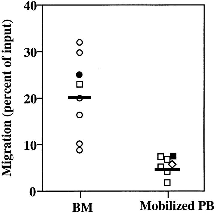
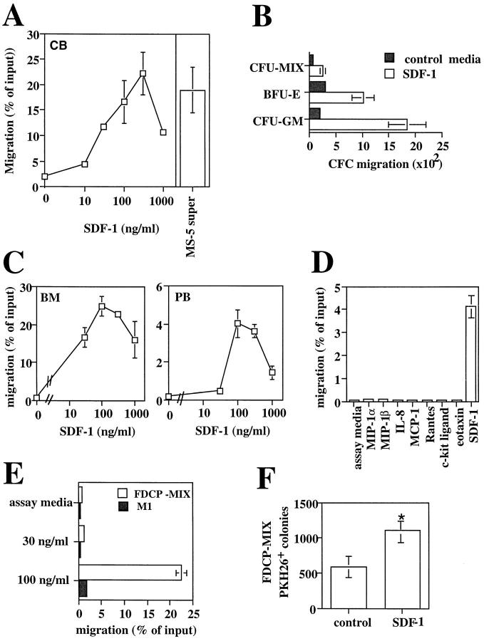
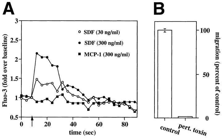
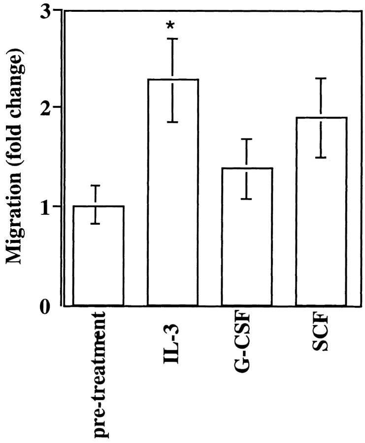
Hematopoietic progenitor cells migrate in vitro and in vivo towards a gradient of the chemotactic factor stromal cell-derived factor-1 (SDF-1) produced by stromal cells. This is the first chemoattractant reported for human CD34+ progenitor cells. Concentrations of SDF-1 that elicit chemotaxis also induce a transient elevation of cytoplasmic calcium in CD34+ cells. SDF-1-induced chemotaxis is inhibited by pertussis toxin, suggesting that its signaling in CD34+ cells is mediated by seven transmembrane receptors coupled to Gi proteins. CD34+ cells migrating to SDF-1 include cells with a more primitive (CD34+/CD38− or CD34+/DR−) phenotype as well as CD34+ cells phenotypically committed to the erythroid, lymphoid and myeloid lineages, including functional BFU-E, CFU-GM, and CFU-MIX progenitors. Chemotaxis of CD34+ cells in response to SDF-1 is increased by IL-3 in vitro and is lower in CD34+ progenitors from peripheral blood than in CD34+ progenitors from bone marrow, suggesting that an altered response to SDF-1 may be associated with CD34 progenitor mobilization.
Overview
The claims and statistics of this paper haven’t been checked yet.
Run the check — free, no account needed. It reads the open-access text and checks the sources it cites.
Integrity signals
Registry
Cites
Statistics
Figures
Verification
Claims the paper makes
The claims and statistics of this paper haven’t been checked yet.
Run the check — free, no account needed. It reads the open-access text and checks the sources it cites.
Claims other papers make about this one
Each is checked against this paper’s own text: does it say what the citing paper says it does?
In the citing paper
“SDF-1, a chemoattractant produced by BM stromal cells, is a ligand for CXCR4, expressed on HSC/P, and is implicated in chemotaxis, homing, and survival of HSC/P[83–85].”
From Finding the needle in the hay stack: Hematopoietic stem cells in Fanconi anemia (2009) · cites this paper as [83]
In the citing paper
“SDF-1, a chemoattractant produced by BM stromal cells, is a ligand for CXCR4, expressed on HSC/P, and is implicated in chemotaxis, homing, and survival of HSC/P[83–85].”
From Finding the needle in the hay stack: Hematopoietic stem cells in Fanconi anemia (2009) · cites this paper as [83]
In the citing paper
“SDF-1, a chemoattractant produced by BM stromal cells, is a ligand for CXCR4, expressed on HSC/P, and is implicated in chemotaxis, homing, and survival of HSC/P[83–85].”
From Finding the needle in the hay stack: Hematopoietic stem cells in Fanconi anemia (2009) · cites this paper as [83]
In the citing paper
“SDF-1, a chemoattractant produced by BM stromal cells, is a ligand for CXCR4, expressed on HSC/P, and is implicated in chemotaxis, homing, and survival of HSC/P[83–85].”
From Finding the needle in the hay stack: Hematopoietic stem cells in Fanconi anemia (2009) · cites this paper as [83]
In the citing paper
“Chemokine-stimulated migration of primitive hematopoietic cells has also been postulated to be a mechanism of mobilization of hematopoietic progenitor cells (16).”
From Rac and Cdc42 GTPases control hematopoietic stem cell shape, adhesion, migration, and mobilization (2001) · cites this paper as [16]
2 more claims about this paper
Create a free account to see every claim other papers make about this one.
Self-check
Percentages, sums and reported differences re-computed from the paper’s own numbers.
The self-check needs the paper’s full text.
Citations
Cites
No references could be resolved yet.
Research works printed but not linked: 57
- Socinski, 1988, Granulocyte–macrophage colony stimulating factor expands the circulating haemopoietic progenitor cell compartment in man, Lancet, 1, 1194, 10.1016/S0140-6736(88)92012-0 · DOI
- Siena, 1989, Circulation of CD34+hematopoietic stem cells in the peripheral blood of high-dose cyclophosphamide-treated patients: enhancement by intravenous recombinant human granulocyte-macrophage colonystimulating factor, Blood, 6, 1905, 10.1182/blood.V74.6.1905.1905 · DOI
- Sheridan, 1992, Effect of peripheral-blood progenitor cells mobilised by filgrastim (G-CSF) on platelet recovery after high-dose chemotherapy, Lancet, 339, 640, 10.1016/0140-6736(92)90795-5 · DOI
- Elias, 1992, Mobilization of peripheral blood progenitor cells by chemotherapy and granulocyte–macrophage colony-stimulating factor for hematologic support after high-dose intensification for breast cancer, Blood, 79, 3036, 10.1182/blood.V79.11.3036.3036 · DOI
- Schofield, 1978, The relationship between the spleen colony-forming cell and the haemopoietic stem cell, Blood Cells, 4, 7
- Mason, 1989, The development of spatial distributions of CFU-S and in-vitro CFC in femora of mice of different ages, Br J Haematol, 73, 455, 10.1111/j.1365-2141.1989.tb00280.x · DOI
- Lord, 1975, The relative spatial distribution of CFU-S and CFU-C in the normal mouse femur, Blood, 46, 65, 10.1182/blood.V46.1.65.65 · DOI
- Uchida, 1993, Heterogeneity of hematopoietic stem cells, Curr Opin Immunol, 5, 177, 10.1016/0952-7915(93)90002-A · DOI
- Goodman, 1962, Evidence for stem cells in the peripheral blood of mice, Blood, 19, 702, 10.1182/blood.V19.6.702.702 · DOI
- Storb, 1977, Demonstration of hemopoietic stem cells in the peripheral blood of baboons by cross circulation, Blood, 50, 537, 10.1182/blood.V50.3.537.537 · DOI
- Springer, 1995, Traffic signals on endothelium for lymphocyte recirculation and leukocyte emigration, Annu Rev Physiol, 57, 827, 10.1146/annurev.ph.57.030195.004143 · DOI
- Butcher, 1991, Leukocyte–endothelial cell recognition: three (or more) steps to specificity and diversity, Cell, 67, 1033, 10.1016/0092-8674(91)90279-8 · DOI
- Lasky, 1992, Selectins: interpreters of cell-specific carbohydrate information during inflammation, Science (Wash DC), 258, 964, 10.1126/science.1439808 · DOI
- Webb, 1996, Kinetics of peripheral blood mononuclear cell mobilization with chemotherapy and/or granulocyte–colonystimulating factor: implications for timing and yield of hematopoietic progenitor cell collections, Transfusion, 36, 160, 10.1046/j.1537-2995.1996.36296181930.x · DOI
- Itoh, 1989, Reproducible establishment of hemopoietic supportive stromal cell lines from murine bone marrow, Exp Hematol, 17, 145
- Friedrich, 1996, Hematopoietic supportive functions of mouse bone marrow- and fetal liver-microenvironment: dissection of granulocyte-, B-lymphocyte-, and hematopoietic progenitorsupport at the stroma cell clone level, Blood, 87, 4596, 10.1182/blood.V87.11.4596.bloodjournal87114596 · DOI
- Bleul, 1996, A highly efficacious lymphocyte chemoattractant, J Exp Med, 184, 1101, 10.1084/jem.184.3.1101 · DOI · Paper page
- Hahne, 1993, Five tumor necrosis factor-inducible cell adhesion mechanisms on the surface of mouse endothelioma cells mediate the binding of leukocytes, J Cell Biol, 121, 655, 10.1083/jcb.121.3.655 · DOI
- Verfaillie, 1990, Purified primitive human hematopoietic progenitor cells with longterm in vitro repopulating capacity adhere selectively to irradiate bone marrow stroma, J Exp Med, 172, 509, 10.1084/jem.172.2.509 · DOI
- Spooncer, 1986, Self renewal and differentiation of IL-3-dependent multipotential stem cells are modulated by stromal cells and serum factors, Differentiation, 31, 111, 10.1111/j.1432-0436.1986.tb00391.x · DOI
- Karasuyama, 1988, Establishment of mouse cell lines which constitutively secrete large quantities of interleukin 2, 3, 4 or 5, using modified cDNA expression vectors, Eur J Immunol, 18, 97, 10.1002/eji.1830180115 · DOI
- Suzuki, 1992, Characterization of murine hemopoietic-supportive (MS-1 and MS-5) and non-supportive (MS-K) cell lines, Leukemia, 6, 452
- Tashiro, 1993, Signal sequence trap: a cloning strategy for secreted proteins and type I, Science (Wash DC), 261, 600, 10.1126/science.8342023 · DOI
- Nagasawa, 1994, Molecular cloning and structure of a pre-B-cell growth-stimulating factor, Proc Nat Acad Sci USA, 91, 2305, 10.1073/pnas.91.6.2305 · DOI
- Jiang, 1994, Molecular cloning of TPAR1, a gene whose expression is repressed by the tumor promoter 12-O-tetradecanoylphorbol 13-acetate (TPA), Exp Cell Res, 215, 284, 10.1006/excr.1994.1344 · DOI
- Graham, 1990, Identification and characterization of an inhibitor of haemopoietic stem cell proliferation, Nature (Lond), 344, 442, 10.1038/344442a0 · DOI
- Broxmeyer, 1989, Myelopoietic enhancing effects of murine macrophage inflammatory proteins 1 and 2 on colony formation in vitro by murine and human bone marrow granulocyte/macrophage progenitor cells, J Exp Med, 170, 1583, 10.1084/jem.170.5.1583 · DOI
- Avalos, 1994, The active monomeric form of macrophage inflammatory protein-1a interacts with high and low affinity classes of receptors on human hematopoietic cells, Blood, 94, 1790, 10.1182/blood.V84.6.1790.1790 · DOI
- Johnston, 1990, On the species specificity of the interaction of LFA-1 with intercellular adhesion molecules, J Immunol, 145, 1181, 10.4049/jimmunol.145.4.1181 · DOI
- Becker-Andre, 1992, Murine endothelial/leukocyte adhesion molecule 1 is a close functional homologue of the human protein, Eur J Biochem, 206, 401, 10.1111/j.1432-1033.1992.tb16940.x · DOI
- Taub, 1996, IP-10 induces mononuclear infiltration in mice and promotes the migration of T lymphocytes in peripheral tissues of hu-SCID mice, Blood, 87, 1423, 10.1182/blood.V87.4.1423.bloodjournal8741423 · DOI
- Miyaura, 1988, Recombinant human interleukin 6 (B-cell stimulatory factor 2) is a potent inducer of differentiation of mouse myeloid leukemia cells (M1), FEBS Lett, 234, 17, 10.1016/0014-5793(88)81293-6 · DOI
- Huang, 1994, Lymphoid and myeloid differentiation of single human CD34+, HLA-DR+, CD38− hematopoietic stem cells, Blood, 83, 1515, 10.1182/blood.V83.6.1515.1515 · DOI
- Andrews, 1989, Precursors of colony-forming cells in humans can be distinguished from colony-forming cells by expression of the CD33 and CD34 antigens and light scatter properties, J Exp Med, 169, 1721, 10.1084/jem.169.5.1721 · DOI
- Shah, 1988, Flow cytometric analysis of human bone marrow. IV. Differential quantitative expression of T-200 common leukocyte antigen during normal hemopoiesis, J Immunol, 140, 1861, 10.4049/jimmunol.140.6.1861 · DOI
- Tjonnfjord, 1993, T lymphocyte differentiation in vitro from adult human prethymic CD34+bone marrow cells, J Exp Med, 177, 1531, 10.1084/jem.177.6.1531 · DOI
- Sutherland, 1989, Characterization and partial purification of human bone marrow cells capable of initiating long-term hematopoiesis in vitro, Blood, 74, 1563, 10.1182/blood.V74.5.1563.1563 · DOI
- Murphy, 1994, The molecular biology of leukocyte chemoattractant receptors, Annu Rev Immunol, 12, 593, 10.1146/annurev.iy.12.040194.003113 · DOI
- Katz, 1992, Subunits beta gamma of heterotrimeric G protein activate beta 2 isoform of phopholipase C, Nature (Lond), 360, 686, 10.1038/360686a0 · DOI
- Gianni, 1989, Granulocyte-macrophage colony-stimulating factor to harvest circulating haemopoietic stem cells for autotransplantation, Lancet, 2, 580, 10.1016/S0140-6736(89)90711-3 · DOI
- Chao, 1993, Granulocyte colony-stimulating factor “mobilized” peripheral blood progenitor cells accelerate granulocyte and platelet recovery after high-dose chemotherapy, Blood, 81, 2031, 10.1182/blood.V81.8.2031.2031 · DOI
- Peters, 1993, Comparative effects of granulocyte-macrophage colony-stimulating factor (GM-CSF) and granulocyte colony-stimulating factor (G-CSF) on priming peripheral blood progenitor cells for use with autologous bone marrow after high-dose chemotherapy, Blood, 81, 1709, 10.1182/blood.V81.7.1709.1709 · DOI
- Lane, 1995, Harvesting and enrichment of hematopoietic progenitor cells mobilized into the peripheral blood of normal donors by granulocyte–macrophage colonystimulating factor (GM-CSF) or G-CSF: potential role in allogeneic marrow transplantation, Blood, 85, 275, 10.1182/blood.V85.1.275.bloodjournal851275 · DOI
- Lee, 1995, Collection and use of circulating hematopoietic progenitor cells, Hematol Oncol Clin North Am, 9, 1, 10.1016/S0889-8588(18)30108-4 · DOI
- Guillaume, 1993, IL-3 and peripheral blood stem cell harvesting, Stem Cells, 11, 173, 10.1002/stem.5530110303 · DOI
- Sato, 1993, In vitro expansion of human peripheral blood CD34+ cells, Blood, 82, 3600, 10.1182/blood.V82.12.3600.3600 · DOI
- Brugger, 1994, Maintenance of transplantation potential in ex vivo expanded CD34+-selected human peripheral blood progenitor cells, Blood, 84, 2898, 10.1182/blood.V84.9.2898.2898 · DOI
- Migliaccio, 1992, Long-term generation of colony-forming cells in liquid culture of CD34+cord blood cells in the presence of recombinant human stem cell factor, Blood, 79, 2620, 10.1182/blood.V79.10.2620.bloodjournal79102620 · DOI
- Flasshove, 1995, Ex vivo expansion and selection of human CD34+peripheral blood progenitor cells after introduction of a mutated dihydrofolate reductase cDNA via retroviral gene transfer, Blood, 85, 566, 10.1182/blood.V85.2.566.566 · DOI
- Shirozu, 1995, Structure and chromosomal localization of the human stromal cell-derived factor 1 (SDF1) gene, Genomics, 28, 495, 10.1006/geno.1995.1180 · DOI
- Okumura, 1996, Chemotactic and chemokinetic activities of stem cell factor on murine hematopoietic progenitor cells, Blood, 87, 4100, 10.1182/blood.V87.10.4100.bloodjournal87104100 · DOI
- Healy, 1994, In vitro proliferation by cells mobilized into the peripheral blood for collection and autologous transplantation, Exp Hematol, 22, 1278
- Haylock, 1992, Ex vivo expansion and maturation of peripheral blood CD34+cells into the myeloid lineage, Blood, 80, 1405, 10.1182/blood.V80.6.1405.bloodjournal8061405 · DOI
- To, 1994, A comparative study of the phenotype and proliferative capacity of peripheral blood (PB) CD34+ cells mobilized by four different protocols and those of steady-phase PB and bone marrow CD34+cells, Blood, 84, 2930, 10.1182/blood.V84.9.2930.2930 · DOI
- Tavassoli, 1991, Enhancement of the grafting efficiency of transplanted marrow cells by preincubation with IL-3 and GM-CSF, Blood, 77, 1599, 10.1182/blood.V77.7.1599.1599 · DOI
- Zanjani, 1993, Liverderived fetal hematopoietic stem cells selectively and preferentially home to the fetal bone marrow, Blood, 81, 399, 10.1182/blood.V81.2.399.399 · DOI
- Nagasawa, 1996, Defects of B-cell lymphopoiesis and bonemarrow myelopoiesis in mice lacking the CXC chemokine PBSF/SDF-1, Nature (Lond), 382, 635, 10.1038/382635a0 · DOI
Cited by
Crump, M. P., Gong, J., Loetscher, P., Rajarathnam, K., Amara, A., Arenzana‐Seisdedos, F., Virelizier, J., Baggiolini, M., Sykes, B. D., & Clark‐Lewis, I. (1997) · The EMBO Journal · doi:10.1093/emboj/16.23.6996 · Cited by 731
Chemokines in the systemic organization of immunity
Campbell, D. J., Kim, C. H., & Butcher, E. C. (2003) · Immunological Reviews · doi:10.1034/j.1600-065x.2003.00067.x · Cited by 330
Stromal cell-derived factor-1α is cardioprotective after myocardial infarction
Saxena, A., Fish, J. E., White, M. D., Yu, S., Smyth, J. W. P., Shaw, R. M., DiMaio, J. M., & Srivastava, D. (2008) · Circulation · doi:10.1161/circulationaha.107.694992 · Cited by 237
Rac and Cdc42 GTPases control hematopoietic stem cell shape, adhesion, migration, and mobilization
Yang, F.-C., Atkinson, S. J., Gu, Y., Borneo, J. B., Roberts, A. W., Zheng, Y., Pennington, J., & Williams, D. A. (2001) · Proceedings of the National Academy of Sciences · doi:10.1073/pnas.101546898 · Cited by 201
The role of chemoattraction in cancer metastases
Moore, M. A. S. (2001) · BioEssays · doi:10.1002/bies.1095 · Cited by 138
Chae, H.-D., Lee, K. E., Williams, D. A., & Gu, Y. (2008) · Blood · doi:10.1182/blood-2007-06-093237 · Cited by 81
Chemokine-protease interactions in cancer
Van Damme, J., Struyf, S., & Opdenakker, G. (2004) · Seminars in Cancer Biology · doi:10.1016/j.semcancer.2003.10.007 · Cited by 71
Sergejeva, S., Malmhäll, C., Lötvall, J., & Pullerits, T. (2005) · Clinical & Experimental Allergy · doi:10.1111/j.1365-2222.2004.02038.x · Cited by 50
Neonatal Osteomacs and Bone Marrow Macrophages Differ in Phenotypic Marker Expression and Function
Mohamad, S. F., Gunawan, A., Blosser, R., Childress, P., Aguilar-Perez, A., Ghosh, J., Hong, J. M., Liu, J., Kanagasabapathy, D., Kacena, M. A., et al. (2020) · Journal of Bone and Mineral Research · doi:10.1002/jbmr.4314 · Cited by 34
Geutskens, S. B., Andrews, W. D., van Stalborch, A.-M. D., Brussen, K., Holtrop-de Haan, S. E., Parnavelas, J. G., Hordijk, P. L., & van Hennik, P. B. (2012) · Laboratory Investigation · doi:10.1038/labinvest.2012.81 · Cited by 34
Finding the needle in the hay stack: Hematopoietic stem cells in Fanconi anemia
Müller, L. U. W., & Williams, D. A. (2009) · Mutation Research - Fundamental and Molecular Mechanisms of Mutagenesis · doi:10.1016/j.mrfmmm.2009.03.010 · Cited by 31
Zhang, X., Tian, L., Majumdar, A., & Scheller, E. L. (2024) · Comprehensive Physiology · doi:10.1002/j.2040-4603.2024.tb00300.x · Cited by 18
Metadata
- Authors
- Aiuti, A., Webb, I. J., Bleul, C., Springer, T., & Gutierrez-Ramos, J. C.
- Affiliations
- From the *Center for Blood Research, Inc., and the ‡Department of Genetics, §Department of Pathology, and ‖Department of Medicine, Harvard Medical School, Boston, Massachusetts 02115; ¶Blood Component Laboratory, Dana-Farber Cancer Institute, Boston, Massachusetts 02115; and **Millennium Pharmaceuticals, Inc., Cambridge, Massachusetts 02139
- Venue
- The Journal of Experimental Medicine
- Publisher
- Rockefeller University Press
- Published
- 1997-01-01
- Volume
- 185
- Issue
- 1
- Pages
- 111-120
- ISSN
- 0022-1007
- 1540-9538
- PMC
- PMC2196104
- Cited by
- 1,158
- References
- 0
- Language
- en
About these checks
Registry, retraction, Cites and statistics results are computed, not judged, with one exception: a printed reference that no index lists is read once by a model before it counts as a citation error. Claims are extracted by a model and kept only when their quote appears verbatim in the text; each cited source is then checked, with a model’s help, for whether it says what the claim relies on. Those verdicts can be wrong — read the claim’s quote and the cited source before relying on one.
Statistics and arithmetic are recomputed, not judged.
Confidence bands are shown only for fields calibrated on our annotated test set. None is calibrated yet, so no band is shown.
What counts as a claim ›How we measure confidence ›How we check this ›
Are you an author, or do you see a problem with this page? Request a review
