

The DOI matches its Crossref record.
Delayed Expulsion of the Nematode Trichinella spiralis In Mice Lacking the Mucosal Mast Cell–Specific Granule Chymase, Mouse Mast Cell Protease-1
Abstract
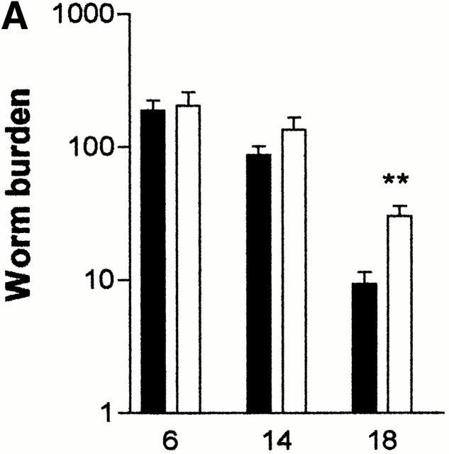
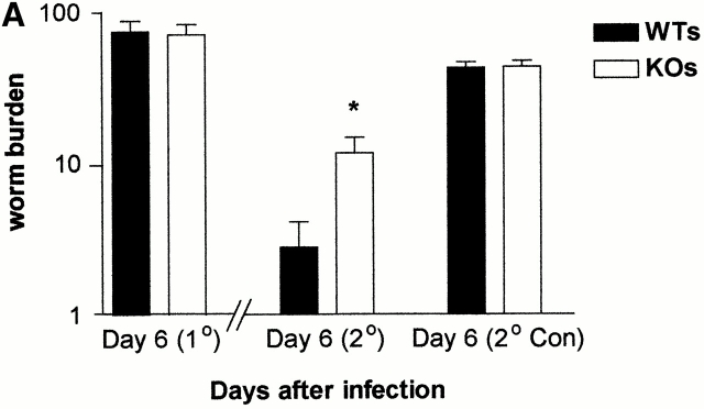
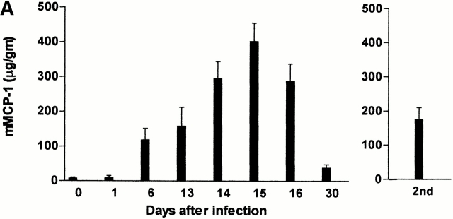
Expulsion of gastrointestinal nematodes is associated with pronounced mucosal mast cell (MMC) hyperplasia, differentiation, and activation, accompanied by the systemic release of MMC granule chymases (chymotrypsin-like serine proteases). The β-chymase mouse mast cell protease-1 (mMCP-1) is expressed predominantly by intraepithelial MMCs, and levels in the bloodstream and intestinal lumen are maximal at the time of worm expulsion in parasitized mice. To address the in vivo functions of MMC-specific β-chymases, we have generated transgenic mice that lack the mMCP-1 gene. They were backcrossed onto a congenic BALB/c background to investigate the response to nematode infection. The deletion of the mMCP-1 gene is associated with significantly delayed expulsion of Trichinella spiralis and increased deposition of muscle larvae in BALB/c mice despite the presence of normal and sometimes increased numbers of MMCs. Neither worm fecundity nor worm burdens were altered in Nippostrongylus-infected mMCP-1−/− BALB/c mice. These data demonstrate, for the first time, that the ablation of an MMC-derived effector molecule compromises the expulsion process.
Overview
The claims and statistics of this paper haven’t been checked yet.
Run the check — free, no account needed. It reads the open-access text and checks the sources it cites.
Integrity signals
Registry
Cites
Statistics
Figures
Verification
Claims the paper makes
The claims and statistics of this paper haven’t been checked yet.
Run the check — free, no account needed. It reads the open-access text and checks the sources it cites.
Claims other papers make about this one
Each is checked against this paper’s own text: does it say what the citing paper says it does?
In the citing paper
“Abnormalities in host responses to certain parasites have been observed in mice that genetically lack IgE [27, 28], mast cells [29–33], or basophils [28, 33] but such studies generally have not included an analysis of the effects of those deficiencies on the overall survival or reproductive success of the infected hosts.”
From Mast cells and IgE in defense against lethality of venoms: Possible “benefit” of allergy (2020) · cites this paper as [30]
Self-check
Percentages, sums and reported differences re-computed from the paper’s own numbers.
The self-check needs the paper’s full text.
Citations
Cites
No references could be resolved yet.
Research works printed but not linked: 24
- Enerback, 1987, Mucosal mast cells in the rat and man, Int. Arch. Allergy Appl. Immunol., 82, 249, 10.1159/000234199 · DOI
- Befus, 1982, Mucosal mast cells 1. Isolation and functional characteristics of rat intestinal mast cells, J. Immunol., 128, 2475, 10.4049/jimmunol.128.6.2475 · DOI
- Miller, 1988, Granule proteases define mast cell heterogeneity in the serosa and gastrointestinal mucosa of the mouse, Immunology., 65, 559
- Stevens, 1994, Strain-specific and tissue specific expression of mouse mast cell secretory granule proteases, Proc. Natl. Acad. Sci. USA., 91, 128, 10.1073/pnas.91.1.128 · DOI
- Woodbury, 1984, Mucosal mast cells are functionally active during spontaneous expulsion of intestinal nematode infections in the rat, Nature., 312, 450, 10.1038/312450a0 · DOI
- Miller, 1996, Mucosal mast cells and the allergic response against nematode parasites, Vet. Immunol. Immunopathol., 54, 331, 10.1016/S0165-2427(96)05696-6 · DOI
- Scudamore, 1995, The release of the mucosal mast cell chymase, rat mast cell protease II, during anaphylaxis is associated with the rapid development of paracellular permeability to macromolecules in rat jejunum, J. Exp. Med, 182, 1871, 10.1084/jem.182.6.1871 · DOI
- Scudamore, 1998, The rat mucosal mast cell chymase, RMCP-II, alters epithelial cell monolayer permeability in association with altered distribution of the tight junction proteins ZO-1 and occludin, Eur. J. Cell. Biol, 75, 321, 10.1016/S0171-9335(98)80065-4 · DOI · Paper page
- Scudamore, 1997, Mast cell heterogeneity in the gastrointestinal tract. Variable expression of mouse mast cell protease-1 (mMCP-1) in intraepithelial mucosal mast cells in nematode-infected and normal BALB/c mice, Am. J. Pathol., 150, 1661
- Huntley, 1990, Distribution of intestinal mast cell protease in blood and tissues of normal and Trichinella-infected mice, Parasite Immunol., 12, 85, 10.1111/j.1365-3024.1990.tb00938.x · DOI
- Wastling, 1998, Histochemical and ultrastructural modification of mucosal mast cell granules in parasitized mice lacking the β-chymase, mouse mast cell protease-1, Am. J. Pathol., 153, 491, 10.1016/S0002-9440(10)65592-7 · DOI
- Urban, 1998, Il-13, IL-4Ra, and Stat6 are required for the expulsion of the gastrointestinal nematode parasite Nippostrongylus brasiliensis, Immunity., 8, 255, 10.1016/S1074-7613(00)80477-X · DOI
- Ha, 1983, Delayed expulsion of adult Trichinella spiralis by mast cell deficient W/Wv mice, Infect. Immun., 41, 445, 10.1128/iai.41.1.445-447.1983 · DOI
- Faulkner, 1997, Interleukin-9 is involved in host protective immunity to intestinal nematode infection, Eur. J. Immunol., 27, 2536, 10.1002/eji.1830271011 · DOI
- Wakelin, 1977, Transfer of immunity to Trichinella spiralis in the mouse with mesenteric lymph node cells in donors and expression of immunity in recipients, Parasitology., 74, 215, 10.1017/S0031182000047843 · DOI
- Miller, 1971, Immune reactions in mucous membranes. 1. Intestinal mast cell responses during helmith expulsion in the rat, Immunology., 20, 227
- Wastling, 1997, Constitutive expression of mouse mast cell protease-1 in normal BALB/c mice and its up-regulation during intestinal nematode infection, Immunology., 90, 308, 10.1046/j.1365-2567.1997.00155.x · DOI
- Lawrence, 1998, IL-4 regulated enteropathy in an intestinal nematode infection, Eur. J. Immunol., 28, 2672, 10.1002/(SICI)1521-4141(199809)28:09<2672::AID-IMMU2672>3.0.CO;2-F · DOI
- Donaldson, 1996, A critical role for stem cell factor and c-kit in host protective immunity to an intestinal helminth, Int. Immunol., 8, 559, 10.1093/intimm/8.4.559 · DOI
- Mitchell, 1983, Kinetics of expulsion of the nematode, Nippostrongylus brasiliensis, in mast-cell deficient W/Wv mice, Parasite Immunol., 4, 1, 10.1111/j.1365-3024.1983.tb00718.x · DOI
- Nawa, 1994, Selective effector mechanisms for the expulsion of intestinal helminths, Parasite Immunol., 16, 333, 10.1111/j.1365-3024.1994.tb00358.x · DOI
- Gurish, 1993, A closely linked complex of mouse mast cell-specific chymase genes on chromosome 14, J. Biol. Chem, 268, 11372, 10.1016/S0021-9258(18)82134-2 · DOI
- Friend, 1996, Mast cells that reside at different locations in the jejunum of mice infected with Trichinella spiralis exhibit sequential changes in their granule ultrastructure and chymase phenotype, J. Cell Biol, 135, 279, 10.1083/jcb.135.1.279 · DOI
- Friend, 2000, Senescent jejunal mast cells and eosinophils in the mouse translocate to the spleen and draining lymph node, respectively, during the recovery phase of helminth infection, J. Immunol., 165, 344, 10.4049/jimmunol.165.1.344 · DOI
Cited by
Sorted by citations
Mast cell tryptases and chymases in inflammation and host defense.
Caughey, G. H. (2007) · Immunological Reviews · doi:10.1111/j.1600-065x.2007.00509.x · Cited by 442
Galli, S. J., & Tsai, M. (2010) · European Journal of Immunology · doi:10.1002/eji.201040559 · Cited by 403
Widespread Immunological Functions of Mast Cells: Fact or Fiction?
Rodewald, H.-R., & Feyerabend, T. B. (2012) · Immunity · doi:10.1016/j.immuni.2012.07.007 · Cited by 240
Mast cells and IgE in defense against venoms: Possible “good side” of allergy?
Galli, S. J., Starkl, P., Marichal, T., & Tsai, M. (2016) · Allergology International · doi:10.1016/j.alit.2015.09.002 · Cited by 73
Mast cells and IgE in defense against lethality of venoms: Possible “benefit” of allergy
Galli, S. J., Metz, M., Starkl, P., Marichal, T., & Tsai, M. (2020) · Allergo Journal International · doi:10.1007/s40629-020-00118-6 · Cited by 57
Metadata
- Authors
- Knight, P. A., Wright, S. H., Lawrence, C. E., Paterson, Y. Y. W., & Miller, H. R. P.
- Venue
- The Journal of Experimental Medicine
- Publisher
- Rockefeller University Press
- Published
- 2000-12-18
- Volume
- 192
- Issue
- 12
- Pages
- 1849-1856
- ISSN
- 0022-1007
- 1540-9538
- PMC
- PMC2213497
- Cited by
- 249
- References
- 0
- Language
- en
About these checks
Registry, retraction, Cites and statistics results are computed, not judged, with one exception: a printed reference that no index lists is read once by a model before it counts as a citation error. Claims are extracted by a model and kept only when their quote appears verbatim in the text; each cited source is then checked, with a model’s help, for whether it says what the claim relies on. Those verdicts can be wrong — read the claim’s quote and the cited source before relying on one.
Statistics and arithmetic are recomputed, not judged.
Confidence bands are shown only for fields calibrated on our annotated test set. None is calibrated yet, so no band is shown.
What counts as a claim ›How we measure confidence ›How we check this ›
Are you an author, or do you see a problem with this page? Request a review
How to cite
Cite CoChat for the checks on this page, and the paper for its own findings.
- This page
- CoChat. (2026). Citation check: Delayed Expulsion of the Nematode Trichinella spiralis In Mice Lacking the Mucosal Mast Cell–Specific Granule Chymase, Mouse Mast Cell Protease-1. https://app.cochat.ai/papers/10.1084/jem.192.12.1849
- The paper
- Knight, P. A., Wright, S. H., Lawrence, C. E., Paterson, Y. Y. W., & Miller, H. R. P. (2000). Delayed Expulsion of the Nematode Trichinella spiralis In Mice Lacking the Mucosal Mast Cell–Specific Granule Chymase, Mouse Mast Cell Protease-1. The Journal of Experimental Medicine. https://doi.org/10.1084/jem.192.12.1849
