

The DOI matches its Crossref record; 31 of the 31 references it cites were found in a registry or an index.
Specific Recruitment of Antigen-presenting Cells by Chemerin, a Novel Processed Ligand from Human Inflammatory Fluids
Open accessAbstract
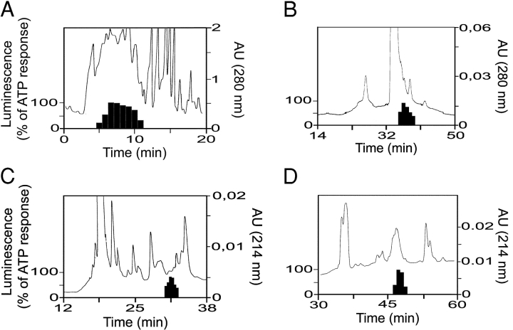
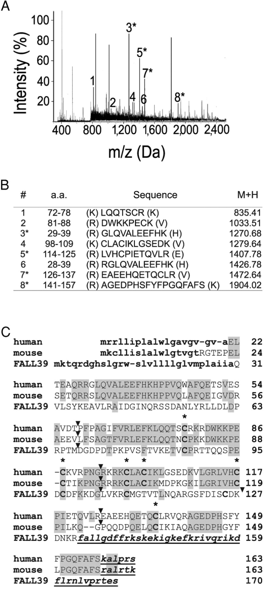
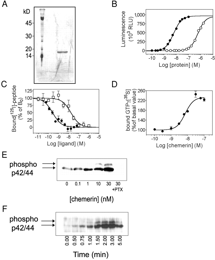
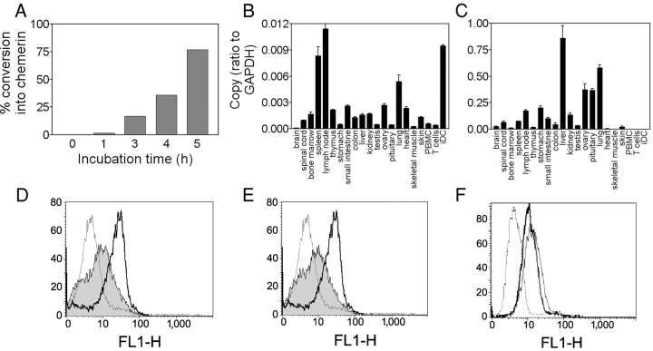
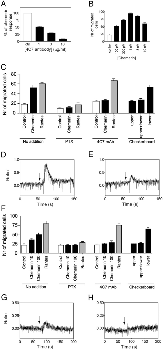
Dendritic cells (DCs) and macrophages are professional antigen-presenting cells (APCs) that play key roles in both innate and adaptive immunity. ChemR23 is an orphan G protein-coupled receptor related to chemokine receptors, which is expressed specifically in these cell types. Here we present the characterization of chemerin, a novel chemoattractant protein, which acts through ChemR23 and is abundant in a diverse set of human inflammatory fluids. Chemerin is secreted as a precursor of low biological activity, which upon proteolytic cleavage of its COOH-terminal domain, is converted into a potent and highly specific agonist of ChemR23, the chemerin receptor. Activation of chemerin receptor results in intracellular calcium release, inhibition of cAMP accumulation, and phosphorylation of p42-p44 MAP kinases, through the Gi class of heterotrimeric G proteins. Chemerin is structurally and evolutionary related to the cathelicidin precursors (antibacterial peptides), cystatins (cysteine protease inhibitors), and kininogens. Chemerin was shown to promote calcium mobilization and chemotaxis of immature DCs and macrophages in a ChemR23-dependent manner. Therefore, chemerin appears as a potent chemoattractant protein of a novel class, which requires proteolytic activation and is specific for APCs.
Overview
The claims and statistics of this paper haven’t been checked yet.
Run the check — free, no account needed. It reads the open-access text and checks the sources it cites.
Integrity signals
Registry
Cites
Statistics
Figures
Verification
Claims the paper makes
The claims and statistics of this paper haven’t been checked yet.
Run the check — free, no account needed. It reads the open-access text and checks the sources it cites.
Claims other papers make about this one
Each is checked against this paper’s own text: does it say what the citing paper says it does?
In the citing paper
“The proinflammatory adipokine chemerin was discovered to promote the chemotaxis of immature dendritic cells and macrophages via its receptor CMKLR1 (36).”
From Circulating Adipokine Levels in Nonobese Women with Polycystic Ovary Syndrome and in Nonobese Control Women: A Systematic Review and Meta-Analysis (2021) · cites this paper as [36]
Self-check
Percentages, sums and reported differences re-computed from the paper’s own numbers.
The self-check needs the paper’s full text.
Citations
Cites
Coussens, L. M., & Werb, Z. (2002) · Nature · doi:10.1038/nature01322 · Cited by 15,111
Mass Spectrometric Sequencing of Proteins from Silver-Stained Polyacrylamide Gels
Shevchenko, A., Wilm, M. S., Vorm, O., & Mann, M. (1996) · Analytical Chemistry · doi:10.1021/ac950914h · Cited by 8,996
International Union of Pharmacology: Approaches to the Nomenclature of Voltage-Gated Ion Channels
Catterall, W. A., Chandy, K. G., Clapham, D. E., Gutman, G. A., Hofmann, F., Harmar, A. J., Abernethy, D. R., & Spedding, M. (2003) · Pharmacological Reviews · doi:10.1124/pr.55.4.5 · Cited by 7,126
Mellman, I., & Steinman, R. M. (2001) · Cell · doi:10.1016/s0092-8674(01)00449-4 · Cited by 2,256
Isolation and structure of the endogenous agonist of opioid receptor-like ORL1 receptor
Meunier, J., Mollereau, C., Toll, L., Suaudeau, C., Moisand, C., Alvinerie, P., Butour, J., Guillemot, J.-C., Ferrara, P., Monsarrat, B., et al. (1995) · Nature · doi:10.1038/377532a0 · Cited by 1,915
Self-citationInternational Union of Pharmacology. XXII. Nomenclature for Chemokine Receptors
Murphy, P. M., Baggiolini, M., Charo, I. F., Hébert, C. A., Horuk, R., Matsushima, K., Miller, L. H., Oppenheim, J. J., & Power, C. A. (2000) · Pharmacological Reviews · doi:10.1016/s0031-6997(24)01439-x · Cited by 1,817
Kotani, M., Detheux, M., Vandenbogaerde, A., Communi, D., Vanderwinden, J.-M., Le Poul, E., Brézillon, S., Tyldesley, R., Suarez-Huerta, N., Vandeput, F., et al. (2001) · Journal of Biological Chemistry · doi:10.1074/jbc.m104847200 · Cited by 1,546
Self-citationYang, D., Chen, Q., Schmidt, A. P., Anderson, G. M., Wang, J., Wooters, J., Oppenheim, J. J., & Chertov, O. (2000) · The Journal of Experimental Medicine · doi:10.1084/jem.192.7.1069 · Cited by 1,238
Regulation of T Cell Immunity by Dendritic Cells
Lanzavecchia, A., & Sallusto, F. (2001) · Cell · doi:10.1016/s0092-8674(01)00455-x · Cited by 997
Chemokines in pathology and medicine
Baggiolini, M. (2001) · Journal of Internal Medicine · doi:10.1046/j.1365-2796.2001.00867.x · Cited by 708
Research works printed but not linked: 3
- 2000, Clin. Exp. Immunol., 119, 486
- 2000, Pharmacol. Rev., 52, 145
- 2002, Genome Biol., 3, 0063
Cited by
Sorted by citations
Items 401–500 of 891
Karampela, Ι., Christodoulatos, G. S., Vallianou, N. G., Tsilingiris, D., Chrysanthopoulou, E., Skyllas, G., Antonakos, G., Μαρίνου, Ι., Vogiatzakis, E., Armaganidis, A., et al. (2022) · Biomolecules · doi:10.3390/biom12020301 · Cited by 33
Plasma chemerin level in metabolic syndrome
Wang, D., Yuan, G., Wang, X.-Z., Jia, J., Di, L., Yang, L. L. H., Chen, X., Qian, F.-F., & Chen, J. J. (2013) · Genetics and Molecular Research · doi:10.4238/2013.november.26.8 · Cited by 33
Joshi, N., Johnson, L. L., Wei, W., Abnet, C. C., Dong, Z., Taylor, P. R., Limburg, P. J., Dawsey, S. M., Hawk, E. T., Qiao, Y., et al. (2006) · Cancer Research · doi:10.1158/0008-5472.can-06-0662 · Cited by 33
Yarahmadi, A., Zare, M. A., Aghayari, M., Afkhami, H., & Jafari, G. A. (2024) · Cell Communication and Signaling · doi:10.1186/s12964-024-01622-w · Cited by 32
Laffranchi, M., Schioppa, T., Sozio, F., Piserà, A., Tiberio, L., Salvi, V., Bosisio, D., Musso, T., Sozzani, S., & Prete, A. D. (2024) · Journal of Leukocyte Biology · doi:10.1093/jleuko/qiae181 · Cited by 32
Self-citationDranse, H. J., Rourke, J. L., Stadnyk, A. W., & Sinal, C. J. (2015) · Physiological Reports · doi:10.14814/phy2.12497 · Cited by 32
CMKLR1 deficiency influences glucose tolerance and thermogenesis in mice on high fat diet
Chen, H., Wang, M., Ren, L., Xiang, L., Chen, J., Li, M., Xiao, T., Ren, P., Xiong, L., & Zhang, J. V. (2016) · Biochemical and Biophysical Research Communications · doi:10.1016/j.bbrc.2016.03.026 · Cited by 32
Kasama, T., Isozaki, T., Takahashi, R., & Miwa, Y. (2016) · International Immunopharmacology · doi:10.1016/j.intimp.2016.03.016 · Cited by 32
Bondue, B., Henau, O. D., Luangsay, S., Devosse, T., de Nadaı̈, P., Springael, J., Parmentier, M., & Vosters, O. (2012) · PLoS ONE · doi:10.1371/journal.pone.0040043 · Cited by 32
Self-citationImai, K., Takai, K., Hanai, T., Shiraki, M., Suzuki, Y., Hayashi, H., Naiki, T., Nishigaki, Y., Tomita, E., Shimizu, M., et al. (2014) · International Journal of Molecular Sciences · doi:10.3390/ijms150711294 · Cited by 32
Lee, M. K., Chu, S. H., Lee, D., An, K. Y., Park, J., Kim, D. I., Kim, J., Hong, S., Im, J. A., Lee, J., et al. (2013) · Clinica Chimica Acta · doi:10.1016/j.cca.2013.02.017 · Cited by 32
Cloning, expression analysis, and regulatory mechanisms of bovine chemerin and chemerin receptor
Song, S., Fukui, K., Nakajima, K., Kozakai, T., Sasaki, S., Roh, S., & Katoh, K. (2010) · Domestic Animal Endocrinology · doi:10.1016/j.domaniend.2010.02.007 · Cited by 32
Meron, M. K., Mazaki‐Tovi, S., Barhod, E., Hemi, R., Haas, J., Gat, I., Zilberberg, E., Yinon, Y., Karasik, A., & Kanety, H. (2013) · Journal of Perinatal Medicine · doi:10.1515/jpm-2013-0166 · Cited by 32
Cheon, D. Y., Kang, J. G., Lee, S. J., Ihm, S. H., Lee, E. J., Choi, M. G., Yoo, H. J., & Kim, C. S. (2017) · Yonsei Medical Journal · doi:10.3349/ymj.2017.58.2.319 · Cited by 32
The Rise and Fall of the Chemoattractant Receptor GPR33
Römpler, H., Schulz, A., Pitra, C., Coop, G., Przeworski, M., Pääbo, S., & Schöneberg, T. (2005) · Journal of Biological Chemistry · doi:10.1074/jbc.m503586200 · Cited by 32
Maguire, C. A., Sapinoro, R., Girgis, N. M., Rodriguez-Colon, S. M., Ramirez, S. H., Williams, J. M., & Dewhurst, S. (2005) · Vaccine · doi:10.1016/j.vaccine.2005.08.038 · Cited by 32
Chemerin/ChemR23 pathway: a system beyond chemokines
Iannone, F., & Lapadula, G. (2011) · Arthritis Research & Therapy · doi:10.1186/ar3273 · Cited by 32
The Role of Adipocytokines in Atherogenesis and Atheroprogression
Carbone, F., Mach, F., & Montecucco, F. (2015) · Current Drug Targets · doi:10.2174/1389450115666141109213439 · Cited by 31
Wen, J., Wang, J., Guo, L. P., Cai, W., Wu, Y., Chen, W., & Tang, X. (2019) · Europe PMC (PubMed Central) · Cited by 31
Druggable Lipid GPCRs: Past, Present, and Prospects
Mizuno, H., & Kihara, Y. (2020) · Advances in experimental medicine and biology · doi:10.1007/978-3-030-50621-6_10 · Cited by 31
The role of Chemerin in human diseases
Yue, G., An, Q., Xu, X., Jin, Z., Ding, J., Hu, Y., Du, Q., Xu, J., & Xie, R. (2022) · Cytokine · doi:10.1016/j.cyto.2022.156089 · Cited by 32
Increased Serum Chemerin Levels in Diabetic Retinopathy of Type 2 Diabetic Patients
Du, J., Li, R., Xu, L., Ma, R., Liu, J., Cheng, J., Zhang, Z., & Sun, H. (2015) · Current Eye Research · doi:10.3109/02713683.2015.1004718 · Cited by 31
Chemerin is present in human cord blood and is positively correlated with birthweight
Mazaki‐Tovi, S., Meron, M. K., Hemi, R., Haas, J., Gat, I., Lantsberg, D., Hendler, I., & Kanety, H. (2012) · American Journal of Obstetrics and Gynecology · doi:10.1016/j.ajog.2012.08.008 · Cited by 31
Blaszak, J., Szołkiewicz, M., Sucajtys-Szulc, E., Konarzewski, M., Lizakowski, S., Świerczyński, J., & Rutkowski, B. (2015) · Renal Failure · doi:10.3109/0886022x.2015.1040707 · Cited by 31
Skin-associated adipocytes in skin barrier immunity: A mini-review
Guan, J., Wu, C., He, Y., & Lu, F. (2023) · Frontiers in Immunology · doi:10.3389/fimmu.2023.1116548 · Cited by 32
Patnaik, K., Pradeep, A. R., Nagpal, K., Karvekar, S., Singh, P., & Raju, A. P. (2015) · Journal of Investigative and Clinical Dentistry · doi:10.1111/jicd.12181 · Cited by 30
Stelmańska, E., Śledziński, T., Turyn, J., Presler, M., Korczyńska, J., & Świerczyński, J. (2013) · Regulatory Peptides · doi:10.1016/j.regpep.2012.12.001 · Cited by 30
Natural killer cells in non-hematopoietic malignancies
Desbois, M., Rusakiewicz, S., Locher, C., Zitvogel, L., & Chaput, N. (2012) · Frontiers in Immunology · doi:10.3389/fimmu.2012.00395 · Cited by 30
Liang, Z., Han, L., Sun, D., Chen, Y., Wu, Q., Zhang, L., Zhou, M., & Chen, D. (2019) · Journal of Neuroinflammation · doi:10.1186/s12974-019-1573-6 · Cited by 31
Li, J., Lu, Y., Li, N., Li, P., Wang, Z., Ting, W., Liu, X., & Wu, W. (2020) · BioMed Research International · doi:10.1155/2020/4574509 · Cited by 32
Chemerin C9 peptide induces receptor internalization through a clathrin-independent pathway
Zhou, J. X., Liao, D., Zhang, S., Cheng, N., He, H.-Q., & Ye, R. D. (2014) · Acta Pharmacologica Sinica · doi:10.1038/aps.2013.198 · Cited by 30
Reduced serum chemerin in patients with more severe liver cirrhosis
Eisinger, K., Krautbauer, S., Wiest, R., Weiß, T. S., & Buechler, C. (2015) · Experimental and Molecular Pathology · doi:10.1016/j.yexmp.2015.01.010 · Cited by 30
Aronis, K. N., Sahin-Efe, A., Chamberland, J. P., Spiro, A., Vokonas, P., & Mantzoros, C. S. (2014) · Metabolism · doi:10.1016/j.metabol.2014.02.013 · Cited by 30
Bekaert, M., Ouwens, D. M., Hörbelt, T., de Velde, F. V., Fahlbusch, P., de Wiza, D. H., Nieuwenhove, Y. V., Calders, P., Praet, M. M., Hoorens, A., et al. (2016) · Obesity · doi:10.1002/oby.21674 · Cited by 30
Zhao, L., Yamaguchi, Y., Ge, X., Robinson, W. H., Morser, J., & Leung, L. L. K. (2018) · Arthritis Research & Therapy · doi:10.1186/s13075-018-1615-y · Cited by 30
Emerging role of adipokines in systemic lupus erythematosus
Li, H., Zhang, T., Leng, R., Li, X.-P., Li, X., Liu, H.-R., Dong-qing, Y., & Pan, H. (2016) · Immunologic Research · doi:10.1007/s12026-016-8808-8 · Cited by 29
Li, L., Sajdyk, T. J., Smith, E., Chang, C. W., Li, C. H., Ho, R. H., Hutchinson, R. J., Wells, E. M., Skiles, J. L., Winick, N. J., et al. (2018) · Clinical Pharmacology & Therapeutics · doi:10.1002/cpt.1324 · Cited by 29
Adipokine Chemerin Stimulates Progression of Atherosclerosis in ApoE−/− Mice
Liu, H., Xiong, W., Luo, Y., Chen, H., He, Y., Cao, Y., & Dong, S. (2019) · BioMed Research International · doi:10.1155/2019/7157865 · Cited by 29
Eicosanoid receptors as therapeutic targets for asthma
Powell, W. S. (2021) · Clinical Science · doi:10.1042/cs20190657 · Cited by 30
Gopal, T., Ai, W., Casey, C. A., Donohue, T. M., & Saraswathi, V. (2021) · Alcoholism Clinical and Experimental Research · doi:10.1111/acer.14698 · Cited by 30
Merlin, J., Park, J., Vandekolk, T. H., Fabb, S. A., Allinne, J., Summers, R. J., Langmead, C. J., & Riddy, D. (2022) · Molecular Pharmacology · doi:10.1124/molpharm.121.000422 · Cited by 30
Metabolic syndrome: A review of emerging markers and management
Singh, B. K., Arora, S., Goswami, B., & Mallika, V. (2009) · Diabetes & Metabolic Syndrome Clinical Research & Reviews · doi:10.1016/j.dsx.2009.04.012 · Cited by 29
Derosa, G., Franzetti, I., Querci, F., Carbone, A., Ciccarelli, L., Piccinni, M. N., Fogari, E., & Maffioli, P. (2013) · Pharmacotherapy The Journal of Human Pharmacology and Drug Therapy · doi:10.1002/phar.1301 · Cited by 31
Rutkowski, P., Śledziński, T., Zielińska, H., Lizakowski, S., Goyke, E., Szrok-Jurga, S., Świerczyński, J., & Rutkowski, B. (2011) · Regulatory Peptides · doi:10.1016/j.regpep.2011.09.005 · Cited by 29
G-Protein Coupled Receptor Targeting on Myeloid Cells in Atherosclerosis
van der Vorst, E. P. C., Peters, L. J. F., Müller, M., Gencer, S., Yan, Y., Weber, C., & Döring, Y. (2019) · Frontiers in Pharmacology · doi:10.3389/fphar.2019.00531 · Cited by 29
Watts, S. W., Darios, E. S., Mullick, A. E., Garver, H. S., Saunders, T. L., Hughes, E. D., Filipiak, W. E., Zeidler, M. G., McMullen, N. M., Sinal, C. J., et al. (2018) · The FASEB Journal · doi:10.1096/fj.201800479 · Cited by 28
Man, A. W. C., Zhou, Y., Reifenberg, G., Camp, A., Münzel, T. F., Daiber, A., Xia, N., & Li, H. (2023) · Cardiovascular Research · doi:10.1093/cvr/cvad164 · Cited by 27
Wolucka, B. A., & Communi, D. (2006) · FEBS Journal · doi:10.1111/j.1742-4658.2006.05443.x · Cited by 28
Self-citationEsteghamati, A., Ghasemiesfe, M., Mousavizadeh, M., Noshad, S., & Nakhjavani, M. (2013) · Journal of Diabetes Investigation · doi:10.1111/jdi.12157 · Cited by 27
Çetin, O., Kurdoğlu, Z., Kurdoğlu, M., & Şahin, H. G. (2016) · Journal of Obstetrics and Gynaecology · doi:10.1080/01443615.2016.1233947 · Cited by 27
Investigation of bioeffects of G protein-coupled receptor 1 on bone turnover in male mice
Li, J., Xiang, L., Jiang, X., Teng, B., Sun, Y., Chen, G., Chen, J., Zhang, J. V., & Ren, P. (2017) · Journal of Orthopaedic Translation · doi:10.1016/j.jot.2017.05.001 · Cited by 27
Gudelska, M., Dobrzyń, K., Kieżun, M., Rytelewska, E., Kisielewska, K., Kamińska, B., Kamiński, T., & Smolińska, N. (2020) · animal · doi:10.1017/s175173112000097x · Cited by 27
Adipokine role in physiopathology of inflammatory and degenerative musculoskeletal diseases
Giardullo, L., Corrado, A., Maruotti, N., Cici, D., Mansueto, N., & Cantatore, F. P. (2021) · International Journal of Immunopathology and Pharmacology · doi:10.1177/20587384211015034 · Cited by 28
Resolvin E1 as a novel agent for the treatment of asthma
Hisada, T., Ishizuka, T., Aoki, H., & Mori, M. (2009) · Expert Opinion on Therapeutic Targets · doi:10.1517/14728220902865622 · Cited by 27
Dayem, S. M. A. E., Battah, A. A., Bohy, A. E. M. E., El‐Shehaby, A., & Ghaffar, E. A. E. (2014) · Journal of Pediatric Endocrinology and Metabolism · doi:10.1515/jpem-2014-0215 · Cited by 27
The predictive value of the first-trimester maternal serum chemerin level for pre-eclampsia
Xu, Q.-L., Zhu, M., Jin, Y., Wang, N., Xu, H.-X., Quan, L., Wang, S., & Li, S.-S. (2014) · Peptides · doi:10.1016/j.peptides.2014.10.002 · Cited by 26
Sawicka, K., Michalska-Jakubus, M. M., Potembska, E., Kowal, M., Pietrzak, A., & Krasowska, D. (2019) · Advances in Dermatology and Allergology · doi:10.5114/ada.2018.79104 · Cited by 26
Impact of Obesity on the Course of Management of Inflammatory Bowel Disease—A Review
Michalak, A., Kasztelan‐Szczerbińska, B., & Cichoż‐Lach, H. (2022) · Nutrients · doi:10.3390/nu14193983 · Cited by 26
Extrinsic and intrinsic modulators of inflammation-resolution signaling in heart failure
Rao, A., Gupta, A., Kain, V., & Halade, G. V. (2023) · American Journal of Physiology-Heart and Circulatory Physiology · doi:10.1152/ajpheart.00276.2023 · Cited by 27
Importance of adipokines in glucose homeostasis
Blüher, M. (2013) · Diabetes Management · doi:10.2217/dmt.13.35 · Cited by 26
Dranse, H. J., Muruganandan, S., Fawcett, J. P., & Sinal, C. J. (2016) · Molecular and Cellular Endocrinology · doi:10.1016/j.mce.2016.07.017 · Cited by 25
Ferland, D. J., Seitz, B. M., Darios, E. S., Thompson, J., Yeh, S. T., Mullick, A. E., & Watts, S. W. (2018) · Journal of Pharmacology and Experimental Therapeutics · doi:10.1124/jpet.117.245456 · Cited by 26
Dynamic and tissue-specific proteolytic processing of chemerin in obese mice
Zhao, L., Yamaguchi, Y., Shen, W., Morser, J., & Leung, L. L. K. (2018) · PLoS ONE · doi:10.1371/journal.pone.0202780 · Cited by 26
Serafín, D., Allyn, B., Sassano, M. F., Timoshchenko, R. G., Mattox, D. E., Brozowski, J. M., Siderovski, D. P., Truong, Y. K.-N., Esserman, D. A., Tarrant, T. K., et al. (2018) · Molecular Immunology · doi:10.1016/j.molimm.2018.12.016 · Cited by 27
Role of cellular immunity in the pathogenesis of autoimmune skin diseases.
Alaibac, M., Berti, E., Chizzolini, C., Fineschi, S., Marzano, A. V., Pigozzi, B., Riboldi, E., Sozzani, S., & Kuhn, A. (2006) · PubMed · Cited by 26
Self-citationChen, Y., Liu, Z., Gong, P., Zhang, H., Chen, Y., Yao, S., Li, W., Zhang, Y., & Yu, Y. (2022) · International Journal of Molecular Sciences · doi:10.3390/ijms23169041 · Cited by 25
Kim, D., Ansari, M. M., Ghosh, M., Heo, Y., Choi, K.-C., & Son, Y. (2025) · Molecular Aspects of Medicine · doi:10.1016/j.mam.2025.101361 · Cited by 26
Derosa, G., Ragonesi, P. D., Carbone, A., Fogari, E., D’Angelo, A., Cicero, A. F. G., & Maffioli, P. (2012) · Expert Opinion on Pharmacotherapy · doi:10.1517/14656566.2012.734499 · Cited by 25
Suzuki, Y., Hong, Y. H., Song, S., Ardiyanti, A., Kato, D., So, K. H., Katoh, K., & Roh, S. G. (2012) · Asian-Australasian Journal of Animal Sciences · doi:10.5713/ajas.2012.12083 · Cited by 25
Gu, P., Wang, W., Yao, Y., Xu, Y., Wang, L., Zang, P., Ma, J., Yang, C., Liang, J., Lü, B., et al. (2019) · International Journal of Endocrinology · doi:10.1155/2019/8693516 · Cited by 25
Adipokines: A Rainbow of Proteins with Metabolic and Endocrine Functions
Parrettini, S., Cavallo, M., Gaggia, F., Calafiore, R., & Luca, G. (2020) · Protein and Peptide Letters · doi:10.2174/0929866527666200505214555 · Cited by 26
A Screened GPR1 Peptide Exerts Antitumor Effects on Triple-Negative Breast Cancer
Huang, C. Y., Dai, X., Cai, J., Chen, J., Wang, B. B., Zhu, W., Wang, E., Wei, W., & Zhang, J. V. (2020) · Molecular Therapy — Oncolytics · doi:10.1016/j.omto.2020.08.013 · Cited by 25
Ligand-binding and -scavenging of the chemerin receptor GPR1
Fischer, T. F., Czerniak, A. S., Weiß, T., Schoeder, C. T., Wolf, P., Seitz, O., Meiler, J., & Beck‐Sickinger, A. G. (2021) · Cellular and Molecular Life Sciences · doi:10.1007/s00018-021-03894-8 · Cited by 25
Derosa, G., Carbone, A., D’Angelo, A., Querci, F., Fogari, E., Cicero, A. F. G., & Maffioli, P. (2012) · Expert Opinion on Pharmacotherapy · doi:10.1517/14656566.2012.730519 · Cited by 25
Regan-Komito, D., Valaris, S., Kapellos, T. S., Recio, C., Taylor, L., Greaves, D. R., & Iqbal, A. J. (2017) · Frontiers in Immunology · doi:10.3389/fimmu.2017.01621 · Cited by 23
Association of Circulating Chemerin With Subclinical Parameters of Atherosclerosis
Zylla, S., Dörr, M., Völzke, H., Schminke, U., Felix, S. B., Nauck, M. A., & Friedrich, N. (2018) · Arteriosclerosis Thrombosis and Vascular Biology · doi:10.1161/atvbaha.118.311219 · Cited by 25
Zhang, H., Lin, A., Gong, P., Chen, Y., Ye, R. D., Qian, F., Zhang, Y., & Yu, Y. (2020) · The Journal of Neuroscience · doi:10.1523/jneurosci.0455-20.2020 · Cited by 24
Zhang, Y., Shen, W., Qiu, S., Yang, P., Dempsey, G., Zhao, L., Zhou, Q., Xiao, H., Dong, D., Stahl, A., et al. (2021) · The FASEB Journal · doi:10.1096/fj.202100156r · Cited by 24
Resolvin E1 Reduces Tumor Growth in a Xenograft Model of Lung Cancer
Kantarcı, A., Kansal, S., Hastürk, H., Stephens, D. N., & Dyke, T. E. V. (2022) · American Journal Of Pathology · doi:10.1016/j.ajpath.2022.07.004 · Cited by 25
Chemokine Regulation in Temporomandibular Joint Disease: A Comprehensive Review
Qiao, Y., Li, J., Yuh, C., Ko, F. C., Mercuri, L. G., Alkhudari, J., Pourzal, R., & Oh, C. (2023) · Genes · doi:10.3390/genes14020408 · Cited by 24
Horn, P., von Loeffelholz, C., Forkert, F., Stengel, S., Reuken, P. A., Aschenbach, R., Stallmach, A., & Bruns, T. (2018) · Scientific Reports · doi:10.1038/s41598-018-27543-6 · Cited by 24
Khasabova, I. A., Golovko, M. Y., Golovko, S. A., Simone, D. A., & Khasabov, S. G. (2020) · Prostaglandins & Other Lipid Mediators · doi:10.1016/j.prostaglandins.2020.106479 · Cited by 25
Consequences of ChemR23 Heteromerization with the Chemokine Receptors CXCR4 and CCR7
Poorter, C. D., Baertsoen, K., Lannoy, V. J., Parmentier, M., & Springael, J. (2013) · PLoS ONE · doi:10.1371/journal.pone.0058075 · Cited by 24
Self-citationMigration of dendritic cells across blood and lymphatic endothelial barriers
Prete, A. D., Locati, M., Otero, K., Riboldi, E., Mantovani, A., Vecchi, A., & Sozzani, S. (2005) · Thrombosis and Haemostasis · doi:10.1160/th05-10-0974 · Cited by 24
Self-citationTargeting leukocyte trafficking to inflamed skin – still an attractive therapeutic approach?
Zollner, T. M., Asadullah, K., & Schön, M. P. (2006) · Experimental Dermatology · doi:10.1111/j.1600-0625.2006.00503.x · Cited by 24
Ren, Q., Wang, H., Zeng, Y., Xia, F., Wang, M., Li, D., Huang, W., & Xu, Y. (2022) · Lipids in Health and Disease · doi:10.1186/s12944-022-01637-7 · Cited by 23
Wang, J., Chen, G., Liao, Q., Lyu, W., Liu, A., Zhu, L., Du, Y., & Ye, R. D. (2023) · Proceedings of the National Academy of Sciences · doi:10.1073/pnas.2214324120 · Cited by 23
- Invited Review - Physiological Roles of Adipokines, Hepatokines, and Myokines in Ruminants
Roh, S., Suzuki, Y., Gotoh, T., Tatsumi, R., & Katoh, K. (2015) · Asian-Australasian Journal of Animal Sciences · doi:10.5713/ajas.16.0001r · Cited by 24
Chua, S., Shyu, K., Lin, Y., Lo, H., Wang, B.-W., Chang, H., & Lien, L. (2016) · PLoS ONE · doi:10.1371/journal.pone.0165613 · Cited by 23
PI3K inhibition protects mice from NAFLD by down-regulating CMKLR1 and NLRP3 in Kupffer cells
Zhang, W., Liu, Y., Wu, M., Zhu, X., Wang, T., He, K., Li, P., & Wu, X. (2017) · Journal of Physiology and Biochemistry · doi:10.1007/s13105-017-0589-6 · Cited by 23
Ali, D. M. M., Al-Fadhel, S. Z., Al-Ghuraibawi, N. H. A., & Al‐Hakeim, H. K. (2020) · Reumatologia/Rheumatology · doi:10.5114/reum.2020.95359 · Cited by 23
Bilska, B., Zegar, A., Slominski, A. T., Kleszczyński, K., Cichy, J., & Pyza, E. (2020) · Experimental Dermatology · doi:10.1111/exd.14229 · Cited by 23
Fischer, T. F., Czerniak, A. S., Weiß, T., Zellmann, T., Zielke, L., Els‐Heindl, S., & Beck‐Sickinger, A. G. (2021) · Cancers · doi:10.3390/cancers13153788 · Cited by 23
Gavrieli, A., & Mantzoros, C. S. (2016) · Endocrinology and Metabolism · doi:10.3803/enm.2016.31.3.361 · Cited by 21
Chou, H., Teng, M., Hsu, L., Er, L., Wu, S., & Ko, Y. (2021) · Clinical Endocrinology · doi:10.1111/cen.14441 · Cited by 22
Bors, K., Kopij, G., Paukszto, Ł., Dobrzyń, K., Kieżun, M., Jastrzebski, J., Kamiński, T., & Smolińska, N. (2022) · Biology of Reproduction · doi:10.1093/biolre/ioac063 · Cited by 22
Lavis, P., Morra, S., Cano, C. O., Albayrak, N., Corbière, V., Olislagers, V., Dauby, N., del Mármol, V., Marchant, A., Decaestecker, C., et al. (2022) · Frontiers in Immunology · doi:10.3389/fimmu.2022.941663 · Cited by 22
Self-citationZhang, L., Wu, Q., Zhu, S., Tang, Y., Chen, Y., Chen, D., & Liang, Z. (2022) · Cells · doi:10.3390/cells11213457 · Cited by 22
Sozio, F., Schioppa, T., Laffranchi, M., Salvi, V., Tamassia, N., Bianchetto-Aguilera, F. M., Tiberio, L., Bonecchi, R., Bosisio, D., Parmentier, M., et al. (2023) · Cancer Immunology Research · doi:10.1158/2326-6066.cir-22-0951 · Cited by 22
Self-citationNMR assignment of human chemerin, a novel chemoattractant
Allen, S. J., Zabel, B. A., Kirkpatrick, J., Butcher, E. C., Nietlispach, D., & Handel, T. M. (2007) · Biomolecular NMR Assignments · doi:10.1007/s12104-007-9047-7 · Cited by 22
Metadata
- Authors
- Wittamer, V., Franssen, J.-D., Vulcano, M., Mirjolet, J.-F., Le Poul, E., Migeotte, I., Brézillon, S., Tyldesley, R., Blanpain, C., Detheux, M., Mantovani, A., Sozzani, S., Vassart, G., Parmentier, M., & Communi, D.
- Affiliations
- 1Institut de Recherche en Biologie Humaine et Moléculaire, B-1070 Brussels, Belgium
- Euroscreen (Belgium) (BE)
- Mario Negri Institute for Pharmacological Research (IT)
- 2Euroscreen s.a., B-6041 Gosselies, Belgium
- 4Micromass Ltd., Manchester M23 9LZ, United Kingdom
- University of Milan (IT)
- Corresponding author
- Parmentier, Marc
- ORCID
- Venue
- The Journal of Experimental Medicine
- Publisher
- Rockefeller University Press
- Published
- 2003-10-06
- Volume
- 198
- Issue
- 7
- Pages
- 977-985
- ISSN
- 0022-1007 (ISSN-L)
- 1540-9538
- PMC
- PMC2194212
- OpenAlex
- W2168015220
- Cited by
- 888
- References
- 31
- Citations by year
- 2026: 22 · 2025: 33 · 2024: 38 · 2023: 40 · 2022: 50 · 2021: 42 · 2020: 65 · 2019: 43 · 2018: 57 · 2017: 44 · 2016: 55 · 2015: 55 · 2014: 63 · 2013: 53 · 2012: 51
- FWCI
- 4.79
- Type
- journal article
- Language
- en
- Open access
- bronze
- doi.org
- Locations
- The Journal of Experimental Medicine (publishedVersion)
- PubMed Central (publishedVersion, cc-by-nc-sa)
- PubMed (publishedVersion)
- Dépôt institutionnel de l'Université libre de Bruxelles (Université Libre de Bruxelles) (submittedVersion)
- Institutional Research Information System (Università degli Studi di Brescia) (submittedVersion)
- IRIS Research product catalog (Sapienza University of Rome) (submittedVersion)
- Funders
- Waalse Gewest
- Fédération Wallonie-Bruxelles
- Associazione Italiana per la Ricerca sul Cancro
- Fondation Médicale Reine Elisabeth
- Topics
- Health Sciences › Medicine › Oncology › Chemokine receptors and signaling
- Life Sciences › Immunology and Microbiology › Immunology › Immune Response and Inflammation
- Life Sciences › Immunology and Microbiology › Microbiology › Antimicrobial Peptides and Activities
- Keywords
- chemerin, ChemR23, G protein-coupled receptors, dendritic cells, macrophages, antigen-presenting cells, chemoattractants, proteolytic processing, intracellular calcium release, cAMP inhibition, MAPK phosphorylation, chemotaxis, kininogen, cystatin
- MeSH terms
- Amino Acid Sequence, Antigen-Presenting Cells, Calcium, Cell Movement, Dendritic Cells, Humans, Molecular Sequence Data, Chemokines, Receptors, Chemokine, Intercellular Signaling Peptides and Proteins
About these checks
Registry, retraction, Cites and statistics results are computed, not judged, with one exception: a printed reference that no index lists is read once by a model before it counts as a citation error. Claims are extracted by a model and kept only when their quote appears verbatim in the text; each cited source is then checked, with a model’s help, for whether it says what the claim relies on. Those verdicts can be wrong — read the claim’s quote and the cited source before relying on one.
Statistics and arithmetic are recomputed, not judged.
Confidence bands are shown only for fields calibrated on our annotated test set. None is calibrated yet, so no band is shown.
What counts as a claim ›How we measure confidence ›How we check this ›
Are you an author, or do you see a problem with this page? Request a review
